Dec 12, 2017 - by using Frenkel-Halsey-Hill (FHH) model. ..... White, J.D.; Groves, F.R., Jr.; Harrison, D.P. Elemental sulfur production during the regeneration ...
Dec 17, 2015 - Huang, H.; Zhang, L.; Kan, J. γ-Fe2O3/polyaniline-lonidamine prepared by doping/dedoping method. Mater. Chem. Phys. 2014, 145, 27â35.
Dec 10, 2015 - Keywords: Fe2O3-Bi2O3-B2O3 glasses; pH electrodes; hydrophobicity. 1. Introduction. Important customer issues in pH measurement are a ...
Jul 26, 2018 - Due to the relatively harsh sputtering conditions of 1 keV O2. + ions, only few ..... to the mander's overlap coefficient. Cytometry A 2010, 77, ...
Nov 2, 2018 - Table 2. Physical properties of portland cement. Specific gravity. Loose bulk density (kg/m3). Compacted bulk density (kg/m3). 3.185. 1162.3.
Ti(OBu)4 was dissolved in 5 mL anhydrous ethanol in a dry atmosphere, and ... added dropwise into another mixture consisting of 20 mL anhydrous ethanol, ...
and α-Fe2O3 magnetic nanoparticles were successfully prepared by a modified hydrothermal method. Ferrous chloride tetrahydrate was solely used as a ...
Iron (III) Oxide Fabrication From Natural Clay With Reference to Phase ..... R. D. McMichael, R. D. Shull, L. J. Swartzendruber, L. H. Bennett, J. Magn. Magn.
Sep 12, 2014 - salts are quite expensive, the preparation of nanoparticles of these ... very cheap in comparison to palladium and gold salts, we envisaged.
May 19, 2018 - Downloaded from http://pubs.acs.org on May 19, 2018. Just Accepted ...... and water were of HPLC grade. All other ... In a typical synthesis, indium chloride (2.21 g) and ammonium metavanadate (1.16 g) were dissolved in two ...
Department of Chemistry, Bar-Ilan University, Ramat-Gan, Israel, 52900. R. Prozorov ..... F.A. Cotton and G. Wilkinson, Advanced Inorganic Chemistry.
Aug 28, 2014 - Structural and magnetic studies of monophasic maghemite (γ-Fe2O3) magnetic nanocrystallites. (MNCs) synthesized by the co-precipitation ...
Department of Chemistry, Bar-Ilan University, Ramat-Gan, Israel, 52900. I. Felner. Racah Institute of Physics, Hebrew University, Jerusalem, Israel. A. Gedanken.
Synthesis of (MgO)x(Fe2O3)1-x nanoparticles via liquid feed flame spray pyrolysis. A non-stoichiometric spinel phase outside the normal phase diagram.
Synthesis and Characterization of γ-Fe2O3 Nanoparticles. L. JAYARATHNA1 ... bands at 627 and 451 cm-1 are typical for ferrihydrite or γ-Fe2O3 nanoparticles.
Department of Chemistry, Bar-Ilan University, Ramat-Gan, Israel, 52900. R. Prozorov ..... F.A. Cotton and G. Wilkinson, Advanced Inorganic Chemistry.
cellulose/Ni,3 cellulose/l-Fe2O3,4 cellulose/Fe3O4/Ag,5 cellulose/ reduced graphene oxide,6 cellulose/Ag3PO4,7 cellulose/CaCO3,8 etc. It was reported that ...
Apr 17, 2008 - arXiv:0804.2870v1 [cond-mat.mtrl-sci] 17 Apr 2008. Interface magnetism in Fe2O3/FeTiO3-heterostructures. Rossitza Pentchevaâ and Hasan ...
using the coarse mortar and followed by the agate mortar. The thermal analysis was carried out on a Setaram SETSYS Evolution 1750 by heating a 15 mg.
where Gslab is the Gibbs free energy of the slab model used in our calculations, ... According to previous literature,1-5 Gibbs free energies in Eqn. (1) can be.
the nanocomposite sensor was higher than the pristine Fe2O3 nanoparticle sensor. In detail, the response of Fe2O3/Co3O4 nanocomposite sensor to 200 ppm ...
X-ray Photoelectron Spectroscopy Measurement. The XPS measurements were done on the ESCALAB 250Xi Thermo Fisher Scientific (Waltham,. MA, USA).
Int. J. Environ. Res. Public Health 2016, 13, 904; doi:10.3390/ijerph13090904
S1 of S3
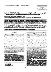
Supplementary Materials: Determination of Histamine in Silages Using Nanomaghemite Core (γ-Fe2O3)-Titanium Dioxide Shell Nanoparticles Off-Line Coupled with Ion Exchange Chromatography Natalia Cernei, Zuzana Lackova, Roman Guran, David Hynek, Jiri Skladanka, Pavel Horky, Ondrej Zitka and Vojtech Adam 1. X-ray Diffraction Measurement X-ray diffraction analyses were performed using SmartLab diffractometer Rigaku (Tokyo, Japan) with copper K alpha X-ray source (wavelength of 1.5406 Å). Standard powder diffraction measurements in Bragg-Brentano configuration were done. 2. X-ray Photoelectron Spectroscopy Measurement The XPS measurements were done on the ESCALAB 250Xi Thermo Fisher Scientific (Waltham, MA, USA). An X-ray beam with power of 200 W (650 μm spot size) was used. The survey spectra were acquired with pass energy of 50 eV and resolution of 1 eV. High-resolution scans were acquired with pass energy of 20 eV and resolution of 0.1 eV. In order to compensate the charges on the surface, electron flood gun was used. Spectra were referenced to the hydrocarbon type C 1s component set at a binding energy of 284.8 eV. The spectra calibration, processing and fitting routines were done using Avantage software (Thermo Fisher Scientific, Waltham, MA, USA). 3. Figures S1–S4
Zero peak
120000
Fe
Intentsity (Counts)
160000
Ti
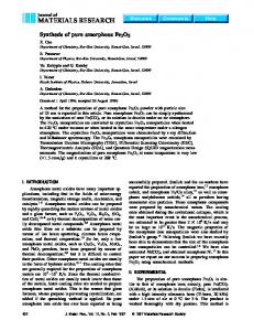
The prepared nanomaghemite core (γ-Fe2O3)-titanium dioxide shell nanoparticles were analyzed and characterized by X-ray fluorescence (Figure S1), X-ray photoelectron spectroscopy (Figures S2 and S3) and X-ray diffraction (Figure S4).
80000
40000
0 0
500
Energy (keV) Figure S1. Characterization of MAN18 using XRF.
Int. J. Environ. Res. Public Health 2016, 13, 904; doi:10.3390/ijerph13090904
Figure S2. XPS survey scan of MAN18.
Figure S3. XPS narrow scan of Fe2p of MAN18.
S2 of S3
Int. J. Environ. Res. Public Health 2016, 13, 904; doi:10.3390/ijerph13090904