Vet. Res. 31 (2000) 277–296 © INRA, EDP Sciences
277
Review article
Porcine antimicrobial peptides: New prospects for ancient molecules of host defense Guolong ZHANG, Christopher R. ROSS, Frank BLECHA* Department of Anatomy and Physiology, Kansas State University, 1600 Denison Avenue, Veterinary Medical Sciences Bldg. 228, Manhattan, KS 66506, USA (Received 15 November 1999; accepted 23 December 1999)
Abstract – Antimicrobial peptides (AMPs) are small, endogenous, polycationic molecules that constitute a ubiquitous and significant component of innate immunity. These natural antibiotics have broad microbicidal activity against various bacteria, fungi, and enveloped viruses. Because most AMPs kill bacteria by physical disruption of cell membranes, which may prevent microorganisms from developing resistance against these agents, they are being explored as possible alternatives to conventional antibiotics. Pigs, like many other mammals, produce an impressive array of AMPs, which are synthesized predominantly by host leukocytic phagocytes or mucosal epithelial cells. Currently, more than a dozen distinct porcine AMPs have been identified and a majority belongs to the cathelicidin family. This review briefly summarizes recent advances in porcine AMP research with an emphasis on the diverse biological functions of each peptide. Mechanisms of action of these AMPs and their role in the resistance to infections are considered. Finally, the current status of pharmaceutical and agricultural uses of AMPs as well as future prospects for their application in the food animal industry is discussed. pig / antimicrobial peptide / cathelicidin / defensin / innate immunity Résumé – Peptides antimicrobiens porcins : nouvelles perspectives pour d’anciennes molécules de défense de l’hôte. Les peptides antimicrobiens sont de petites molécules endogènes, polycationiques, qui représentent un élément important et ubiquitaire des défenses immunes naturelles. Ces antibiotiques naturels ont un effet antimicrobien à large spectre, à la fois contre des bactéries, des moisissures et des virus enveloppés. Comme la plupart de ces peptides antimicrobiens tuent les bactéries par altération physique de leur membrane, ce qui peut éviter le développement de souches microbiennes résistantes à ces agents, ils représentent des alternatives possibles à l'emploi des antibiotiques classiques. Le porc, comme de nombreux autres mammifères, produit une gamme importante de peptides antimicrobiens, synthétisés pour l'essentiel par les leucocytes à activité phagocytaire, ou par les cellules épithéliales des muqueuses. Il y a actuellement plus d'une douzaine de peptides antimicrobiens identifiés chez le porc, dont la majorité fait partie de la famille des cathélicidines. Cet article résume les progrès récents accomplis dans le domaine des peptides antimicrobiens du porc,
* Correspondence and reprints Tel.: (1) 785 532 4537; fax: (1) 785 532 4557; e-mail:
[email protected]
278
G. Zhang et al.
en insistant particulièrement sur les effets biologiques variés de chaque peptide, sur leurs mécanismes d'action et leur rôle anti-infectieux. Enfin, cette revue discute de l'état actuel des utilisations pharmaceutiques et agronomiques de ces peptides antimicrobiens, ainsi que des perpectives de leur utilisation pour l’élevage industriel. porc / peptide antimicrobien / cathélicidine / défensine / immunité naturelle
Table of contents 1. Introduction ............................................................................................................................. 278 2. Arsenal of porcine AMPs........................................................................................................ 279 2.1. Defensins, a superfamily of conserved cysteine-rich AMPs.......................................... 280 2.2. Cathelicidins, a family of AMP precursors with a common prosequence ..................... 283 2.2.1. PR-39, a multifunctional proline-arginine-rich peptide...................................... 285 2.2.2. Protegrins, a group of compact AMPs with broad-spectrum microbicidal activity 285 2.2.3. Prophenins, proline-phenylalanine-rich AMPs with repeated decamers ............ 286 2.2.4. PMAP-23, -36, and –37, three novel porcine cathelicidins ................................ 286 2.3. NK-lysin, a new effector molecule of cytotoxic T and NK cells ................................... 287 2.4. Cecropin P1, a mammalian homolog of insect cecropins .............................................. 287 3. Synergistic interactions among porcine AMPs ....................................................................... 288 4. Mechanisms of action of AMPs .............................................................................................. 288 5. Role of AMPs in natural resistance to infections.................................................................... 289 6. Prospects of AMPs for the food animal industry .................................................................... 290
1. INTRODUCTION The discovery and development of antibiotics have led to a dramatic improvement in the ability to treat infectious diseases and significant increases in food animal production during the past five decades and are among the major advances of the 20th century. In the food animal industry, the use of antibiotics as growth enhancers has been a common practice for more than 30 years and is estimated to constitute more than half of the total antimicrobial use worldwide [123]. Unfortunately, widespread and sometimes indiscriminate use of antibiotics has been accompanied by the emergence of microorganisms that are resistant to these agents. Antibiotic-resistant bacteria have been posing increasingly serious concerns to the public, health specialists, and food animal producers [53, 75, 123]. To overcome antibiotic resistance and to retain consumer confidence in a safe food supply, health spe-
cialists and food animal producers are searching for alternative, yet effective, means of preventing and treating emerging and re-emerging diseases. Antimicrobial peptides (AMPs, also known as peptide antibiotics or natural antibiotics) constitute a ubiquitous and broadly effective component of innate immunity of hosts [15, 16, 34, 66]. Since the first discoveries of plant thionins in 1972 [28] and insect cecropins in 1981 [108], nearly 400 AMPs have been identified in bacteria, plants, invertebrates, and vertebrates [16, 37, 40, 97]. They appear to be an ancient but effective component of host defense and are being explored as possible alternatives to conventional antibiotics [31, 38, 47–49, 60, 80, 124]. Unlike conventional antibiotics, which are synthesized enzymatically by microorganisms, each AMP is encoded by a distinct gene and made from an mRNA template. Most AMPs appear to kill bacteria by a
Porcine antimicrobial peptides
common mechanism, which involves direct electrostatic interaction with negatively charged microbial cell membranes, followed by physical disruption [9, 15, 84]. In contrast, most traditional antibiotics act by inhibiting enzymes involved in synthesis of bacterial cell walls, proteins, or DNA and usually have a single or limited type of target molecule(s), which can be mutated easily by bacteria to gain resistance [49, 60]. Because specific receptors are not involved, AMPs are capable of killing a broad spectrum of microorganisms without development of resistance [47–49, 60]. All AMPs share common features, such as small size (12-100 amino acid residues), polycationic charge, and amphipathic structure [9, 15, 47]. Based on structural similarities, they can be classified into two broad groups, i.e., linear and cyclic peptides. The first group includes linear peptides with amphipathic α-helical structures or extended helices with a high proportion of certain residues, and the second group consists of peptides containing one or more disulfide bridges with loop or β-sheet structures [9, 15, 16, 47]. Many cells in the immune system or on mucosal surfaces have the potential to produce AMPs and protect hosts against pathogen invasion. Granules of polymorphonuclear neutrophils (PMNs), macrophages, eosinophils, T lymphocytes, and natural killer (NK) cells are equipped with an impressive array of AMPs [36, 42, 66, 70]. Upon cell activation and degranulation, these granule-associated peptides are either fused intracellularly with pathogencontaining vacuoles or secreted extracelluarly and exert their effects through nonoxidative killing mechanisms [25]. Interestingly, mucosal epithelial cells, which do not have granules, also express and secrete AMPs [24, 39]. More and more peptides probably will be identified from additional cell types and added to the AMP superfamily. Although much of the early work on mammalian AMPs was conducted in pigs, investigations in other species have been
279
extensive, and readers are referred to several in-depth reviews for comparative information [37, 40, 55, 56, 66]. This review summarizes recent advances in AMP research in pigs, with an emphasis on the diverse biological functions of each porcine AMP. Mechanisms of action of these AMPs and their role in the resistance to infections are considered. Finally, the current status of pharmaceutical and agricultural uses of AMPs as a new generation of antibiotics as well as future prospects for their application in the food animal industry is highlighted.
2. ARSENAL OF PORCINE AMPS More than a dozen distinct AMPs have been identified in pigs. All of these peptides adopt diverse spatial structures, are relatively small with a molecular weight of less than 10 kDa, but are broadly effective against various species of microorganisms (Tab. I). Either they were isolated as mature peptides from PMNs, lymphocytes, and the small intestine, or their amino acid sequences were deduced from cDNA or gene sequences. Cecropin P1 was the first porcine AMP isolated from the upper part of the small intestine by Boman’s group in 1989 [64]. Another proline-arginine-rich 39-amino acid peptide, PR-39, also was purified by this group from the small intestine 2 years later [1]. Protegrins 1 to 5 constitute a group of broad-spectrum AMPs of pig myeloid origin identified by Lehrer’s group [61, 136, 137]. That group also isolated two proline-phenylalanine-rich AMPs, prophenin-1 and -2, from porcine neutrophils [50]. Three porcine myeloid antimicrobial peptides, PMAP-23, -36, and -37, were identified by cDNA cloning [113, 120, 130]. A novel AMP, termed NK-lysin, was isolated from porcine small intestine and has been shown to be a new effector molecule of cytotoxic T and NK cells [5]. Recently, a porcine β-defensin, pBD-1 was cloned and found to be expressed throughout
280
G. Zhang et al.
Table I. Summary and characteristics of porcine cationic antimicrobial peptides.
epithelia of the respiratory and gastrointestinal tracts [134]. All of these AMPs, except cecropin P1 and NK-lysin, belong to either the defensin or the cathelicidin family, which are the two major groups of AMPs found in most mammalian species [37-39, 66]. Cathelicidin-related AMPs represent the majority of those identified in pigs so far. Although α-defensins are the most abundant AMPs in granules of PMNs or intestinal paneth cells in many mammalian species [36, 67, 85], they have not been found in pigs. 2.1. Defensins, a superfamily of conserved cysteine-rich AMPs Defensins constitute a large family of endogenous cysteine-rich peptide antibi-
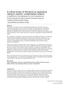
otics with broad-spectrum activity against various bacteria, fungi, and enveloped viruses [35, 67, 85]. In addition to their microbicidal activity, defensins also are chemotactic for monocytes, T lymphocytes, and dendritic cells [22, 118, 126]; inhibit the binding of ACTH to its receptors (hence the name “corticostatins”) [119]; suppress the activation of the classical pathway of complement [121]; induce histamine release from mast cells [14]; and promote the binding of lipoprotein(a) to the vascular matrix [13, 54]. All defensins are polycationic peptides of 3–5 kDa and are characterized by the presence of six or eight conserved cysteine residues forming three or four intramolecular disulfide bridges. Five families of defensins have been reported in eukaryotes ranging from plants, insects, and mammals [18, 24, 55, 57, 65, 116] (Fig. 1).
Porcine antimicrobial peptides
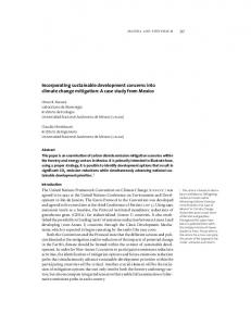
Figure 1. Conserved disulfide bridge patterns of mammalian α- and β-defensins, insect defensins, and plant defensins. Each group is characterized by the number, spacing, and linkage of cysteine (C) residues. Each dash represents an amino acid residue. Rhesus θ-defensin-1 represents a third group of mammalian defensins, and its unique cyclic structure is formed by the head-to-tail ligation of two α-defensin-like propeptides [116]. Therefore, it can be considered a subgroup of α-defensins.
Based on the positions of cysteine residues and linkages of the disulfide bridges, mammalian defensins are divided further into three groups: α-, β-, and θ-defensins. Rhesus θ-defensin-1 currently is the only θ-defensin and was isolated recently from leukocytes of rhesus monkeys [116]. However, it also can be considered a subgroup of α-defensin, because it is formed by the headto-tail ligation of two α-defensin-like propeptides via a posttranscriptional processing pathway [116]. Despite a lack of similarity in amino acid sequences, the threedimensional structures of all eukaryotic defensins are rather similar, consisting of two or three antiparallel β-sheets with or without an α-helix [57], except that rhesus θ-defensin-1 adopts a cyclic structure [116]. Furthermore, all mammalian α- and β-defensin genes are clustered in close proximity on the same chromosome, as demonstrated in humans, mice, cattle, and sheep [65]. Conserved structures and homologous chromosomal locations point to a common ancestry of these defensins and probably the system of innate immunity [57]. Mammalian defensins are synthesized by either bone marrow myeloid cells or mucosal epithelial cells as prepro-peptides that are composed of a signal sequence, a
281
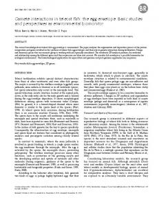
prosequence, and a mature biologically active peptide [35, 67]. Upon microbial invasion, mature active defensins are released quickly by proteolytic processing from precursor peptides. All α-defensins are expressed in granule-containing granulocytic leukocytes or intestinal paneth cells, whereas most β-defensins are synthesized by epithelial cells lining the respiratory, gastroenteric, and urogenital tracts, which do not contain storage granules [24, 65]. Therefore, α-defensins are considered traditionally as important mediators involved in systemic host defense, whereas β-defensins may be involved more in mucosal immunity. However, cattle are the only species examined to date, in which β-defensins are expressed abundantly in both PMNs and macrophages in addition to mucosal epithelial cells [96, 100]. Based on gene structure, length and homology of amino acid sequences, and sites of expression, β-defensins are classified further into two subgroups [24, 135] (Fig. 2). The first group contains precursor peptides of 61-65 amino acid residues, and each has a compact gene with a shorter intron of less than 2 kb (Fig. 3). Many are expressed abundantly in oral and airway epithelia. The second group contains precursor peptides that are generally 3-10 amino acids longer, each with a relatively large gene containing an intron of greater than 6.5 kb. Besides the epithelia of respiratory and digestive tracts, kidney and genitourinary epithelia appear to be the major expression sites. Another striking difference between these two groups is that expression of most β-defensin genes in the first group is induced upon exposure to inflammatory and infectious agents, whereas those in the second group have a constitutive expression pattern. Examination of the promoter sequences of genes for inducible β-defensins revealed the presence of several consensus binding sites for nuclear factor-κB (NF-κB) and NF-interleukin (IL)-6, which may explain their inducibility upon inflammation and infection [24].
282
G. Zhang et al.
Figure 2. Classification of mammalian β-defensins. Based on length and homology of amino acid sequences, gene structure, and sites of expression, β-defensins are classified into two groups. Group 1 contains precursor peptides of 61-65 amino acid residues, and each has a compact gene with a shorter intron of less than 2 kb. Group 2 contains precursor peptides that are generally 3-10 amino acids longer, each with a relatively large gene containing an intron of greater than 6.5 kb. Abbreviations: BD, β-defensin; TAP, tracheal antimicrobial peptide; LAP, lingual antimicrobial peptide; EBD, enteric β-defensin; BNBD, bovine neutrophil β-defensin; p, porcine; h, human; s, sheep; r, rat; m, mouse; GAL, Gallinacin; THP, turkey heterophil peptide. (Modified from Ref. [135].)
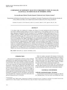
pBD-1, a porcine form of epithelial β-defensin Recently, we cloned the full-length cDNA for pBD-1, which is the only member of the defensin family identified in pigs thus far [134]. The pBD-1 mRNA is expressed abundantly in tongue epithelia and to a lesser extent throughout the respiratory and digestive tracts. The pBD-1 gene spans approximately 1.9 kb and, like its congeners in other mammals, consists of two short exons separated by a 1.5-kb intron [135] (Fig. 3). Exon 1 encodes the 5'-untranslated region (UTR) and signal sequence of the 64-amino acid prepro-pBD-1, and exon 2 encodes the prosequence, mature peptide, and the 3'-UTR. Despite its resemblance to many inducible β-defensins in amino acid sequence, gene structure, and sites of expres-
sion, the pBD-1 gene failed to upregulate in response to both in vitro stimulation of tongue epithelial cells with lipopolysaccharide (LPS), tumor necrosis factor (TNF)-α, or IL-1β and in vivo infection of pigs with Salmonella typhimurium or Actinobacillus pleuropneumoniae [135]. In addition, direct transfection of the pBD-1 gene promoter into mouse embryonic fibroblast NIH/3T3 cells showed no difference in reporter gene activity upon stimulations with LPS and IL-1β [135]. Thus, pBD-1 appears to be the only β-defensin that can be classified structurally into the inducible group but exhibits a constitutive expression pattern. The constant expression of pBD-1 in airway and oral mucosa, which also is consistent with a lack of consensus binding sites for NF-κB or NF-IL-6 in its promoter region, suggests that it may play a surveillance role in
Porcine antimicrobial peptides
283
Figure 3. Structural organization of β-defensin genes and a list of encoded precursor peptides. Each β-defensin gene contains two short exons interrupted by a large intron. The encoded prepro-peptide is composed of a signal sequence, a proregion, and a mature peptide with antibacterial activity. Exon 1 encodes the 5'-untranslated region (UTR) and signal sequence, and exon 2 encodes the prosequence, mature peptide, and the 3'-UTR. The map is drawn to scale. Abbreviations: BD, β-defensin; TAP, tracheal antimicrobial peptide; LAP, lingual antimicrobial peptide; EBD, enteric β-defensin; BNBD, bovine neutrophil β-defensin; p, porcine; h, human; s, sheep; g, goat; m, mouse; r, rat; GAL, Gallinacin; THP, turkey heterophil peptide.
maintaining the steady state of microflora on mucosal surfaces. Fluorescence in situ hybridization mapped the pBD-1 gene to porcine chromosome 15q14-q15.1 within a region of conserved synteny to the chromosomal locations of human α- and β-defensins, further supporting the notion that defensins are highly conserved, innate, defense molecules with a common ancestry [135]. Consistent with its abundant expression of transcripts in tongue epithelial cells, the pBD-1 peptide was immunolocalized to the cornified mature epithelial cells in filiform papillae (but not in fungiform papillae) of the dorsal tongue [105]. Recombinant pBD-1 peptide has potent antibacterial activity against both gram-positive and -negative bacteria as well as fungi, including Escherichia coli, S. typhimurium, Listeria monocytogenes, and Candida albicans [105]. Killing of microbes by pBD-1, how-
ever, is pH, salt, and serum dependent; either low pH (5.5), high salt (100-150 mM NaCl), or serum inactivates its microbicidal activity [105], as they do for other defensins [35, 65].
2.2. Cathelicidins, a family of AMP precursors with a common prosequence Cathelicidins constitute a group of AMPs sharing a conserved N-terminal prosequence followed by highly heterogeneous 12-79amino acid C-terminal mature peptides [131, 132] (Fig. 4). The C-terminal peptides of cathelicidins in various mammalian species have extremely diverse amino acid sequences and subsequent spacial structures ranging from an α-helix to a β-sheet. They are named cathelicidins for the high homology of their prosequences to cathelin,
284
G. Zhang et al.
a 96-amino acid polypeptide originally purified from porcine PMNs [94]. These peptides are synthesized as prepro-peptides by bone marrow myeloid cells, then constitutively stored in peripheral PMN granules as propeptides, from which mature active peptides are cleaved by endogenous elastase upon PMN activation and degranulation [88, 131, 132]. In some cases, the mature molecules are modified further by C-terminal amidation. Porcine cathelicidins include PR-39; protegrins 1-5; prophenins 1-2; and PMAP-23, -36, and -37. They all are derived from bone marrow myeloid cells and constitutively stored as pro-peptides in peripheral PMN granules, where few or no transcripts are expressed [131, 132]. LL-37/hCAP-18, the only cathelicidin found in humans, appears to be the sole exception; it also is synthesized inducibly by skin keratinocytes and airway epithelial cells [11, 30].
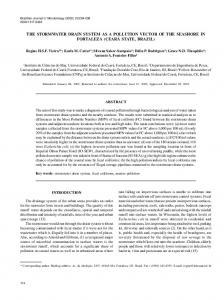
Gene structures for cathelicidins, as exemplified by PR-39, protegrins, and prophenins, are all compact and organized in the same manner with four exons and three introns [46, 137, 138] (Fig. 4). Exons 1-3 encode the prepro-sequence, and exon 4 encodes several final residues of the prosequence followed by the mature peptide sequence. Promoter regions of cathelicidin genes are conserved and contain several binding sites for NF-κB, NF-IL-6, and acute phase response factor, suggesting that cytokines generated early in infections may upregulate cathelicidin gene expression, similar to inducible β-defensins. All porcine cathelicidin genes are clustered densely on chromosome 13 [137, 138]. Their homology and nearby chromosomal locations indicate that this family may have evolved through gene duplications [138]. To date, nearly 30 cathelicidins have been identified in at least eight mammalian species, includ-
Figure 4. Structural organization of cathelicidin genes and a list of encoded precursor peptides. Each cathelicidin gene contains four exons separated by three introns. The prepro-peptide is composed of a conserved signal sequence and a proregion, followed by highly heterogeneous mature peptide with antibacterial activity. Exons 1-3 encode the 5'-untranslated region (UTR) and the prepro-sequence, and exon 4 encodes several final residues of the proregion and the mature peptide sequence as well as 3'-UTR. The map is drawn to scale. Abbreviations: PR-39, proline-arginine-rich 39-amino acid peptide; PG, protegrin; PF, prophenin; PMAP, BMAP, SMAP, porcine, bovine, sheep myeloid antimicrobial peptide; Bac, bactenecin; CAP, cationic antimicrobial peptide; LL-37, leucine-leucine 37-amino acid peptide; eCATH, equine cathelicidin; CRAMP, cathelin-related antimicrobial peptide.
Porcine antimicrobial peptides
ing humans, pigs, cattle, sheep, rabbits, mice, guinea pigs, and horses, either by cDNA cloning of bone marrow cells or by direct purification from peripheral PMNs [33, 82, 99, 131, 132]. 2.2.1. PR-39, a multifunctional proline-arginine-rich peptide PR-39 is a linear cathelicidin of 39 amino acid residues with high contents of proline (49%) and arginine (26%) adopting a polyproline type II structure [1, 19]. It was isolated originally from bulk homogenates of porcine small intestines [1], but later cloning of PR-39 cDNA in myeloid cells from porcine bone marrow suggested that the enteric PR-39 may be derived from resident leukocytes in the intestine rather than from intestinal epithelia [112]. Indeed, the PR39 peptide has been purified from porcine PMNs [102], but PR-39 mRNA could not be detected by reverse-transcriptase-polymerase chain reaction in small intestines of pigs at any age [125]. PR-39 is active mainly against gram-negative bacteria [1, 103] and increases significantly in sera of pigs during the onset of salmonellosis [133], further demonstrating the in vivo involvement of this peptide in host defense. In addition to its antibacterial activity, PR-39 has several other important functions. It is a specific PMN chemoattractant [58], accumulates in wound fluid and induces the expression of syndecan-1 and -4, which are important heparan sulfate proteoglycans on cell surfaces involved in wound repair [32]. It also is capable of suppressing invasive and mobile activities of human hepatocellular carcinoma cells [83]. More strikingly, PR-39 potently inhibits the assembly of the phagocyte NADPH oxidase complex by binding to Src homology 3 (SH3) domains of p47phox, thereby limiting the production of reactive oxygen species (ROS) [104]. Consistent with its function as a potent NADPH oxidase inhibitor, PR-39 has been shown to block ischemia- and high K +induced ROS production in isolated per-
285
fused rat lungs [4]. In vivo studies showed that a single intravenous injection of PR-39 completely abolished postischemic ROS production, neutrophil adhesion, and transvascular emigration in rat mesenteric venules subjected to ischemia-reperfusion [62]. Furthermore, pretreatment with PR-39 significantly increased the survival rate and abrogated the liver injury of galactosamine-sensitized mice following a potentially lethal endotoxic shock (C.R. Ross and F. Blecha, unpublished results). These findings suggest that PR-39 may be therapeutically useful as a potent anti-inflammatory drug to prevent neutrophil adhesion and activation as well as excessive tissue injury during postischemic and other inflammatory responses. Nevertheless, the prevailing function of PR-39 in vivo remains unclear. However, we can speculate that all of the above activities may be tightly integrated and finely tuned in pigs during injury, infection, and wound healing. 2.2.2. Protegrins, a group of compact AMPs with broad-spectrum microbicidal activity Protegrins constitute a group of small AMPs that contain 16-18 amino acid residues with four cysteines forming two disulfide bridges (C1-C2 and C3-C4), which stabilize an antiparallel β-sheet structure [10, 51]. Among five known congeners, protegrins 1-3 have been purified from porcine PMNs [61], and the other two, protegrins 4-5, were derived from a bone marrow cDNA and a gene sequence, respectively [136, 137]. Protegrins exhibit potent, broadspectrum, microbicidal activity against various gram-positive and -negative bacteria, mycobacteria, fungi, and enveloped viruses, including several sexually transmitted human pathogens, Chlamydia trachomatis, Neisseria gonorrhoeae, and Haemophilus ducreyi; periodontopathic bacteria, Actinobacillus actinomycetemcomitans, Capnocytophaga spp., Porphyromonas gingivalis, Prevotella intermedia, and
286
G. Zhang et al.
Fusobacterium nucleatum; and HIV type I virions [23, 79, 80, 91, 115]. In contrast to defensins, antimicrobial activities of protegrins are maintained at physiological NaCl concentrations and are not inhibited by extracellular cations or serum components [52]. Intramolecular disulfide bridges for maintaining the antiparallel β-sheet structure are believed to be required for the peptides to form pores in the membrane, as demonstrated in Xenopus laevis oocytes [76]. In fact, the antimicrobial activity of a linearized protegrin variant is decreased remarkably [52]. The minimal structure of protegrin-1 for activity against N. gonorrhoeae and C. trachomatis is defined within the central region of the molecule (residues 5-16), and optimal activity requires both intramolecular disulfide bridges [52, 76, 92, 128]. The small size, stability, and overall effectiveness, combined with a substantial lack of cytotoxicity to host cells, make protegrins promising topical therapeutics to prevent and treat human sexually transmitted diseases and periodontal infections. 2.2.3. Prophenins, prolinephenylalanine-rich AMPs with repeated decamers Prophenin-1 and -2 are two other members of the cathelicidin family purified from porcine leukocytes [50]. They contain 79 amino acid residues with an unusually high content of proline (53.2%) and considerable amounts of phenylalanine (19%) and arginine (7.6%). Their precursors were identified independently as c6 and c12, two cDNA clones isolated from a porcine bone marrow cDNA library [114]. The primary structures of prophenins are remarkable for the six nearly perfect tandem repeats of a proline-rich decamer, FPPPNFPGPR. Under low ionic strength conditions (10 mM PBS), prophenin-1 is effective against gram-negative bacteria but not against gram-positive. The antibacterial activity of prophenin-1 is abolished under physiological NaCl condi-
tions (10 mM PBS + 0.1 M NaCl) [50]. The repeated decamer and the hydrophobic tail exhibit no activity against E. coli even under low ionic conditions, leaving their potential functions unknown. If the prosequence were cleaved after the valine, which is the common cleavage site, as in the case of cathelin and several other cathelicidins [90], prophenin precursors would be processed into a 97-residue mature peptide. However, the actual mature products are prophenins-1 and -2, which contain only the C-terminal 79 residues. Surprisingly, a 13-amino acid peptide between the common cleavage site and the N-terminus of prophenin-1, termed tritrpticin, has been synthesized and shows strong antibacterial activity against both gram-negative and positive bacteria [63]. However, whether this 13-residue peptide exists in nature remains to be determined. If it does exist, it presents the intriguing possibility that a precursor of cathelicidin can be processed into two functionally active mature AMPs. 2.2.4. PMAP-23, -36, and -37, three novel porcine cathelicidins Identification of porcine bone marrow cDNAs that encode peptides with cathelinlike pro-sequence places three additional members into the rapidly expanding family of cathelicidins. They are porcine myeloid antimicrobial peptides (PMAP) of 23, 36, and 37 amino acid residues, i.e., PMAP-23, -36, and -37 [113, 120, 130]. Structure prediction analysis and circular dichroism spectra suggest that PMAP-36, and -37 adopt an amphipathic α-helix, whereas PMAP-23 assumes a hairpin-like structure (an antiparallel β-sheet connected by a loop at center). Sequence comparison reveals that PMAP-23 shows no significant similarity to any known AMPs, whereas PMAP-36 (1-20) has a moderate homology of 35% to a rabbit cathelicidin CAP18. Although PMAP-37 shows negligible identity to porcine cecropin P1, it has more than 50% similarity to two insect AMPs, cecropins A and B. However,
Porcine antimicrobial peptides
no evolutionary relationship seems to exist between them, because they are generated from totally different precursors. Although none of these three peptides has been purified from natural sources, synthetic peptides exhibit remarkable in vitro antibacterial activity against gram-negative and -positive bacteria. PMAP-37 is the most potent membrane-active agent and causes the permeabilization of the bacterial inner membrane at 0.2-1 µM, whereas PMAP-23 and PMAP-36 kill bacteria at 1-10 µM and 10-50 µM, respectively. However, PMAP-37 shows hemolytic activity to human erythrocytes at 10-50 µM, whereas no lysis occurs with PMAP-23 or PMAP-36 at concentrations up to 100 µM [113, 120, 130]. 2.3. NK-lysin, a new effector molecule of cytotoxic T and NK cells NK-lysin is a 78 amino acid cationic AMP that recently has been purified from the porcine small intestine based on its antibacterial activity and has been shown to be produced by cytotoxic T and NK cells [5]. A corresponding cDNA clone also has been identified in a porcine bone marrow cDNA library [5]. NK-lysin contains six cysteines that form three intramolecular disulfide bridges (C1-C6, C2-C5, C3-C4). The three-dimensional structure of NK-lysin consists of five amphipathic α-helices folded into a single globular domain with a hydrophobic core and a hydrophilic surface [74]. Its amino acid sequence exhibits 43% identity and 67% similarity to granulysin, a protein present in cytotoxic granules of activated human T and NK cells [109]. Like granulysin, NK-lysin is an additional component of the cytotoxic arsenal of activated lymphocytes in addition to perforin and FasFas ligand-mediated apoptosis [89, 110]. Comparison of NK-lysin’s structure with those of saposin-like proteins, including saposins, surfactant-associated protein B, plant aspartic proteinases, and amoebapores
287
(protozoan pore-forming AMPs), reveals 18-27% identities, including positions of six conserved cysteines, disulfide bridging patterns, and secondary structures [6, 68]. Thus, placement of NK-lysin in the saposinlike protein family, all members of which appear to interact with lipids, has been proposed [6]. Indeed, NK-lysin renders lipid bilayers permeable in a nonspecific manner [95]. Furthermore, it can directly bind the lipid A portion of lipopolysaccharide and protects galactosamine-sensitized mice from lethal endotoxic shock [8]. It also shows activity against various bacteria and fungi, including E. coli, Bacillus megaterium, Acinetobacter calcoaceticus, Streptococcus pyogeneis, and C. albicans [5]. It lyses certain tumor cells, but not erythrocytes [5]. Antimicrobial and tumorolytic activities of NK-lysin, like those of other saposin-like proteins, are believed to arise from its interaction with lipids and ability to form pores in the cell membrane because of its α-helical structure. Intact disulfide bridges are required to maintain its activity, and the peptide is inactivated when reduced by thioredoxin reductase, which is present on PMN cytoplasmic membranes [7]. Unexpectedly, NK-lysin also has been found to stimulate potent insulin secretion from rat pancreatic islets and to exert its direct effect on β-cells, but the stimulatory activity was independent of the concomitant changes in cytosolic free Ca2+ concentration [129].
2.4. Cecropin P1, a mammalian homolog of insect cecropins Cecropin P1 was the first porcine AMP isolated, and its name reflects the identity (33%) to its insect congeners [64]. Although most of the features typical of insect cecropins are conserved in cecropin P1, some slight differences in structure do exist. For example, cecropin P1 has a continuous amphipathic α-helical structure over its entire length, which is different from the helix-hinge-helix structure of its insect
288
G. Zhang et al.
homologs [106]. Porcine cecropin is amidated at the C-terminus, which is not true for insect homologs. Cecropin P1 shows much more potent activity against gramnegative bacteria than gram-positives, and amidation does not affect its antibacterial activity [64, 122]. However, neither the gene nor cell types that are responsible for the production of this peptide have been identified. Merrifield et al. [122] compared the antibacterial activities of the D-enantiomer and the retro isomer of cecropin P1 and demonstrated that chirality (L/D enantiomer) had no marked effect on the antibacterial activity of the peptide, suggesting that no stereo-specific protein receptors were involved. However, the primary sequence is the determining factor, because the reverse sequence was inactive against the bacteria tested. Cecropin P1 is also capable of uncoupling oxidative phosphorylation in the mitochondria, but this activity is not correlated with its antibacterial activity [59].
3. SYNERGISTIC INTERACTIONS AMONG PORCINE AMPS Because PMN degranulation results in the release of a vast array of AMPs as well as reactive oxygen and nitrogen intermediates, it is not surprising that these neutrophil granule-derived substances work in concert under in vivo conditions. Synergistic interactions appear to be beneficial for the host’s efficient use of these weapons to fight microbial invasions. Synergism has been shown for human defensins with each other or with ROS and for rabbit bactericidal/permeability-increasing protein with p15s or defensins [26, 71-73]. However, little is known about how porcine PMN-derived protegrins, prophenins, PR-39, and/or ROS work in concert or how enteric NK-lysin, PR-39 and cecropin P1 affect each other on the porcine intestinal surface. Antagonism also has been found in some cases. For example, PR-39 inhibits the production of ROS by inhibiting
the proper assembly and activation of phagocyte NADPH oxidase [104], and NK-lysin is inactivated by the PMN cytoplasmic membrane-bound thioredoxin reductase [7]. We recently demonstrated that porcine epithelial-derived pBD-1 is remarkably synergistic with neutrophil-derived peptide antibiotics, PR-39 and protegrin 3, in killing bacteria even at physiological NaCl concentrations, which otherwise would render pBD-1 totally inactive [105]. This scenario may occur in vivo and be functionally significant, because PMNs will be recruited to mucosal epithelial surfaces and interact with local epithelial cells under inflammatory conditions. In some cases, serum components and extracellular concentrations of cations and NaCl also influence the microbicidal activities of AMPs. Human α- and β-defensins are inactivated by serum components and higher NaCl concentrations [86, 87], which also is the case for porcine pBD-1 [105]. However, protegrins maintain their activities in the presence of cations, serum or at physiological NaCl concentrations in vitro [52]. Therefore, caution must be used when an in vitro activity is assigned to an individual AMP.
4. MECHANISMS OF ACTION OF AMPS The exact killing mechanisms of various microorganisms by AMPs with diverse structures are not understood clearly. However, peptide-lipid interactions leading to membrane permeabilization, rather than receptor-mediated recognition processes, apparently are a common mechanism of their lytic action [9, 15, 84]. Cytoplasmic membranes of cells are the targets in most cases. The cationic property of AMPs assists in electrostatic interactions with negatively charged phospholipids on target cell membranes. Membrane permeabilization by amphipathic peptides with α-helical or β-sheet structures can proceed through either
Porcine antimicrobial peptides
of the following two mechanisms: transmembrane pore formation via a “barrelstave” mechanism and membrane disruption and solubilization via a “carpet-like” mechanism [84]. In the barrel-stave model, the peptides tend to form bundles and penetrate membranes, thereby forming voltagedependent transmembrane pores or channels, in which hydrophobic surfaces of peptides interact with the lipid core of the target membrane and hydrophilic surfaces point inward. In the carpet-like model, the peptides bind the membrane with their hydrophobic surfaces facing the membrane and their hydrophilic surfaces facing the solvent. When a threshold concentration of peptide monomers is reached, the membrane is solubilized into pieces and transient pores are formed. Such pores are different from those in the barrel-stave model in that the lipid bends back on itself. AMPs with linear α-helical structures, including cecropin P1 and probably PMAP-23, -36, and -37, exert their antimicrobial activity via the carpetlike mechanism, whereas cyclic peptides with β-sheet structures, such as pBD-1, protegrins, and NK-lysin may utilize the barrel-stave mechanism as demonstrated with insect cecropins, neutrophil α-defensins, and protegrins [35, 41, 67]. PR-39 is a notable exception to the mechanisms described above in that it does not lyse bacteria directly but seems to induce the degradation of proteins required for bacteria DNA replication [17]. It requires a lag time of 8-10 min to penetrate the outer membrane of E. coli, then kills bacteria without lysis [17]. Consistent with this, a recent study found that PR-39 rapidly penetrates into cells without permeabilizing the plasma membrane and binds a number of SH3-containing cytoplasmic proteins, including Lck, Src, p13k, and p130Cas, which are either protein kinases or adaptor molecules involved in diverse signaling pathways [21]. Perhaps PR-39 inhibits bacterial protein and DNA syntheses by binding to certain intracellular receptor(s). This also may explain the pleiotropic effects of PR-39 on mam-
289
malian cell behaviors. For instance, PR-39 exerts its anti-inflammatory function at least partly by binding to SH3 domain-containing p47phox, thereby interfering with the assembly of the NADPH oxidase enzyme complex [104]. All AMPs appear to have selectivity toward target cells, i.e., they lyse microbes vigorously but with little or no cytotoxicity to host cells. The unique high content of anionic phospholipids, the absence of cholesterol, and large transmembrane potentials across prokaryotic cell membranes explain the difference in their preference of killing prokaryotic over eukaryotic cells [15, 47, 48].
5. ROLE OF AMPS IN NATURAL RESISTANCE TO INFECTIONS AMPs constitute an ancient but not obsolete system of host defense. Although it is difficult to demonstrate the contribution of any single AMP to disease resistance because of the extreme complexity and redundancy of host-defense mechanisms, accumulating evidence suggests that AMPs are involved actively in inflammatory and infectious processes. The broad antimicrobial spectrum and strategic locations of AMPs in leukocytic phagocytes or mucosal epithelial cells provide the most obvious indirect argument for their participation in inflammation and infection. Several AMPs, particularly cathelicidins and defensins, have been found in blisters and wound fluid, some at antimicrobial concentrations [29, 101]. Increased levels of AMPs also are associated with animal or human infections [2, 133]. Prominent induction of genes for many epithelial β-defensins occurs during skin injuries and airway and enteric infections [98, 111, 117]. In addition, administration of AMPs to mice has been shown to provide significant protection against lethal endotoxic shock or experimental infections [3, 45, 107].
290
G. Zhang et al.
Moreover, transgenic animals and plants that overexpress AMPs show enhanced resistance to bacterial or fungal infections [20, 27, 93], and fruit flies deficient in the Toll-signaling pathway, which controls synthesis of the antifungal peptide drosomycin, exhibit dramatically reduced survival following a fungal infection [69]. The recurrent bacterial infections in the airways of cystic fibrosis (CF) patients were believed to be due to impairment of the antimicrobial activity of salt-sensitive β-defensins and cathelicidin LL-37/hCAP-18 by the highsalt airway surface fluid in CF patients [44]. However, recent evidence showed that NaCl concentrations in airways of both CF patients and normal controls are comparably low, leaving the functions of AMPs on the airway surface unclear [77, 78]. Nevertheless, other pathophysiological functions of AMPs provide evidence to support their important roles in infection and inflammation. For example, PR-39 limits tissue injury by diminishing excessive ROS production via inhibition of NADPH oxidase assembly and PMN recruitment [4, 104]. At the same time, PR-39 may aid in wound repair by inducing syndecan expression and presumably altering cell division characteristics [32].
6. PROSPECTS OF AMPS FOR THE FOOD ANIMAL INDUSTRY The use of antibiotics to treat infectious diseases in both human and veterinary medicine has been tremendously beneficial to societies worldwide. However, the efficacy of these agents has been compromised increasingly by the appearance of drug-resistant microbes. The widespread use of antibiotics as growth enhancers in the food animal industry is posing serious concerns to the public because of the potential development of resistant pathogens and the resultant public health risk [123]. Thus, new approaches to the problem of antimicrobial resistance and development of novel classes of antimi-
crobial agents with less likelihood to gain resistance are needed. AMPs show a low level of resistance development in vitro and other highly desirable properties, such as the ability to kill rapidly a broad spectrum of microorganisms including drug-resistant bacteria and often fungi, to protect animals against both topical and systemic infections, the capacity to neutralize endotoxin, and synergy with conventional antibiotics [47-49]. Moreover, AMPs demonstrate an equal activity against drug-resistant bacterial strains both in vivo and in vitro. For example, pBD-1 in combination with PR-39 or protegrin-3 caused a reduction of 4-5 log units CFU within 45 min of S. typhimurium Definitive Type 104 [105], which is resistant to at least five common antibiotics [43]. Protegrin-1 similarly decreased the CFU of either methicillin-resistant Staphylococcus aureus or Pseudomonas aeruginosa by more than 3 log units in less than 15 min [107]. Resistance to protegrin-1 did not develop after 18 serial passages of MRSA or 11 passages of P. aeruginosa under conditions of cultivation, which increased the minimal inhibitory concentration for norflozacin by a factor of 85 and 10, respectively. A single injection of protegrin-1 significantly protected mice from infections with antibioticresistant pathogens, including methicillinresistant S. aureus, P. aeruginosa, and vancomycin-resistant Enterococcus faecium [107]. Several AMPs and their synthetic homologs currently are being tested in clinical trials mainly to treat topical infections [47-49, 80, 124]. Some of these peptides undoubtedly will be approved in the near future as new antimicrobials, particularly to treat drug-resistant pathogens. However, use of chromatographically purified, chemically or recombinantly synthetic AMPs in the food animal industry may be limited by their cost of production. Alternative cost-effective strategies would include immunomodulation, gene transfer, and transgenic approaches. Using an immunomodulator(s) to enhance the in vivo
Porcine antimicrobial peptides
expression, synthesis, and release of AMPs during or preceding the outbreak of a disease appears to be the simplest approach. However, finding such modulators selective for the enhancement of AMP expression might be difficult. On the other hand, attempts to transfer AMP gene(s) to specific tissues and to develop transgenic animals with controlled expression of AMPs have yielded some promising results. Recently, adenovirus-mediated transfer of human LL-37/hCAP-18 to human bronchial CF xenografts increased the expression of this peptide by three- to fourfold above normal levels in airway surface fluid and more importantly restored killing of P. aeruginosa and S. aureus by airway surface fluid [12]. Transgenic mice expressing an α-helical AMP under the control of the IL-2 promoter showed significant resistance to Brucella abortus [93], and overexpression of AMPs in transgenic plants conferred enhanced resistance to bacterial and fungal infections [20, 27]. Moreover, some reports indicate the use of animals or plants as bioreactors to produce large amounts of AMPs for pharmaceutical use [81, 127]. Clearly, AMPs that bear broad-spectrum microbicidal activity with less likelihood for microbial resistance may offer new therapeutic options in the control of infectious diseases. The next decade probably will see the use of gene transfer and/or transgenic animals with controlled AMP expression in the food animal industry. Less reliance on traditional antibiotics will reduce potential public health concerns, while increasing both consumers’ confidence in the food supply and the profits of food animal producers.
ACKNOWLEDGMENTS The work conducted in our laboratories was supported by the United States Department of Agriculture National Research Initiative Competitive Grants 95-37204-2141 and 98-352046397 and the American Heart Association (Kansas Affiliate) Grants KS-96-GS-4 and KS97-GS-4. We thank Bernard Charley (INRA,
291
Jouy-en-Josas, France) for the French translation of the abstract. This is contribution no. 00155-J of the Kansas Agricultural Experiment Station.
REFERENCES [1]
[2]
[3]
[4]
[5]
[6]
[7]
[8]
[9]
[10]
Agerberth B., Lee J., Bergman T., Carlquist M., Boman H.G., Mutt V., Jörnvall H., Amino acid sequence of PR-39. Isolation from pig intestine of a new member of the family of proline-arginine-rich antibacterial peptides, Eur. J. Biochem. 202 (1991) 849-854. Agerberth B., Grunewald J., Castanos-Velez E., Olsson B., Jörnvall H., Wigzell H., Eklund A., Gudmundsson G.H., Antibacterial components in bronchoalveolar lavage fluid from healthy individuals and sarcoidosis patients, Am. J. Respir. Crit. Care Med. 160 (1999) 283-290. Ahmad I., Perkins W.R., Lupan D.M., Selsted M.E., Janoff A.S., Liposomal entrapment of the neutrophil-derived peptide indolicidin endows it with in vivo antifungal activity, Biochim. Biophys. Acta 1237 (1995) 109-114. Al-Mehdi A.B., Zhao G., Dodia C., Tozawa K., Costa K., Muzykantov V., Ross C., Blecha F., Dinauer M., Fisher A.B., Endothelial NADPH Oxidase as the source of oxidants in lungs exposed to ischemia or high K+, Circ. Res. 83 (1998) 730-737. Andersson, M., Gunne H., Agerberth B., Boman A., Bergman T., Sillard R., Jörnvall H., Mutt V., Olsson B., Wigzell H., Dagerlind A., Boman H.G., Gudmundsson G.H., NK-lysin, a novel effector peptide of cytotoxic T and NK cells. Structure and cDNA cloning of the porcine form, induction by interleukin 2, antibacterial and antitumour activity, EMBO J. 14 (1995) 1615-1625. Andersson M., Curstedt T., Jörnvall H., Johansson J., An amphipathic helical motif common to tumourolytic polypeptide NK-lysin and pulmonary surfactant polypeptide SP-B, FEBS Lett. 362 (1995) 328-332. Andersson, M., Holmgren A., Spyrou G., NKlysin, a disulfide-containing effector peptide of T-lymphocytes, is reduced and inactivated by human thioredoxin reductase, J. Biol. Chem. 271 (1996) 10116-10120. Andersson M., Girard R., Cazenave P.-A., Interaction of NK Lysin, a peptide produced by cytotoxic lymphocytes, with endotoxin, Infect. Immun. 67 (1999) 201-205. Andreu D., Rivas L., Animal antimicrobial peptides: an overview, Biopolymers 47 (1999) 415433. Aumelas A., Mangoni M., Roumestand C., Chiche L., Despaux E., Grassy G., Calas B.,
292
[11]
[12]
[13]
[14]
[15]
[16]
[17]
[18]
[19]
[20]
[21]
[22]
G. Zhang et al. Chavanieu A., Synthesis and solution structure of the antimicrobial peptide protegrin-1, Eur. J. Biochem. 237 (1996) 575-583. Bals R., Wang X., Zasloff M., Wilson J.M., The peptide antibiotic LL-37/hCAP-18 is expressed in epithelia of the human lung where it has broad antimicrobial activity at the airway surface, Proc. Natl Acad. Sci. USA 95 (1998) 9541-9546. Bals R., Weiner D.J., Meegalla R.L., Wilson J.M., Transfer of a cathelicidin peptide antibiotic gene restores bacterial killing in a cystic fibrosis xenograft model, J. Clin. Invest. 103 (1999) 1113-1117. Bdeir K., Cane W., Canziani G., Chaiken I., Weisel J., Koschinsky M.L., Lawn R.M., Bannerman P.G., Sachais B.S., Kuo A., Hancock M.A., Tomaszewski J., Raghunath P.N., Ganz T., Higazi A.A., Cines D.B., Defensin promotes the binding of lipoprotein(a) to vascular matrix, Blood 94 (1999) 2007-2019. Befus A.D., Mowat C., Gilchrist M., Hu J., Solomon S., Bateman A., Neutrophil defensins induce histamine secretion from mast cells: mechanisms of action, J. Immunol. 163 (1999) 947-953. Boman H.G., Peptide antibiotics and their role in innate immunity, Annu. Rev. Immunol. 13 (1995) 61-92. Bomam H.G., Gene-encoded peptide antibiotics and the concept of innate immunity: an updated review, Scand. J. Immunol. 48 (1998) 15-25. Boman H.G., Agerberth B., Boman A., Mechanism of action on Escherichia coli of cecropin P1 and PR-39, two antibacterial peptides from pig intestine, Infect. Immun. 61 (1993) 29782984. Broekaert W.F., Terras F.R., Cammue B.P., Osborn R.W., Plant defensins: novel antimicrobial peptides as components of the host defense system, Plant Physiol. 108 (1995) 1353-1358. Cabiaux V., Agerberth B., Johansson J., Homble F., Goormaghtigh E., Ruysschaert J., Secondary structure and membrane interaction of PR-39, a pro-arg-rich antibacterial peptide, Eur. J. Biochem. 224 (1994) 1019-1027. Carmona M.J., Molina A., Fernandez J.A., Lopez-Fando J.J., Garcia-Olmedo F., Expression of the α-thionin gene from barley in tobacco confers enhanced resistance to bacterial pathogens, Plant J. 3 (1993) 457-462. Chan Y.R., Gallo R.L., PR-39, a syndecaninducing antimicrobial peptide, binds and affects p130 Cas, J. Biol. Chem. 273 (1998) 2897828985. Chertov O., Michiel D.F., Xu L., Wang J.M., Tani K., Murphy W.J., Longo D.L., Taub D.D., Oppenheim J.J., Identification of defensin-1, defensin-2, and CAP37/azurocidin as T-cell
[23]
[24]
[25]
[26]
[27]
[28]
[29]
[30]
[31]
[32]
[33]
[34] [35]
chemoattractant proteins released from interleukin-8-stimulated neutrophils, J. Biol. Chem. 271 (1996) 2935-2940. Cho Y., Turner J.S., Dinh N.N., Lehrer R.I., Activity of protegrins against yeast-phase Candida albicans, Infect. Immun. 66 (1998) 2486-2493. Diamond G., Bevins C.L., β-Defensins: endogenous antibiotics of the innate host defense response, Clin. Immunol. Immunopathol. 88 (1998) 221-225. Elsbach P., Weiss J., Oxygen-independent antimicrobial systems of phagocytes, in: Gallin J.I., Goldstein I.M., Snyderman R. (Ed.), Inflammation: basic principles and clinical correlates, Raven, New York, 1992, pp. 603-636. Elsbach P., Weiss J., Levy O., Integration of antimicrobial host defenses: role of the bactericidal/ permeability-increasing protein, Trends Microbiol. 2 (1994) 324-328. Epple P., Apel K., Bohlmann H., Overexpression of an endogenous thionin enhances resistance of Arabidopsis against Fusarium oxysporum, Plant Cell 9 (1997) 509-520. Fernandez de Caleya R., Gonzales-Pasqual B., García-Olmedo F., Carbonero P., Susceptibility of phytopathogenic bacteria to wheat purothionins in vitro, Appl. Microbiol. 23 (1972) 9981000. Frohm M., Gunne H., Bergman A.C., Agerberth B., Bergman T., Boman A., Liden S., Jörnvall H., Boman H.G., Biochemical and antibacterial analysis of human wound and blister fluid, Eur. J. Biochem. 237 (1996) 86-92. Frohm M., Agerberth B., Ahangari G., Stahle-Backdahl M., Liden S., Wigzell H., Gudmundsson G.H., The expression of the gene coding for the antibacterial peptide LL-37 is induced in human keratinocytes during inflammatory disorders, J. Biol. Chem. 272 (1997) 15258-15263. Gallo R.L., Huttner K.M., Antimicrobial peptides: an emerging concept in cutaneous biology, J. Invest. Dermatol. 111 (1998) 739-743. Gallo R.L., Ono M., Povsic T., Page C., Eriksson E., Klagsbrun M., Bernfield M., Syndecans, cell surface heparan sulfate proteoglycans, are induced by a proline-rich antimicrobial peptide from wounds, Proc. Natl Acad. Sci. USA 91 (1994) 11035-11039. Gallo R.L., Kim K.J., Bernfield M., Kozak C.A., Zanetti M., Merluzzi L., Gennaro R., Identification of CRAMP, a cathelin-related antimicrobial peptide expressed in the embryonic and adult mouse, J. Biol. Chem. 272 (1997) 1308813093. Ganz T., Defensins and host defense, Science 286 (1999) 420-421. Ganz T., Lehrer R.I., Defensins, Pharmacol. Ther. 66 (1995) 191-205.
Porcine antimicrobial peptides [36]
[37]
[38]
[39]
[40]
[41]
[42]
[43]
[44]
[45]
[46]
[47] [48]
[49]
[50]
[51]
Ganz T., Lehrer R.I., Antimicrobial peptides of leukocytes, Curr. Opin. Hematol. 4 (1997) 53-58. Ganz T., Lehrer R.I., Antimicrobial peptides of vertebrates, Curr. Opin. Immunol. 10 (1998) 41-44. Ganz T., Lehrer R.I., Antibiotic peptides from higher eukaryotes: biology and applications, Mol. Med. Today, 5 (1999) 292-297. Ganz T., Weiss J., Antimicrobial peptides of phagocytes and epithelia, Semin. Hematol. 34 (1997) 343-354. García-Olmedo F., Molina A., Alamillo J.M., Rodríguez-Palenzuéla P., Plant defense peptides, Biopolymers 47 (1998) 479-491. Gazit E., Miller I.R., Biggin P.C., Sansom M.S., Shai Y., Structure and orientation of the mammalian antibacterial peptide cecropin P1 within phospholipid membranes, J. Mol. Biol. 258 (1996) 860-870. Gleich G.J., Adolphson C.R., Leiferman K.M., The biology of the eosinophilic leukocyte, Annu. Rev. Med. 44 (1993) 85-101. Glynn M.K., Bopp C., Dewitt W., Dabney P., Mokhtar M., Angulo F.J., Emergence of multidrug-resistant Salmonella enterica serotype typhimurium DT104 infections in the United States, N. Engl. J. Med. 338 (1998) 1333-1338. Goldman M.J., Anderson G.M., Stolzenberg E.D., Kari U.P., Zasloff M., Wilson J.M., Human beta-defensin-1 is a salt-sensitive antibiotic in lung that is inactivated in cystic fibrosis, Cell 88 (1997) 553-560. Gough M., Hancock R.E., Kelly N.M., Antiendotoxin activity of cationic peptide antimicrobial agents, Infect. Immun. 64 (1996) 4922-4927. Gudmundsson G.H., Magnusson K.P., Chowdhary B.P., Johansson M., Andersson L., Boman H.G., Structure of the gene for porcine peptide antibiotic PR-39, a cathelin gene family member: comparative mapping of the locus for the human peptide antibiotic FALL-39, Proc. Natl Acad. Sci. USA 92 (1995) 7085-7089. Hancock R.E., Peptide antibiotics, Lancet 349 (1997) 418-422. Hancock R.E., Host defence (cationic) peptides: what is their future clinical potential? Drugs 57 (1999) 469-473. Hancock R.E., Lehrer R., Cationic peptides: a new source of antibiotics, Trends Biotechnol. 16 (1998) 82-88. Harwig S.S., Kokryakov V.N., Swiderek K.M., Aleshina G.M., Zhao C., Lehrer R.I., Prophenin1, an exceptionally proline-rich antimicrobial peptide from porcine leukocytes, FEBS Lett. 362 (1995) 65-69. Harwig S.S., Swiderek K.M., Lee T.D., Lehrer R.I., Determination of disulphide bridges in PG-2, an antimicrobial peptide from porcine leukocytes, J. Peptide Sci. 1 (1995) 207-215.
[52]
[53]
[54]
[55]
[56]
[57]
[58]
[59]
[60]
[61]
[62]
[63]
[64]
[65]
293
Harwig S.S., Waring A., Yang H.J., Cho Y., Tan L., Lehrer R.I., Intramolecular disulfide bonds enhance the antimicrobial and lytic activities of protegrins at physiological sodium chloride concentrations, Eur. J. Biochem. 240 (1996) 352-357. Hawkey P.M., Action against antibiotic resistance: no time to lose, Lancet 351 (1998) 1298-1299. Higazi A.A., Lavi E., Bdeir K., Ulrich A.M., Jamieson D.G., Rader D.J., Usher D.C., Kane W., Ganz T., Cines D.B., Defensin stimulates the binding of lipoprotein (a) to human vascular endothelial and smooth muscle cells, Blood 89 (1997) 4290-4298. Hoffmann J.A., Hetru C., Insect defensins: inducible antibacterial peptides, Immunol Today 13 (1992) 411-415. Hoffmann J.A., Reichhart J.M., Hetru C., Innate immunity in higher insects, Curr. Opin. Immunol. 8 (1996) 8-13. Hoffmann J.A., Kafatos F.C., Janeway C.A., Ezekowitz R.A., Phylogenetic perspectives in innate immunity, Science 284 (1999) 1313-1318. Huang H., Ross C.R., Blecha F., Chemoattractant properties of PR-39, a neutrophil antibacterial peptide, J. Leukoc. Biol. 61 (1997) 624629. Hugosson M., Andreu D., Boman H.G., Glaser E., Antibacterial peptides and mitochondrial presequences affect mitochondrial coupling, respiration and protein import, Eur. J. Biochem. 223 (1994) 1027-1033. Kelley K.J., Using host defenses to fight infectious diseases, Nature Biotechnol. 14 (1996) 587-590. Kokryakov V.N., Harwig S.S., Panyutich E.A., Shevchenko A.A., Aleshina G.M., Shamova O.V., Korneva H.A., Lehrer R.I., Protegrins: leukocyte antimicrobial peptides that combine features of corticostatic defensins and tachyplesins, FEBS Lett. 327 (1993) 231-236. Korthuis R.J., Gute D.C., Blecha F., Ross C.R., PR-39, a proline/arginine-rich antimicrobial peptide, prevents postischemic microvascular dysfunction, Am. J. Physiol. 277 (1999) H1007H1013. Lawyer C., Pai S., Watabe M., Borgia P., Mashimo T., Eagleton L., Watabe K., Antimicrobial activity of a 13 amino acid tryptophanrich peptide derived from a putative porcine precursor protein of a novel family of antibacterial peptides, FEBS Lett. 390 (1996) 95-98. Lee J., Boman A., Sun C., Andersson M., Jörnvall H., Mutt V., Boman H.G., Antibacterial peptides from pig intestine: isolation of a mammalian cecropin, Proc. Natl Acad. Sci. USA 86 (1989) 9159-9162. Lehrer R.I., Ganz T., Endogenous vertebrate antibiotics. Defensins, protegrins, and other
294
[66]
[67]
[68]
[69]
[70]
[71]
[72]
[73]
[74]
[75]
[76]
[77]
[78]
[79]
G. Zhang et al. cysteine-rich antimicrobial peptides, Ann. N. Y. Acad. Sci. 797 (1996) 228-239. Lehrer R.I., Ganz T., Antimicrobial peptides in mammalian and insect host defense, Curr. Opin. Immunol., 11 (1999) 23-27. Lehrer R.I., Lichtenstein A.K., Ganz T., Defensins: Antimicrobial and cytotoxic peptides of mammalian cells, Annu. Rev. Immunol. 11 (1993) 105-128. Leippe M., Ancient weapons: NK-lysin, is a mammalian homolog to pore-forming peptides of a protozoan parasite, Cell 83 (1995) 17-18. Lemaitre B., Nicolas E., Michaut L., Reichhart J.M., Hoffmann J.A., The dorsoventral regulatory gene cassette spatzle/Toll/cactus controls the potent antifungal response in Drosophila adults, Cell 86 (1996) 973-983. Levy, O., Antibiotic protein of polymorphonuclear leukocytes, Eur. J. Haematol. 56 (1996) 263-277. Levy O., Ooi C.E., Weiss J., Lehrer R.I., Elsbach P., Individual and synergistic effects of rabbit granulocyte proteins on Escherichia coli, J. Clin. Invest. 94 (1994) 672-682. Levy O., Ooi C.E., Elsbach P., Doerfler M.E., Lehrer R.I., Weiss J., Antibacterial proteins of granulocytes differ in interaction with endotoxin: comparison of bactericidal/permeabilityincreasing protein, p15s, and defensins, J. Immunol. 154 (1995) 5403-5410. Lichtenstein A.K., Ganz T., Selsted M.E., Lehrer R.I., Synergistic cytolysis mediated by hydrogen peroxide combined with peptide defensins, Cell Immunol. 114 (1988) 104-116. Liepinsh E., Andersson M., Ruysschaert J.M., Otting G., Saposin fold revealed by the NMR structure of NK-lysin, Nature Struct. Biol. 4 (1997) 793-795. MacGowan A.P., Bowker K.E., Bennett P.M., Lovering A.M., Surveillance of antimicrobial resistance, Lancet 352 (1998) 1783. Mangoni O.V., Abdalla M.E., Aumelas A., Charnet P., Roumestand C., Chiche L., Despaux E., Grassy G., Calas B., Chavanieu A., Change in membrane permeability induced by protegrin 1: implication of disulphide bridges for pore formation, FEBS Lett. 383 (1996) 93-98. Matsui H., Grubb B.R., Tarran R., Randell S.H., Gatzy J.T., Davis C.W., Boucher R.C., Evidence for periciliary liquid layer depletion, not abnormal ion composition, in the pathogenesis of cystic fibrosis airways disease, Cell 95 (1998) 1005-1015. McCray P.B. Jr., Zabner J., Jia H.P., Welsh M.J., Thorne P.S., Efficient killing of inhaled bacteria in ∆F508 mice: role of airway surface liquid composition, Am. J. Physiol. 277 (1999) L183-L190. Miyakawa Y., Ratnakar P., Rao A.G., Costello M.L., Mathieu-costello O., Lehrer R.I., Catan-
[80]
[81]
[82]
[83]
[84]
[85]
[86]
[87]
[88]
[89]
[90]
[91]
[92]
zaro A., In vitro activity of the antimicrobial peptides human and rabbit defensins and porcine leukocyte protegrin against Mycobacterium tuberculosis, Infect. Immun. 64 (1996) 926932. Miyasaki K.T., Lehrer R.I., β-sheet antibiotic peptides as potential dental therapeutics, Int. J. Antimicrob. Agents 9 (1998) 269-280. Mourgues F., Brisset M.N., Chevreau E., Strategies to improve plant resistance to bacterial diseases through genetic engineering, Trends Biotechnol. 16 (1998) 203-210. Nagaoka I., Someya A., Iwabuchi K., Yamashita T., Characterization of cDNA clones encoding guinea pig neutrophil cationic peptides, FEBS Lett. 280 (1991) 287-291. Ohtake T., Fujimoto Y., Ikuta K., Saito H., Ohhira M., Ono M., Kohgo Y., Proline-rich antimicrobial peptide, PR-39 gene transduction altered invasive activity and actin structure in human hepatocellular carcinoma cells, Br. J. Cancer 81 (1999) 393-403. Oren Z., Shai Y., Mode of action of linear amphipathic α-helical antimicrobial peptides, Biopolymers 47 (1998) 451-463. Ouellette A.J., Selsted M.E., Paneth cell defensins: endogenous peptide components of intestinal host defense, FASEB J. 10 (1996) 1280-1289. Panyutich A.V., Szold O., Poon P.H., Tseng Y., Ganz T., Identification of defensin binding to C1 complement, FEBS Lett. 356 (1994) 169173. Panyutich A.V., Hiemstra P.S., van Wetering S., Ganz T., Human neutrophil defensin and serpins form complexes and inactivate each other, Am. J. Resp. Cell Mol. Biol. 12 (1995) 351-357. Panyutich A., Shi J., Boutz P.L., Zhao C., Ganz T., Porcine polymorphonuclear leukocytes generate extracellular microbicidal activity by elastase-mediated activation of secreted proprotegrins, Infect. Immun. 65 (1997) 978-985. Pena S.V., Krensky A.M., Granulysin, a new human cytolytic granule-associated protein with possible involvement in cell-mediated cytotoxicity, Semin. Immunol. 9 (1997) 117-125. Pungercar J., Strukelj B., Kopitar G., Renko M., Lenarcic B., Gubensek F., Turk V., Molecular cloning of a putative homolog of proline/arginine-rich antimicrobial peptides from porcine bone marrow, FEBS Lett. 336 (1993) 284-288. Qu X., Harwig S.S., Oren A.M., Shafer W.M., Lehrer R.I., Susceptibility of Neisseria gonorrhoeae to protegrins, Infect. Immun. 64 (1996) 1240-1245. Qu, X., Harwig S.S., Shafer W.M., Lehrer R.I., Protegrin structure and activity against Neisseria gonorrhoeae, Infect. Immun. 65 (1997) 636639.
Porcine antimicrobial peptides [93] Reed W.A., Elzer P.H., Enright F.M., Jaynes J.M., Morrey J.D., White K.L., Interleukin 2 promoter/enhancer controlled expression of a synthetic cecropin-class lytic peptide in transgenic mice and subsequent resistance to Brucella abortus, Transgenic Res. 6 (1997) 337-347. [94] Ritonja A., Kopitar M., Jerala R., Turk V., Primary structure of a new cysteine proteinase inhibitor from pig leukocytes, FEBS Lett. 255 (1989) 211-214. [95] Ruysschaert J.M., Goormaghtigh E., Homble F., Andersson M., Liepinsh E., Otting G., Lipid membrane binding of NK-lysin, FEBS Lett. 425 (1998) 341-344. [96] Ryan L.K., Rhodes J., Bhat M., Diamond G., Expression of β-defensin genes in bovine alveolar macrophages, Infect. Immun. 66 (1998) 878-881. [97] Sahl H.G., Bierbaum G., Lantibiotics: biosynthesis and biological activities of uniquely modified peptides from gram-positive bacteria, Annu. Rev. Microbiol. 52 (1998) 41-79. [98] Schonwetter B.S., Stolzenberg E.D., Zasloff M.A, Epithelial antibiotics induced at sites of inflammation, Science 267 (1995) 1645-1648. [99] Scocchi M., Bontempo D., Boscolo S., Tomasinsig L., Giulotto E., Zanetti M., Novel cathelicidins in horse leukocytes, FEBS Lett. 457 (1999) 459-464. [100] Selsted M.E., Tang Y.Q., Morris W.L., McGuire P.A., Novotny M.J., Smith W., Henschen A.H., Cullor J.S., Purification, primary structures, and antibacterial activities of β-defensins, a new family of antimicrobial peptides from bovine neutrophils, J. Biol. Chem. 268 (1993) 6641-6648. [101] Shi J., Ganz T., The role of protegrins and other elastase-activated polypeptides in the bactericidal properties of porcine inflammatory fluids, Infect. Immun. 66 (1998) 3611-3617. [102] Shi J., Ross C.R.,. Chengappa M.M, Blecha F., Identification of a proline-arginine-rich antibacterial peptide from neutrophils that is analogous to PR-39, an antibacterial peptide from the small intestine., J. Leukoc. Biol. 56 (1994) 807-811. [103] Shi J., Ross C.R., Chengappa M.M., Sylte M.J., McVey D.S., Blecha F., Antimicrobial activity of a synthetic peptide (PR-26) derived from PR-39, a proline-arginine-rich neutrophil antimicrobial peptide, Antimicrob. Agents Chemother. 40 (1996) 115-121. [104] Shi J., Ross C.R., Leto T.L., Blecha F., PR-39, a proline-rich antibacterial peptide that inhibits phagocyte NADPH oxidase activity by binding to Src homology 3 domains of p47phox, Proc. Natl Acad. Sci. USA 93 (1996) 6014-6018. [105] Shi J., Zhang G., Wu H., Ross C.R., Blecha F., Ganz T., Porcine epithelial β-defensin-1 is expressed in the dorsal tongue at antimicrobial
[106]
[107]
[108]
[109]
[110]
[111]
[112]
[113]
[114]
[115]
[116]
295
concentrations, Infect. Immun. 67 (1999) 31213127. Sipos D., Andersson M., Ehrenberg A., The structure of the mammalian antibacterial peptide cecropin P1 in solution, determined by proton-NMR, Eur. J. Biochem. 209 (1992) 163-169. Steinberg D.A., Hurst M.A., Fujii C.A., Kung A.H., Ho J.F., Cheng F.C., Loury D.J., Fiddes J.C., Protegrin-1: a broad-spectrum, rapidly microbicidal peptide with in vivo activity, Antimicrob. Agents Chemother. 41 (1997) 1738-1742. Steiner H., Hultmark D., Engstrom A., Bennich H., Boman H.G., Sequence and specificity of two antibacterial proteins involved in insect immunity, Nature 292 (1981) 246-248. Stenger S., Hanson D.A., Teitelbaum R., Dewan P., Niazi K.R., Froelich C.J., Ganz T., ThomaUszynski S., Melian A., Bogdan C., Porcelli S.A., Bloom B.R., Krensky A.M., Modlin R.L., An antimicrobial activity of cytotoxic T cells mediated by granulysin, Science 282 (1998) 121-125. Stenger S., Rosat J.P., Bloom B.R., Krensky A.M., Modlin R.L., Granulysin: a lethal weapon of cytolytic T cells, Immunol. Today 20 (1999) 390-394. Stolzenberg E.D., Anderson G.M., Ackermann M.R., Whitlock R.H., Zasloff M., Epithelial antibiotic induced in states of disease, Proc. Natl Acad. Sci. USA 94 (1997) 8686-8690. Storici P., Zanetti M., A cDNA derived from pig bone marrow cells predicts a sequence identical to the intestinal antibacterial peptide PR-39, Biochem. Biophys. Res. Commun. 196 (1993) 1058-1065. Storici P., Scocchi M., Tossi A., Gennaro R., Zanetti M., Chemical synthesis and biological activity of a novel antibacterial peptide deduced from a pig myeloid cDNA, FEBS Lett. 337 (1994) 303-307. Strukelj B., Pungercar J., Kopitar G., Renko M., Lenarcic B., Berbic S., Turk V., Molecular cloning and identification of a novel porcine cathelin-like antibacterial peptide precursor, Biol. Chem. Hoppe-Seyler 376 (1995) 507-510. Tamamura H., Murakami T., Horiuchi S., Sugihara K., Otaka A., Takada W., Ibuka T., Waki M., Yamamoto N., Fujii N., Synthesis of protegrin-related peptides and their antibacterial and anti-human immunodeficiency virus activity, Chem. Pharm. Bull. (Tokyo) 43 (1995) 853-858. Tang Y.-Q., Yuan J., Ösapay G., Ösapay K., Tran D., Miller C.J., Ouellette A.J., Selsted M.E., A cyclic antimicrobial peptide produced in primate leukocytes by the ligation of two truncated-defensins, Science 286 (1999) 498502.
296
G. Zhang et al.
[117] Tarver A.P., Clark D.P., Diamond G., Russell J.P., Erdjument-Bromage H., Tempst P., Cohen K.S., Jones D.E., Sweeney R.W., Wines M., Hwang S., Bevins C.L., Enteric beta-defensin: molecular cloning and characterization of a gene with inducible intestinal epithelial cell expression associated with Cryptosporidium parvum infection, Infect. Immun. 66 (1998) 1045-1056. [118] Territo M.C., Ganz T., Selsted M.E., Lehrer R., Monocyte-chemotactic activity of defensins from human neutrophils, J. Clin. Invest. 84 (1989) 2017-2020. [119] Tominaga T., Fukata J., Naito Y., Nakai Y., Funakoshi S., Fujii N., Imura H., Effects of corticostatin-I on rat adrenal cells in vitro, J. Endocrinol. 125 (1990) 287-292. [120] Tossi A., Scocchi M., Zanetti M., Storici P., Gennaro R., PMAP-37, a novel antibacterial peptide from pig myeloid cells cDNA cloning, chemical synthesis and activity, Eur. J. Biochem. 228 (1995) 941-946. [121] van den Berg R.H., Faber-Krol M.C., van Wetering S., Hiemstra P.S., Daha M.R., Inhibition of activation of the classical pathway of complement by human neutrophil defensins, Blood 92 (1998) 3898-3903. [122] Vunnam S., Juvvadi P., Merrifield R.B., Synthesis and antibacterial action of cecropin and proline-arginine-rich peptides from pig intestine, J. Peptide Res. 49 (1997) 59-66. [123] Wegener H.C., Aarestrup F.M., Jensen L.B., Hammerum A.M., Bager F., Use of antimicrobial growth promoters in food animals and Enterococcus faecium resistance to therapeutic antimicrobial drugs in Europe, Emerg. Infect. Dis. 5 (1999) 329-335. [124] Weinberg A., Krisanaprakornkit S., Dale B.A., Epithelial antimicrobial peptides: review and significance for oral applications, Crit. Rev. Oral Biol. Med. 9 (1998) 399-414. [125] Wu H., Zhang G., Ross C.R., Blecha F., Cathelicidin gene expression in porcine tissues: roles in ontogeny and tissue specificity, Infect. Immun. 67 (1999) 439-442. [126] Yang D., Chertov O., Bykovskaia S.N., Chen Q., Buffo M.J., Shogan J., Anderson M., Schroder J.M., Wang J.M., Howard O.M., Oppenheim J.J., β-defensins: linking innate and adaptive immunity through dendritic and T cell CCR6, Science 286 (1999) 525-528. [127] Yarus S., Rosen J.M., Cole A.M., Diamond G., Production of active bovine tracheal antimi-
[128]
[129]
[130]
[131]
[132]
[133]
[134]
[135]
[136]
[137]
[138]
crobial peptide in milk of transgenic mice, Proc. Natl Acad. Sci. USA 93 (1996) 14118-14121. Yasin B., Lehrer RI., Harwig S.S., Wagar E.A., Protegrins: structural requirements for inactivating elementary bodies of Chlamydia trachomatis, Infect. Immun. 64 (1996) 4863-4866. Zaitsev S.V., Andersson M., Efanov A.M., Efanova I.B., Östenson C.-G., Juntti-Berggren L., Berggren P.-O., Mutt V., Efendi_ S., An endogenous peptide isolated from the gut, NKlysin, stimulates insulin secretion without changes in cytosolic free Ca2+ concentration, FEBS Lett. 439 (1998) 267-270. Zanetti M., Storici P., Tossi A., Scocchi M., Gennaro R., Molecular cloning and chemical synthesis of a novel antibacterial peptide derived from pig myeloid cells, J. Biol. Chem. 269 (1994) 7855-7858. Zanetti M., Gennaro R., Romeo D., Cathelicidins: a novel protein family with a common proregion and a variable C-terminal antimicrobial domain, FEBS Lett. 374 (1995) 1-5. Zanetti M., Gennaro R., Romeo D., The cathelicidin family of antimicrobial peptide precursors: a component of the oxygen-independent defense mechanisms of neutrophils, Ann. N. Y. Acad. Sci. 832 (1997) 147-162. Zhang G., Ross C.R., Dritz S.S., Nietfeld J.C., Blecha F., Salmonella infection increases porcine antibacterial peptide concentrations in serum, Clin. Diag. Lab. Immunol. 4 (1997) 774777. Zhang G., Wu H., Shi J., Ganz T., Ross. C.R., Blecha F., Molecular cloning and tissue expression of pBD-1, a porcine β-defensin, FEBS Lett. 424 (1998) 37-40. Zhang G., Hiraiwa H., Yasue H., Wu H., Ross C.R., Troyer D., Blecha F., Cloning and characterization of the gene for a new epithelial β-defensin: genomic structure, chromosomal localization, and evidence for its constitutive expression, J. Biol. Chem. 274 (1999) 2403124037. Zhao C., Liu L., Lehrer R.I., Identification of a new member of the protegrin family by cDNA cloning, FEBS Lett. 346 (1994) 285-288. Zhao C., Ganz T., Lehrer R.I., The structure of porcine protegrin genes, FEBS Lett. 368 (1995) 197-202. Zhao C., Ganz T., Lehrer R.I., Structures of genes for two cathelin-associated antimicrobial peptides: prophenin-2 and PR-39, FEBS Lett. 376 (1995) 130-134.