Title: Oocytes express an endogenous red fluorescent protein in a stony coral, Euphyllia. 2 ... millepora (AAU06854), A. hyacinthus (AAU06855), Discosoma sp.
1
1
Supplementary Information
2
Title: Oocytes express an endogenous red fluorescent protein in a stony coral, Euphyllia
3
ancora: a potential involvement in coral oogenesis.
4
Authors: Shinya Shikina, Yi-Ling Chiu, Yi-Jou Chung, Chieh-Jhen Chen, Yan-Horn
5
Lee, and Ching-Fong Chang
6 7
Supplementary Table S1, Table S2, Figures, and Figure Legends
8
1
2
9
2
3
10 11
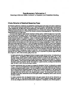
Supplementary Fig. 1
12
Deduced amino acid sequences of clone #2-6 (EaRFP) and phylogenetic tree
13
comparing with RFP sequences from other anthozoans in Cnidaria. (a) The deduced
14
amino acid sequences of clone #2-6 (EaRFP). Black box, chromophore-forming residues.
15
Sequence data are available from GenBank, under accession number KT452623. (b) A
3
4
16
phylogenetic tree comparing the amino acid sequences of RFP from various anthozoans.
17
The number at each node represents the bootstrap probability (%); the branches shown
18
correspond to values of 50 % and higher. The corresponding GenBank accession
19
numbers of the proteins analyzed are as follows: Acropora aculeus (AAU06856), A.
20
millepora (AAU06854), A. hyacinthus (AAU06855), Discosoma sp. SSAL-2000
21
(AAG16224), Discosoma sp. (AAF03369), Porites porites (ABB17953), Echinopora
22
forskaliana (ACD13196), Euphyllia ancora (KT452623), Ricordea florida (AAK71339),
23
Montastraea
24
Trachyphyllia geoffroyi (BAC20344), Catalaphyllia jardinei (ABN41777), Scolymia
25
cubensis (AAU06843), Lobophyllia hemprichii (AAV54099), Mycedium elephantotus
26
(ABB17959), Verrillofungia concinn (BAD24721), A. millepora (AAU06852),
27
Zoanthus sp. (AAL23574), Montipora efflorescens (ABB17952), Cerianthus sp.
28
(AAP55761), Entacmaea quadricolor (AAN05449).
cavernosa
(AAO61598),
Echinophyllia
29
4
echinata
(ABB17960),
5
30 31
Supplementary Fig. 2
32
Fluorescence microscopic observation of the isolated E. ancora tissues. a-d) Isolated
33
tentacles (Ten), e-h) Isolated ovary (Ov), i-l) Isolated mesenterial filament (Mf). a, e,
34
and i) Bright view. b, f, and j) U-MWIG 2 (RFP) filter view. c, g, and k) U-MWIB 2
35
(GFP) filter view. d, h, and l) U-MWU2 (CFP) filter view. All bars = 500 µm.
5
6
36 37
Supplementary Fig. 3
38
Fluorescence microscopic observation of EaRFP in the ovarian tissues with
39
mesenterial filaments. (a,b) The sample collected in August (Aug). (c,d) The sample
40
collected in December (Dec). (e,f) The sample collected in March (Mar). a, c, and e:
41
bright views; b, d, and f: U-MWIG 2 (RFP) filter-fluorescent views. All bars = 500 µm.
42
6
7
43 44
Supplementary Fig. 4
45
Fluorescence microscopic observation of EaRFP in the released egg and the
46
embryos. (a,b) The released egg. (c,d) The planula larva (e,f) The larva after settlement
47
and metamorphosis. a, c, and e: bright views; b, d, and f: U-MWIG 2 (RFP)
48
filter-fluorescent views. All bars = 500 µm.
49
7