Are you looking for a fully featured web host that gives you a lot of room to grow? Your search is finally over. Once you sign up for a free account you will get;.
0031-3998/00/4806-0725 PEDIATRIC RESEARCH Copyright © 2000 International Pediatric Research Foundation, Inc.
Vol. 48, No. 6, 2000 Printed in U.S.A.
The Developing Nervous System: A Series of Review Articles The following article is the first in an important series of review articles which discuss the developmental biology of the nervous system and its relation to diseases and disorders that are found in new born infants and children. This review, by Pierre Gressens, describes neuronal migration in the developing brain and abnormalities in humans resulting from aberrations in neuronal migration. Alvin Zipursky Editor-in-Chief
Mechanisms and Disturbances of Neuronal Migration PIERRE GRESSENS Service de Neurologie Pédiatrique and INSERM E 9935, Hôpital Robert-Debré, F-75019 Paris, France
ABSTRACT of migration of neurons destined to the neocortex. New insights gained from the analysis of animal models as well as from the study of human diseases will be included. (Pediatr Res 48: 725–730, 2000)
Neuronal migration appears as a complex ontogenic step occurring early during embryonic and fetal development. Control of neuronal migration involves different cell populations including Cajal-Retzius neurons, subplate neurons, neuronal precursors or radial glia. The integrity of multiple molecular mechanisms, such as cell cycle control, cell-cell adhesion, interaction with extracellular matrix protein, neurotransmitter release, growth factor availability, platelet-activating factor degradation or transduction pathways seems to be critical for normal neuronal migration. The complexity and the multiplicity of these mechanisms probably explain the clinical, radiologic and genetic heterogeneity of human disorders of neuronal migration. The present review will be focused on mechanisms and disturbances
During neocorticogenesis, neurons derive from the primitive neuroepithelium and migrate to their appropriate position in the cerebral mantle. In humans, migration of neocortical neurons occurs mostly between the 12th and the 24th weeks of gestation (1). In laboratory rodents, including mice, rats and hamsters, this ontogenic step roughly extends from embryonic day twelve to the first postnatal days, although interspecies differences do exist (2– 4). The first postmitotic neurons produced in the periventricular germinative neuroepithelium will migrate to
Abbreviations: BDNF, brain-derived neurotrophic factor EGF, epidermal growth factor NMDA, N-methyl-D-aspartate NT, neurotrophin PAF, platelet-activating factor Tish, telencephalic internal structural heterotopia
Received May 26, 2000; accepted June 29, 2000 Correspondence and reprint requests: Pierre Gressens, Service de Neurologie Pédiatrique and INSERM E 9935, Hôpital Robert-Debré, 48 Boulevard Sérurier, F-75019 Paris, France. This work was supported by the INSERM, the European Community (Biomed grant) and the Fondation Grace de Monaco.
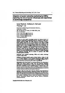
form a subpial preplate or primitive plexiform zone (Fig. 1A). Subsequently produced neurons, which will form the cortical plate, migrate into the preplate and split it into the superficial molecular layer [or layer I or marginal zone containing CajalRetzius neurons (5)] and the deep subplate (Fig. 1A). Schematically, the successive waves of migratory neurons will pass the subplate neurons and end their migratory pathway below layer I, forming successively (but with substantial overlap) cortical layers VI, V, IV, II, and II (inside-out pattern) (Fig. 1). The analysis of neuronal migration disorders occurring in humans and animal models has expanded our knowledge of migratory mechanisms (6, 7). Because of a failure of normal migration, neurons may accumulate in unusual areas (heterotopias). Neuronal heterotopias can be focal (nodular heterotopias) and located at all levels of the migration pathway, from
725
726
GRESSENS
the lateral ventricle up to the cortical plate itself. Sometimes, over migration is observed with neurons present in the molecular layer or even in the meninges. On the other hand, migratory disorders can appear as diffuse band heterotopias in the white matter (pachygyrias/lissencephalies, double cortex syndrome). Radial glia and migratory pathways. During neocorticogenesis, migrating neurons can adopt different types of trajectories (Fig. 1B): 1) a large proportion of neurons migrate radially, along radial glial guides, from the germinative zone to the cortical plate (8). Radial glia are specialized glial cells present in the neocortex during neuronal migration; these cells display a radial shape with a nucleus located in the germinative zone, a basal process attached on the ventricular surface and a radial apical process reaching the pial surface. Rakic (9) has postulated that these radially arranged glial guides keep a topographical correspondence between a hypothesized protomap present in the germinative zone and the cortical areas. 2) An important group of neuronal precursors initially adopt a tangential trajectory at the level of the periventricular germinative zone (10) before adopting a classical radial migrating pathway along radial glia. This tangential migration could permit some dispersion at the level of the cortical plate of neurons originating from a single clone in the germinative neuroepithelium, increasing the clonal heterogeneity within a given cortical area. 3) Tangentially migrating neurons have also been described at the level of the intermediate zone (prospective white matter) (11). Recent studies (12, 13) suggest that most of these neuronal cells displaying a migrating pathway orthogonal to radial glia originate in the ganglionic eminence. GABA-expressing interneurons seem to be produced by this mechanism.
The phenotype of radial glia seems to be determined both by migrating neurons and by intrinsic factors expressed by glial cells. Among the latter, Götz et al. (14) recently demonstrated that the transcription factor Pax6, which is specifically localized in radial glia during cortical development, is critical for the morphology, number, function and cell cycle of radial glia. From the appearance of the cortical plate, the radial glial fibers are grouped in fascicles of five to eight fibers in the intermediate zone (15, 16). The final cortical location of the neurons, which could be determined by the guiding glial fascicle (see above), partly determines the connections the neuron will be able to establish. The fascicles of glial fibers, filled with glycogen could also provide energy supply for migrating neurons which are far away from developing blood vessels. The ontogenic unit made of the radial glial fascicle is very similar in the mouse, rat, hamster, cat, and in the human (16). Because this glial unit is constant throughout the mammalian species studied, it could represent the basic developmental module of the developing cortex: the unit remains stable while the number of adjacent units gradually increases to permit brain expansion in the evolution of mammalian species. Knowledge of the genetic and environmental factors that control the organization, number and function of these glial fascicles could, therefore, improve our understanding of cortical development and evolution of the brain (17, 18). Effects of neurotransmitters and growth factors on neuronal migration. Komuro and Rakic (19) first reported that specific inhibitors of the N-methyl-D-aspartate (NMDA) glutamate receptor subtype slowed down the rate of in vitro neuronal migration. Further demonstration that glutamate plays a role in neuronal migration was given by Marret et al. (20).
Figure 1. (A) Schematic illustration of mammalian neocortical formation. GZ, germinative zone; IZ, intermediate zone (prospective white matter); PPZ, primitive plexiform zone; SP, subplate; I, cortical layer I or molecular layer; II to VI, cortical layers II to VI. Arrows and light gray circles indicate migrating neurons while black circles and triangles represent postmigratory neurons. (B) Coronal neopallial section at 15 wk of gestation showing a schematic representation of the different migratory pathways adopted by neurons. 1: radial migration along radial glial cells of neurons originating from the periventricular germinative zone (GZ). 2: Tangential migration in the germinative zone (GZ) followed by a radial migration along glial guides. 3: Tangential migration in the intermediate zone (IZ) of neurons originating from the ganglionic eminence. M, meninges; I, layer I or molecular layer; V-VI, cortical layers V and VI; LV, lateral ventricule.
NEURONAL MIGRATION
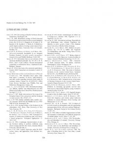
They performed focal injections of ibotenate, a glutamate analog mainly acting on NMDA receptors, into the brain of hamsters on the day of birth, a stage where neurons destined to the superficial cortical layers are migrating. The resulting pattern was a laminar band heterotopia (see below), focal periventricular nodular heterotopias or intracortical arrests of migrating neurons. Ibotenate, a unique molecular trigger of the excitotoxic cascade, produces a wide spectrum of abnormal neuronal migration patterns encountered in the human brain such as nodular heterotopias, lissencephalies and double cortex syndromes. GABA is also involved in neuronal migration modulation as it stimulates, in tissue culture, neuronal migration via calciumdependent mechanisms (21). Growth factors have been recently implicated in the control of neuronal migration. Intraventricular injection of neurotrophin-4 (NT-4) (22) or over expression of brain-derived neurotrophic factor (BDNF) (23) produce heterotopic accumulation of neurons in the molecular layer (layer I), mimicking some aspects observed in human status verrucosus deformans. X-linked periventricular heterotopia. The human X-linked dominant periventricular heterotopia (Fig. 2A) is characterized by neuronal nodules lining the ventricular surface. Hemizygous males die during the embryonic period and affected females have epilepsy which can be accompanied by other manifestations such as patent ductus arteriosus and coagulopathy. The gene responsible for this disease has been recently identified (24): filamin 1 (FLN1) encodes an actin-crosslinking phosphoprotein which transduces ligand-receptor binding into actin reorganization. Filamin 1 is necessary for locomotion of several cell types and is present at high levels in the developing neocortex. Rakic et aI. (25) had previously shown the polarity of microtubule assemblies during migration of rodent cerebellar neurons and proposed that the dynamics of
727
slow polymerization combined with fast disintegration of oriented microtubules was critical for displacement of the nucleus and cytoplasm within the membrane cylinder of the leading process of the migrating neuron. Type I lissencephaly and platelet-activating factor (PAF). Lissencephaly and agyria-pachygyria are the terms used to describe brains with absent or poor sulcation. Agyria refers to brains or portions of brains with absence of gyri and sulci whereas pachygyria refers to brains or portions thereof with few broad, flat gyri and shallow sulci. Lissencephaly (“smooth brain”) is another term used to describe such brains. Complete lissencephaly is synonymous with agyria, whereas incomplete lissencephaly refers to brains with shallow sulci and a relatively smooth surface; incomplete lissencephaly is often use synonymously with agyria-pachygyria. Type I lissencephaly (Fig. 2B) usually has both agyric and pachygyric regions (26). The histologic appearance of the cortex varies according to the brain area; the majority of the neocortex is that of a “4-layered” cortex, composed of a molecular layer, a disorganized outer cellular layer, a cell sparse layer, and an inner cellular layer (probably composed of neurons whose migration has been prematurely arrested). The migratory defect is postulated to occur between 12 and 16 gestational weeks. Miller-Dieker syndrome is a malformative syndrome associating classical type I lissencephaly and a characteristic facies. Clinically, the head size is normal to small at birth but progressive microcephaly over the first few years of life is common. Patients are hypotonic at birth and develop progressive spasticity. Seizures usually begin within the first few months of life. Miller-Dieker syndrome and up to 40% of cases of isolated lissencephaly sequence (27) result from a hemideletion or mutations of the LIS1 gene. LIS1 encodes the beta subunit of
Figure 2. (A) X-linked periventricular heterotopias. Arrows indicate nodules of heterotopic neurons which failed to migrate to the cortical plate and remained blocked in the periventricular zone. (B) Type 1 lissencephaly. CT-scan showing the typical 8-shape picture. Note the smooth (agyric) surface of the brain, the presence of heterotopic neurons (arrows) in the white matter and the enlarged lateral ventricles. (C) Double cortex syndrome. Arrows point to the normally localized cortical plate while stars indicate the laminar band heterotopia. (A and C: courtesy of Dr. Thierry Duprez, Service de Radiologie, St-Luc Hospital, University of Louvain Medical School at Brussels, Belgium).
728
GRESSENS
a brain acetylhydrolase which degrades platelet-activating factor (PAF). Accordingly, in vitro stimulation of PAF receptor disrupts neuronal migration (28) and mice with one inactive LIS1 allele display cortical and hippocampal neuronal migration delay (29). PAF is an ether phospholipid acting in the brain on receptors present on synaptic endings and intracellular membranes. As PAF increases intracellular calcium concentration and increases NMDA receptor currents, it is tempting to speculate that some of brain abnormalities observed in MillerDieker syndrome are secondary to an excess of glutamatergic transmission, potentially linking the above described hamster model of migration disorder to human lissencephaly. An alternative hypothesis is that PAF acetylhydrolase could be a link to the microtubule network and motility apparatus in migrating neurons (7). Studying human fetuses, Clarck et al. (30) have shown that LIS gene products are localized in Cajal-Retzius neurons, some subplate neurons, thalamic neurons and in the periventricular germinative zone. The expression of LIS in Cajal-Retzius cells strongly support the previously suspected role of these cells in the neuronal migration and in the cytoarchitectonic organization of the cortical plate. Previous studies have suggested that subplate neurons could interact with migrating neurons (31), although the precise effects of these subplate neurons on neuronal migration have not yet been reported. Finally, the presence of LIS gene product in the germinative zone is in agreement with other studies which have shown that laminar distribution of cortical neurons is influenced by events occurring during the mitotic cycle of neuronal precursors in the germinative zone (32). Double cortex syndrome. Diffuse subcortical laminar heterotopias (Fig. 2C) have been described pathologically as well as on clinical and imaging basis under the names of “double cortex,” “bicortical lissencephalies,” “partial lissencephalies,” or “laminar band heterotopias” (26). In this brain malformation, two cortical plates, separated by white matter, co-exist: one cortical plate is in a normal superficial position while the other one is located in a deep heterotopic position and is separated from the lateral ventricle by another area of white matter. It seems that different clinical conditions have been described under these names: reported patients have usually moderate to severe developmental delay and an early onset of medically refractory seizures but other patients can have relatively mild clinical manifestations. Des Portes et al. (33) and Gleeson et al. (34) have recently identified a novel gene (DCX or XLIS gene) involved in X-linked neuronal migration disorder where females display subcortical laminar heterotopia (or double cortex syndrome) while lissencephaly is found in males. The gene encodes a protein, doublecortin, which is a microtubule-associated protein expressed by migrating and differentiating neurons (35, 36), again emphasizing the importance of the cytoskeleton for neuronal migration (7, 25). From a clinical point of view, a recent report has proposed that deletions or mutations of DCX and LIS1 genes account for 76% of isolated type I lissencephalies (37). Two animal models of laminar band heterotopias are currently available: the ibotenate-induced laminar heterotopias in the newborn hamster (see above) and the tish (telencephalic
internal structural heterotopia) rat. The tish rat is a spontaneously mutant animal exhibiting a bilateral laminar band heterotopia which predominates in the frontal and parietal cortex (38). The heterotopic cortical plate could be secondary to the combination of abnormal neuronal migration and heterotopic neurogenesis. Affected rats exhibit spontaneous recurrent electrographic and behavioral seizures. Zellweger syndrome. The Zellweger cerebro-hepato-renal syndrome is a fatal autosomic recessive disease caused by an absence of functional peroxisomes. One hallmark of this human disease is the presence of heterotopic neurons in the neocortex, the cerebellum and the inferior olivary complex. Patients are characterized by the absence of psychomotor development or a rapid regression, a dysmorphic facies and a severe hypotonia; they generally die within a few months. Animal models of this human disease have been produced by inactivation of a gene critically involved in peroxisomal assembly (39, 40). Homozygous animals for the gene inactivation have no functional peroxisomes, die around birth and display heterotopic neurons in the subcortical white matter and dysplastic olivary complex. The study of radial glia cells did not reveal any significant morphologic change although the available data do not permit one to exclude a functional defect. Analysis of mutant mice revealed that the migration defect was caused by altered N-methyl-D-aspartate (NMDA) glutamate receptor-mediated calcium mobilization (41). This NMDA receptor dysfunction was linked to a deficit in PAF, a phenomenon related to peroxisome impairment. These findings confirm NMDA receptor and PAF involvement in neuronal migration and suggest a link between peroxisome metabolism and NMDA receptor efficacy. Effects of environmental factors on neuronal migration. Neuronal migration disorders have been described in humans and/or in animal models following in utero exposure to several environmental factors, including infection with cytomegalovirus (42) or toxoplasmosis (Evrard and Gadisseux, unpublished observation), ethanol (3, 43, 44), cocaine (45, 46) or ionizing radiation (47). In most cases, the mechanisms by which these factors disturb neuronal migration remain unclear. Prenatal cocaine administration to pregnant mice or monkeys induces abnormal addressing of neurons in the neocortical plate (45, 46). This abnormal neuronal migration pattern is probably linked to the observed abnormalities of radial glia density and disturbances of neuronal proliferation in the germinative zone.
CONCLUSION A few human and rodent genes and factors linked to neuronal migration disorders have been recently identified (Table 1). The elucidation of their function should help to clarify the mechanisms of neuronal migration as well as the pathophysiology of some brain malformations. Furthermore, these diseases can now benefit from a genetic diagnosis which can also be applied in some instances to prenatal cases. Improvement of imaging techniques has provided new classifications of neuronal migration disorders as well
NEURONAL MIGRATION
729
Table 1. Summary of the established or hypothetical links between genes and gene products and neuronal migration disorders in human and rodent neocortex (adapted and modified from Gressens, 1998) LIS1 gene, PAF DCX or XLIS gene Filamin 1 (FLN1) Tish gene Reelin p35/cdk5 kinase Neurotrophins GABAergic system Glutamatergic system Peroxysomal apparatus Pax6 gene
Miller-Dieker syndrome and some isolated type I lissencephalies X-linked subcortical laminar heterotopia and lissencephaly syndrome (or double cortex syndrome) X-linked dominant periventricular heterotopia Laminar band heterotopias, double cortex syndrome Inverted cortical layers Inverted cortical layers Status verrucosus, molecular ectopias, inverted cortical layers Neuronal migration disorders Neuronal heterotopias, molecular ectopias Zellweger syndrome Abnormalities of radial glia
as their diagnosis in the developing fetal brain. On the other hand, basic research has led to the identification of some critical molecular factors involved in normal and abnormal neuronal migration and has also emphasized the critical interactions between genetic and environmental/epigenetic factors. The long term consequences in terms of behavior, learning and motor abilities or electrophysiological properties of animals with migration disorders are being extensively studied, opening new avenues in the understanding of human disorders such as epilepsy, learning disabilities, mental retardation and some psychiatric diseases.
REFERENCES 1. Sidman RL, Rakic P 1973 Neuronal migration, with special reference to developing human: a review. Brain Res 62:1–35 2. Caviness VS 1982 Neocortical histogenesis in normal and reeler mice: a developmental study based upon [H]thymidine autoradiography. Brain Res 256:293–302 3. Miller MW 1986 Effects of ethanol on the generation and migration of cerebral cortical neurons. Science 233:1308 –1311 4. Shimada M, Langman J 1970 Cell proliferation, migration and differentiation in the cerebral cortex of the golden hamster. J Comp Neurol 139:227–244 5. Meyer G, Goffinet AM, Fairen A 1999 What is a Cajal-Retzius cell? A reassessment of a classical cell type based on recent observations in the developing cortex. Cereb Cortex 9:765–775 6. Gressens P 1998 Mechanisms of cerebral dysgenesis. Curr Opin Pediatr 10:556 –560 7. Walsh CA 1999 Genetic malformation of the cerebral cortex. Neuron 23:19 –29 8. Rakic P 1971 Guidance of neurons migrating to the fetal monkey neocortex. Brain Res 33:471– 476 9. Rakic P 1988 Specification of cerebral cortical areas. Science 241:170 –176 10. Austin CP, Cepko CL 1990 Cellular migration patterns in the developing mouse cerebral cortex. Development 110:713–732 11. O’Rourke NA Dailey ME, Smith SJ, McConnell SK 1992 Diverse migratory pathways in the developing cerebral cortex. Science 258:299 –302 12. Anderson SA, Eisenstat DD, Shi L, Rubenstein JL 1997 Interneuron migration from basal forebrain to neocortex: dependence on Dlx genes. Science 278:474 – 476 13. Zhu Y, Li HS, Zhou L, Wu JY, Rao Y 1999 Cellular and molecular guidance of GABAergic neuronal migration from an extracortical origin to neocortex. Neuron 23:473– 485 14. Götz M, Stoykova A, Gruss P 1998 Pax6 controls radial glia differentiation in the cerebral cortex. Neuron 21:1031–1044 15. Gadisseux JF, Evrard P 1985 Glial-Neuronal relationship in the developing central nervous system. Dev Neurosci 7:12–32 16. Gressens P, Evrard P 1993 The glial fascicle: a developmental unit guiding, supplying and organizing mammalian cortical neurons. Dev Brain Res 76:272–277 17. Gressens P, Gofflot F, Van Maele-Fabry G, Misson JP, Gadisseux JF, Picard JJ, Evrard P 1992 Early neurogenesis and teratogenesis in whole mouse embryo cultures. J Neuropathol Exp Neurol 51:206 –219 18. Kadhim HJ, Gadisseux JF, Evrard P 1988 Topographical and cytological evolution of the glial phase during prenatal development of the human brain: histochemical and electron microscopic study. J Neuropathol Exp Neurol 47:166 –188 19. Komuro H, Rakic P 1993 Modulation of neuronal migration by NMDA receptors. Science 260:95–97 20. Marret S, Gressens P, Evrard P 1996 Neuronal migration disorders induced by ibotenate in the neocortex. Proc Natl Acad Sci USA 93:15463–15468
21. Behar TN, Li YX, Tran HT, Dunlap V, Scott V, Barker JL 1996 GABA stimulates chemotaxis and chemokinesis of embryonic cortical neurons via calcium-dependent mechanisms. J Neurosci 16:1808 –1818 22. Brunstrom JE, Gray-Swain MR, Osborne PA, Pearlman AL 1997 Neuronal heterotopias in the developing cerebral cortex produced by neurotrophin-4. Neuron 18:505– 517 23. Ringstedt T, Linnarsson S, Wagner J, Lendhal U, Kokaia Z, Arenas E, Ernfors P, Ibanez CF 1998 BDNF regulates reelin expression and Cajal-Retzius cell development in the cerebral cortex. Neuron 21:305–315 24. Fox JW, Lamperti ED, Eksioglu YZ, Hong SE, Feng Y, Graham DA, Scheffer IE, Dobyns WB, Hirsch BA, Radtke RA, Berkovic SF, Huttenlocher PR, Walsh CA 1998 Mutations in filamin 1 prevent migration of cerebral cortical neurons in human periventricular heterotopia. Neuron 21:1315–1325 25. Rakic P, Knyihar-Csillik E, Csillik B 1996 Polarity of microtubule assemblies during neuronal cell migration. Proc Natl Acad Sci USA 17:9218 –9222 26. Barkovich AJ, Gressens P, Evrard P 1992 Formation, maturation, and disorders of neocortex. Am J Neuroradiol 13:423– 446 27. Lo Nigro C, Chong CS, Smith AC, Dobyns WB, Carrozzo R, Ledbetter DH 1997 Point mutations and an intragenic deletion of LIS1, the lissencephaly causative gene in isolated lissencephaly sequence and Miller-Dieker syndrome. Human Mol Genet 6:157–164 28. Bix GJ, Clarck GD 1998 Platelet-activating factor receptor stimulation disrupts neuronal migration in vitro. J Neurosci 18:307–318 29. Hirotsune S, Fleck MW, Gambello MJ, Bix GJ, Chen A, Clark GD, Ledbetter DH, McBain CJ, Wynshaw-Boris A 1998 Graded reduction of Pafah1b1 (Lis1) activity results in neuronal migration defects and early embryonic lethality. Nature Genet 19:333–339 30. Clarck GD, Mizuguchi M, Antalffy B, Barnes J, Armstrong D 1997 Predominant localization of the LIS family gene products to Cajal-Retzius cells and ventricular neuroepithelium in the developing human cortex. J Neuropathol Exp Neurol 56:1044 –1052 31. Shatz CJ, Chun LLM, Luskin MB 1988 The role of the subplate in the development of the mammalian telencephalon. In: Peters A, Jones EG (eds) Development and Maturation of Cerebral Cortex. Plenum, New York, pp 35–58 32. McConnell SK, Kaznowski CE 1991 Cell cycle dependence of laminar determination in developing neocortex. Science 254:282–285 33. Des Portes V, Pinard JM, Billuart P, Vinet MC, Koulakoff A, Carrie A, Gelot A, Dupuis E, Motte J, Berwald-Netter Y, Catala M, Kahn A, Beldjord C, Chelly J 1998 A novel CNS gene required for neuronal migration and involved in X-linked subcortical laminar heterotopia and lissencephaly syndrome. Cell 92:51– 61 34. Gleeson JG, Allen KM, Fox JW, Lamperti ED, Berkovic S, Scheffer I, Cooper EC, Dobyns WB, Minnerath SR, Ross ME, Walsh CA 1998 Doublecortin, a brain-specific gene mutated in human X-linked lissencephaly and double cortex syndrome, encodes a putative signaling protein. Cell 92:63–72 35. Francis F, Koulakoff A, Boucher D, Chafey P, Schaar B, Vinet MC, Friocourt G, McConnell N, Reiner O, Kahn A, McConnell SK, Berwald-Netter Y, Denoulet P, Chelly J 1999 Doublecortin is a developmentally regulated, microtubule-associated protein expressed in migrating and differentiating neurons. Neuron 23:247–256 36. Gleeson JG, Lin PT, Flanagan LA, Walsh CA 1999 Doublecortin is a microtubuleassociated protein and is widely expressed by migrating neurons. Neuron 23:257–271 37. Pilz DT, Matsumoto N, Minnerath S, Mills P, Gleeson JG, Allen KM, Walsh CA, Barkovich JA, Dobyns WB, Ledbetter DH, Ross ME 1998 LIS1 and XLIS (DCX) mutations cause most classical lissencephaly, but different patterns of malformation. Hum Mol Genet 7:2029 –2037 38. Lee KS, Schotter F, Collins JL, Lanzino G, Couture D, Rao A, Hiramatsu KI, Goto Y, Hong SC, Caner H, Yamamoto H, Chen ZF, Bertram E, Berr S, Omary R, Scrable H, Jackson T, Goble J, Eisenman L 1997 A genetic animal model of human neocortical heterotopia associated with seizures. J Neurosci 17:6236 – 6242 39. Baes M, Gressens P, Baumgart E, Casteels M, Fransen M, Carmeliet P, Evrard P, Fahimi D, Declercq PE, Collen D, Van Veldhoven PP, Mannaerts GP 1997 Peroxisome deficiency induces abnormal brain development and intrauterine growth retardation in Zellweger mice. Nature Genet 17:49 –57
730
GRESSENS
40. Faust PL, Hatten ME 1997 Targeted deletion of the PEX2 peroxisome assembly gene in mice provides a model for Zellweger syndrome, a human neuronal migration disorder. J Cell Biol 139:1293–1305 41. Gressens P, Baes M, Leroux P, Lombet A, Van Veldhoven P, Janssen A, Vamecq J, Marret S, Evrard P 2000 Neuronal migration in Zellweger mice is secondary to glutamate receptor dysfunction. Ann Neurol 48:336 –343 42. Shinmura Y, Kosugi I, Aiba-Masago S, Baba S, Yong LR, Tsutsui Y 1997 Disordered migration and loss of virus-infected neuronal cells in developing mouse brains infected with murine cytomegalovirus. Acta Neuropathol 93:551–557 43. Gressens P, Lammens M, Picard JJ, Evrard P 1992 Ethanol-induced disturbances of gliogenesis and neurogenesis in the developing murine brain: an in vitro and in vivo
44. 45. 46. 47.
immunohistochemical, morphological, and ultrastructural study. Alcohol Alcohol 27:219 –226 Wisniewski K, Dambska M, Sher JH, Qazi Q 1983 A clinical neuropathological study of the fetal alcohol syndrome. Neuropediatr 14:197–201 Gressens P, Kosofsky BE, Evrard P 1992 Cocaine-induced disturbances of neurogenesis in the developing murine brain. Neurosci Lett 140:113–116 Lidow MS 1995 Prenatal cocaine exposure adversely affects development of the primate cerebral cortex. Synapse 21:332–341 Sun XZ, Takahashi S, Fukui Y, Hisano S, Kuboda Y, Sato H, Inouye M 1999 Different patterns of abnormal neuronal migration in the cerebral cortex of mice prenatally exposed to irradiation. Dev Brain Res 114:99 –108