CARBALLO ET AL. Biol Res 39, 2006, 331-340
Biol Res 39: 331-340, 2006
BR331
The G2 checkpoint activated by DNA damage does not prevent genome instability in plant cells JESÚS A. CARBALLO1, 2, JUANA PINCHEIRA 3 and CONSUELO DE LA TORRE 1* 1
Centro de Investigaciones Biológicas, CSIC, Ramiro de Maeztu, 9, 28040-Madrid, Spain. The National Institute for Medical Research, MRC, The Ridgeway, Mill Hill, London, NW7 1AA, United Kingdom. 3 Programa de Genética Humana, ICBM, Facultad de Medicina, Universidad de Chile, Santiago, Chile. 2
ABSTRACT
Root growth, G2 length, and the frequency of aberrant mitoses and apoptotic nuclei were recorded after a single X-ray irradiation, ranging from 2.5 to 40 Gy, in Allium cepa L. root meristematic cells. After 72 h of recovery, root growth was reduced in a dose-dependent manner from 10 to 40 Gy, but not at 2.5 or 5 Gy doses. Flow cytometry plus TUNEL (TdT-mediated dUTP nick end labeling) showed that activation of apoptosis occurred only after 20 and 40 Gy of X-rays. Nevertheless, irrespective of the radiation dose, conventional flow cytometry showed that cells accumulated in G2 (4C DNA content). Simultaneously, the mitotic index fell, though a mitotic wave appeared later. Cell accumulation in G2 was transient and partially reversed by caffeine, thus it was checkpoint-dependent. Strikingly, the additional G2 time provided by this checkpoint was never long enough to complete DNA repair. Then, in all cases, some G2 cells with stillunrepaired DNA underwent checkpoint adaptation, i.e., they entered into the late mitotic wave with chromatid breaks. These cells and those produced by the breakage of chromosomal bridges in anaphase will reach the G1 of the next cell cycle unrepaired, ensuring the appearance of genome instability. Key terms: apoptosis, checkpoint adaptation, G2 DNA damage checkpoint, genome instability, X-rays.
INTRODUCTION
Evolution in plants has preserved most of the pathways that repair DNA damage in other eukaryotes (Tuteja et al., 2001) and also the mitogenic and antimitogenic mechanisms that advance and restrain cell proliferation, respectively. The positive control of proliferation depends on the activation of a set of cyclin-dependent kinases (CDK) (Nurse, 1990). CDK activation is responsible for the promotion of the unidirectional advancement of the cell cycle. Moreover, there are checkpoint or feedback mechanisms incorporated into the cell cycle that survey whether a particular cell is duly prepared for the transition from a cycle phase to the subsequent one. The checkpoint transmits antimitogenic signals when the cell is not
yet ready for such irreversible transition. These signals restrain cycle progression, usually by preventing the removal of the inhibitory phosphorylations from the CDK catalytic subunit (Walworth et al., 1993; Sánchez et al., 1997). The activation of checkpoints also may induce the initiation of a new gene expression program, leading to apoptosis that allows the tissue to be free from those cells possessing a damaged genome (Wang, 2001). The main checkpoint pathways in the eukaryotic cells are based on either the ATM (Ataxia-Telangiectasia Mutated kinase)-CHK2 or the ATR (ATM and Rad53-related kinase)-CHK1 kinase pathways (Marcelain et al., 2005). CHK1 and 2 are the effector kinases in the checkpoint pathways.
Corresponding author: Consuelo de la Torre, CIB-CSIC, Ramiro de Maeztu, 9, E-28040-Madrid, Spain, Tel: (34-91) 837-3112 ext. 4307, Fax: (34-91) 536-0432, E-mail:
[email protected] Received: November 4, 2005, In revised form: January 16, 2006, Accepted: January 17, 2006
332
CARBALLO ET AL. Biol Res 39, 2006, 331-340
As checkpoint activation is transcriptionindependent, these mechanisms provide a fast cellular response to unfavorable intracellular conditions. The precise checkpoint targets for specific DNA damage are unknown in plant cells. In mammalian cells, the ATM-CHK2 checkpoint pathway is mostly responsible for G1 and G2 checkpoints activated by DNA damage as that induced by X-rays (Falck et al., 2001). On the other hand, the ATR-CHK1 is responsible for checkpoints activated by replication blocks within the S phase of the cycle, such as those induced by hydroxyurea (Xiao et al., 2003) or by ultraviolet irradiation (O’Connell and Cimprich, 2005). However, when one of these two checkpoint pathways fails, the other can serve as a substitute (Boddy et al., 1998). The efficiency of any checkpoint mechanism to stop a cycle transition is diminished with the presence of caffeine (Weingartner et al., 2003), because this purine analogue blocks both the ATM and ATR kinase activity (Blasina et al., 1999; Sarkaria et al., 1999). The present study shows that the additional time provided by checkpoints is not large enough to complete DNA repair. This fact, observed in plants, questions the well-established belief that checkpoints prevent the appearance of genome instability.
METHODS
Culture conditions The root meristems of the growing roots of Allium cepa L. cv. “Francesa” bulbs were used. Onion bulbs were allowed to sprout their roots and later to grow in the dark, at a constant temperature of 25ºC, in a Refritherm Struers incubator (Copenhagen, Denmark), in cylindrical glass receptacles (85 cm3 capacity and 11 cm in height), using filtered tap water (Milli RO-4 from Millipore, Bedford MA, USA). Water was renewed every 24 h and aerated continuously by bubbling air using an aquarium pump (René Super, Lyon, France), through an hermetic flask containing distilled water, so
that the air reached each of the culture tubes saturated with humidity. The rate of air bubbles was kept constant so that a 15 to 20 cm3 min-1 oxygen tension was maintained in the culture. The lengths of the six to ten longest roots in each bulb were measured daily with a flexible ruler. All measurements were repeated six times. X-ray irradiation Bulbs without their dry cover layers whose roots had been growing for 72 h in glass tubes containing water were irradiated with an X-ray machine (Philips M6-102 model, Eindhoven, The Netherlands). It was operated at 80 kV and 15 mA, with a large exposition window and at room temperature. The distance between the source and the samples was 23 cm. The irradiation doses (2,5, 5, 10, 20, and 40 Gy) were estimated from the exposure time to X-rays (1.5, 3, 6, 12, and 24 min, respectively) through the calibration curves obtained from the Radiological Protection Service of the Centro de Investigaciones Biológicas. The unirradiated bulbs were maintained outside the X-ray machine, also at room temperature, during the corresponding times of irradiation. After the single irradiation dose, the roots attached to the bulbs were relocated in tubes containing fresh tap water and kept in the incubator until the corresponding recovery times. For statistical analysis, mean values, standard deviations, and number of recorded DNA lesions were taken into account to estimate the significance level among means. The univariate analysis was carried out by using one- or two-way ANOVA with the SPSS statistical program for Windows. Analysis of variance and Bonferroni adjustments were made by multiple pairwise comparison between the data obtained under the different irradiation doses and the control (unirradiated) cells. The minimal significance was at a maximal P value equal to 0.05. Caffeine treatment Solutions of 5 mM caffeine (Sigma, St. Louis, Missouri, USA) were prepared in
CARBALLO ET AL. Biol Res 39, 2006, 331-340
filtered tap water from a stock solution (50 mM caffeine in distilled water). Roots growing from bulbs that had reached more than 2 cm in length were immersed in the caffeine solution, without altering the other environmental conditions. Cytological procedures The roots were always fixed for about one week in a 3: 1 mixture (v/v) of ethanol and acetic acid. Root fixations were carried out, at every 2 h, during the first 24 h after irradiation. Cell nuclei were Feulgenstained after 1 h of hydrolysis in 5 N HCl, at 20º C. Squashes were prepared from the apical 2 mm of the growing roots, after their staining. Coverslips were removed by the dry ice method. For recording mitotic index (MI), at least four root meristems from five different bulbs were studied at each fixation time. About 2,000 cells per root were sampled to estimate the mitotic index as well as the frequency of aberrant mitoses, estimated as abnormal anatelophases. Apoptotic DNA cleavage determination and flow cytometry Nuclei were isolated (Pelayo et al., 2001) and resuspended in a lysis buffer (15 mM Tris, 2 mM EDTA, 80 mM KCl, 20 mM NaCl, and 0.1% Triton X-100, pH 7.5) in which ice-cooled ethanol was added until a 70% final concentration was reached. Samples were subjected to post-fixation processing at -20ºC overnight. The ethanol was removed by spinning down the nuclei at 260 g for 10 min at 4°C. The pellets were washed once with lysis buffer, and resuspended in PBS. To estimate the fraction of cells undergoing apoptotic-like DNA cleavage, nuclei were incubated at 37ºC with the TUNEL detection kit (Roche, Mannheim, Germany), as described by the manufacturer, for 2 h. After incubation, samples were washed twice with the lysis buffer and resuspended in it. To estimate the DNA content by flow cytometry, samples were incubated with 30 µg/ml RNase A (BoehringerMannheim, Germany) and 100 µg/ml
333
propidium iodide (Sigma, USA), for 30 min at room temperature. Flow cytometry analysis was carried out with an EPICS XL analyzer (Coulter, Florida, USA). 25,000 nuclei were scored for each sample at each fixation time. The EXPOTM32 v 1.2. Analysis Software (Beckman Coulter Inc., USA) was used for the analysis of the data obtained.
RESULTS
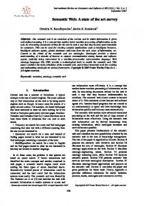
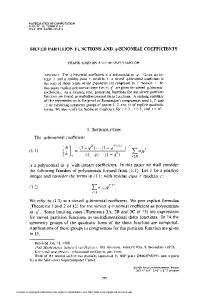
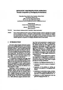
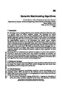
Effect of X-rays on root growth The rate of root growth depends on cell production brought about by the proliferative activity of the cells in meristems. The response of root growth to ionizing radiation was evaluated after a single dose of X-ray irradiation ranging from 2.5 to 40 Gy in Allium cepa L. roots that had been growing under steady state kinetics up to the irradiation time (Fig. 1). After 48h of recovery from the 2.5 Gy dose, root growth was not inhibited. The 5 Gy should still be considered a low dose, since it only decreased root growth after 96 h of recovery. However, root growth decreased with the other irradiation doses (10, 20, and 40 Gy) and with the time of recovery, although to different extents. The doseresponse curve (Fig. 1) showed that 31.5 Gy was the dose that halved root growth, 96 h after irradiation. On the other hand, after 48 h of recovery from a single dose of 40 Gy, root growth was minimal. Therefore, other evaluations were made during the first 24 h of recovery. Chromosomal damage in mitosis after Xrays After irradiation, broken chromatids were observed in some metaphases, anaphases and telophases. Acentric chromosomal fragments also were present in some metaphases (Fig. 2B) and in ana- and telophases (Fig. 2D, F). Double minute chromosomes (arrowhead in Fig. 2D) appeared in some anaphases. Their midzone position and their orientation towards both mitotic poles suggest that they correspond
334
CARBALLO ET AL. Biol Res 39, 2006, 331-340
to acentric telomeric fragments of chromosomes, which sometimes become segregated without interaction with spindle microtubules (see the single-minute contiguous to the double-minute marked by the arrowhead in Fig. 2D). In some telophases, chromosomal bridges were evident (Fig. 2F). These bridges resulted from the migration of dicentric chromosomes towards opposite spindle poles. The dicentrics should have been formed by previous ectopic nonhomologous recombination between either two replicated or unreplicated chromosomes of the complement. The chromosomal bridges are, then, resolved by the growing cytokinetic plate in a process that gives rise to nuclei with unbalanced genomes. The cytokinesis-induced DNA
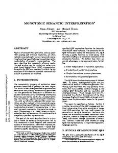
breaks will arrive unrepaired to G1 of the next cell cycle. This ensures the initiation of genome instability, mediated by the formation of novel dicentric chromosomal bridges in the anaphases of the subsequent cycles. In other words, the DNA breakagefusion-bridge cycle (McClintock, 1984) will be initiated. The kinetics of cell progression throughout interphase after X-rays The behavior of the interphasic cell population in response to ionizing radiation was followed by flow cytometry (Fig. 3). During radiation recovery, cells accumulated with 4C DNA content, i.e., they accumulated in G2. This accumulation was dose-dependent, being nearly undetectable after 5 Gy (Fig. 3A), while at higher irradiation doses, the frequency of cells that accumulated in G2 increased progressively (Fig. 3 C, E, G). Restoration of G2 cell frequencies to control levels took place after 24 h of irradiation with 10 Gy (Fig. 3C), while there was only a moderate reversal after 40 Gy (Fig. 3 G). Fate of the cells accumulated in G2 after irradiation
Figure 1: Length of the Allium cepa L. roots measured at 24-hour intervals during recovery from different X-ray doses, considering zero as the length they had when irradiated.
As cells with unrepaired DNA accumulated in G2 may activate an apoptosis-like gene program, the TUNEL technique was performed to label the 3’OH-DNA breaks that characterize apoptotic nuclei. After that, the labeled nuclei were detected by flow cytometry. There was hardly any increase in the basal level of apoptosis after 5 or 10 Gy of irradiation (Fig. 3B, D). After 20 Gy, apoptotic cells increased from 16 h after irradiation (Fig. 3 F), while at 40 Gy, the presence of apoptotic cells was evident from 4 h onwards (Fig. 3H). Thus, apoptosis was a response to irradiation that started to be operative only at high irradiation doses in plant cells. Nevertheless, there was a basal level of apoptosis in the unirradiated cells, probably linked to a renewal of the cells being kept proliferating in plant meristems.
CARBALLO ET AL. Biol Res 39, 2006, 331-340
335
Figure 2: Effect of the X-rays on the mitotic chromosomes. Unirradiated (A, C, and E) and 20 Gyirradiated (B, D, and F) metaphase, anaphase, and telophase. The irradiated ones correspond to mitoses observed in the post-irradiation mitotic wave. Acentric chromosome fragments (black arrows); double minute chromosomes (arrowhead); chromosomal bridges (empty arrow). Bar in F, also valid from A to E = 10 µm.
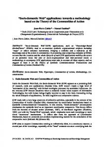
Effect of X-rays on the mitotic index In the proliferating meristems of roots growing under steady-state kinetics, the frequency of any cycle phase remains constant with time (Giménez-Abián et al. 1987). The behavior of the proliferating cells was followed by recording the evolution of the frequency of cells in mitosis (MI) in relation to the irradiation dose and recovery time. Irrespective of the irradiation dose, there was a consistent fall in the mitotic index, as shown in Figure 4 (left panels). Moreover, these panels show that the higher the irradiation dose, the later the recovery of the mitotic index. The left panels in Figure 4 also show the frequency of G2 cells estimated from the flow cytometry data in Figure 3. There is an inverse relationship between the fall in the mitotic index and G2 accumulation. This shows that interphase cells response to irradiation is an inverse relationship between the fall in the mitotic index and G2
accumulation. Thus, the interphase cells respond to irradiation by accumulating in G2 and by later progressing into a delayed mitotic wave. The right panels of Figure 4 show the distribution of aberrant mitoses (AM) similar to those displayed in Figures 2B, D, and F, during recovery from different irradiation doses. The highest AM level was after 8h of recovery from 5 Gy, which declined to about 50% of all observed mitoses at later time points. This finding is consistent with our previous data on the presence of aberrant mitoses after exposure to only 2.5 Gy, where the level of AM 24 h after irradiation was roughly 25% of that observed after 5 Gy (Pérez-Talavera et al., 2003). The histograms corresponding to 10, 20, and 40 Gy in the right panels of Figure 4, show no AM at the middle time points because there were no cells undergoing mitosis at these times (shown by a MI of 0 in the left panels).
336
CARBALLO ET AL. Biol Res 39, 2006, 331-340
Figure 3: Cell cycle phase distribution after ionizing radiation. DNA content (A, C, E, and G as well as TUNEL signal (B, D, F, H) at different X-ray doses (5, 10, 20, and 40 Gy) were recorded by flow cytometry. Arrows in G label the propidium iodide signals that correspond to nuclei with DNA content below 2C.
In the late mitotic wave, the higher the irradiation dose, the higher the frequency of abnormal mitoses. This relationship is evident when comparing the responses to 5 and 10 Gy of ionizing radiation. Around 90% of all mitoses were aberrant after 20 and 40 Gy, between 20 and 24 h of recovery after a single irradiation dose. Post-irradiation caffeine effects Caffeine selectively attenuates the blocks in cycle progression produced by checkpoint because this purine inhibits the ATM and ATR checkpoint kinases, as commented
earlier (Blasina et al., 1999; Sarkaria et al., 1999). Both kinases are responsible for the transmission of antimitogenic signals to the cell-cycle machinery. Flow cytometry showed that, during the first 12 h of recovery from either 10 or 20 Gy of X-ray irradiation, the presence of caffeine reduced the number of cells accumulated in G2. In parallel, the fall in MI also was reduced (Fig. 5). These observations reinforce the idea that the transient G2 accumulation that ends in a delayed mitotic wave, which appears during recovery from irradiation, is in fact a normal checkpoint response.
CARBALLO ET AL. Biol Res 39, 2006, 331-340
337
Figure 4: Mitotic index (MI) and frequency of cells accumulated in G2 (4C DNA content) after different irradiation doses (left column). Frequency of aberrant mitoses (AM) during the 24 hours after irradiation, at different X-ray doses (right column).
338
CARBALLO ET AL. Biol Res 39, 2006, 331-340
Figure 5: Caffeine effect on the G2 cell population (open circles) and on the mitotic index (closed circles) during the first 12 hours after irradiation with 10 and 20 Gy of X-rays.
DISCUSSION
When the genome of a cell is damaged by X-rays in G2, it has various options. The best option in terms of genomic integrity is to properly repair the damage induced. Nature has provided the cell with checkpoints that may help in the repair task by providing additional time to the cycling cell, before it undergoes the irreversible transition to the next cell-cycle phase. Some of the irradiated cells that activate the G2 DNA damage checkpoint may stop proliferation and initiate an
apoptotic-like program (Wang, 2001). This last option obviously diminishes the growth fraction or frequency of cells that are proliferating in the meristem and should growth. Alternatively, the cell may go on proliferating despite its inability to complete DNA repair. This last option, called “checkpoint adaptation” (Paulovich et al., 1997), does not necessarily diminish the growth (or proliferative) fraction, at least in the short run, although it does increase the length of the cell cycle and slow root growth.
CARBALLO ET AL. Biol Res 39, 2006, 331-340
Our present results show that the apoptotic option is only taken by these plant-proliferating cells during recovery from high irradiation doses (20 and 40 Gy). Nevertheless, some cells from the meristems that had received high irradiation doses, as well as most of the cells recovering from lower irradiation doses (5 and 10 Gy) choose to follow the full checkpoint pathway. These cells initially are retained in G2, but eventually undergo checkpoint adaptation, i.e., they enter into mitosis, even though they still have broken chromatids (Weingartner et al., 2003). The choice of this option is reinforced by the fact that caffeine shortens the duration of the G2 checkpoint block by advancing checkpoint adaptation (Andreassen and Margolis, 1992; Pelayo et al., 2001). There are other situations that modulate the differential caffeine efficiency in some of the plant checkpoint pathways, as the undue persistence of the plant mitotic cyclin B (Weingartner et al., 2003). The data presented here does not provide information on the way G2 timing is modulated. However, inhibition of the putative functional homolog of the Cdc25 phosphatase (Reichheld et al., 1999) is probably involved in these cells. Such phosphatase would activate the CDK • mitotic cyclin complex, which licenses the entry into mitosis. Why is the G2 checkpoint block not large enough to allow repair of all DNA lesions? It was reasonable to think that, especially in the presence of low DNA damage, the time provided by the G2 checkpoint would be enough to allow full recovery from the induced DNA damage. However, the present study shows that this is not the case. Checkpoint adaptation is a misleading way of saying that a still-unprepared cell finally faces an irreversible cycle transition (Weinert and Hartwell, 1988). This premature transition into the next cycle phase compromises the fate of the cell and its progeny, as McClintock (1984) has shown. Thus, the induction of a single chromatid break incorporated in a postmitotic nucleus activates the breakage-
339
fusion-bridge cycle that maintains genome instability throughout subsequent cell cycles. By leaving some DNA damage unrepaired, checkpoint pathways open the way to the clonal initiation of microevolution processes in some of the plant proliferating cells. This unexpected finding suggests that the development of genome instability in some cells of a population should not be considered a deadly risk. The use of knowledge about checkpoints to deal with uncontrolled cell proliferation Negative regulatory controls have raised much interest as targets of potential antineoplasic agents. Proliferation may be reduced if the checkpoints are made more robust. Reinforcement of the checkpoint may be achieved at least in three different ways: first, by stimulating the transcriptiondependent synthesis of the physiological CDK inhibitors (CKIs) (Harper and Elledge, 1996); second, by the pharmacological use of chemical CDK inhibitors such as roscovitin (Meijer, 1996); and third, by blocking the expression of some of the genes responsible for checkpoint undue override (checkpoint adaptation) (Bennet et al., 2001). There is, however, a more radical way to stop undesired proliferation through the checkpoint pathways. In this case, checkpoints would be cancelled completely, so that cells enter the next cycle phase with no delay and without the DNA repaired. They would then become inviable and unable to transmit their defective gene information to the cells derived clonally from them. While the present study does not invalidate these approaches to reduce unwanted proliferation in a tissue, the data questions one of the basic assumptions on checkpoints. We have shown that, in plants, the additional time provided by checkpoints is not long enough to complete DNA repair, even in the presence of a low level of DNA damage. Therefore, the behavior of checkpoints in other cell systems should be approached to know whether or not they prevent genomic instability.
340
CARBALLO ET AL. Biol Res 39, 2006, 331-340
ACKNOWLEDGEMENTS
We thank Dr. R.S. Cha and Ms. C. Erp for critical reading of the manuscript and English corrections. Dr. Susana PérezTalavera’s encouragement during the first part of this work is greatly acknowledged. We also thank Mr. J.L Marcilla and M. Carrascosa for technical assistance and Mrs. B.L. Walker and Ms. N. Hashash for their help with the written English. This work has been supported by the Dirección General de Investigación, MEC, Spain (Project BFU2004-03071) and the Departamento de Investigación, Universidad de Chile.
REFERENCES ANDREASSEN PR, MARGOLIS RL (1992) 2Aminopurine overrides multiple cell cycle checkpoints in BHK cells. Proc Natl Acad Sci USA 89: 2272-2276 BENNET CB, SNIPE JR, WESTMORELAND JW, RESNIK MA (2001) SIR functions are required for the toleration of an unrepaired double-strand break in a dispensable yeast chromosome. Mol Cell Biol 21: 5359-5373 BLASINA A, BRENDAN DP, GAETAN AT, MCGOWAN CH (1999) Caffeine inhibits the checkpoint kinase ATM. Curr Biol 9: 1135-1138 BODDY MN, FURNARI B, MONDESERT O, RUSSELL P (1998) Replication checkpoint enforced by kinases Cds1 and Chk1. Science 280: 909-912 FALCK J, MAILAND N, SYLJUASEN RG, BARTEK J, LUKAS J (2001) The ATM-Chk2-Cdc25A checkpoint pathway guards against radioresistant DNA synthesis. Nature 410: 842-847 GIMÉNEZ-ABIÁN MI, DE LA TORRE C, LÓPEZ-SÁEZ JF (1987) Growth and cell proliferation in Allium roots at different oxygen tensions. Environ Exp Bot 27: 233237 HARPER JW, ELLEDGE SJ (1996) Cdk inhibitors in development and cancer. Curr Opin Genet Dev 6: 5664 MARCELAIN K, DE LA TORRE C, GONZÁLEZ P, PINCHEIRA J (2005) Roles of nibrin and ATM/ATR kinases on the G2 checkpoint under endogenous or radio-induced DNA damage. Biol Res 38: 179-185
MCCLINTOCK B (1984) The significance of responses of the genome to challenge. Science 226: 792-801 MEIJER L (1996) Chemical inhibitors of cyclin-dependent kinases. Prog Cell Cycle Res 1, 351-363 NURSE P (1990) Universal control mechanism regulating onset of M-phase. Nature 344: 503-508 O’CONNELL MJ, CIMPRICH KA (2005) G2 damage checkpoints: What is the turn-on? J Cell Sci 118: 1-6 PAULOVICH AG, TOCZYSKI DP, HARTWELL LH (1997) When checkpoints fail. Cell 88: 315-321 PELAYO HR, LASTRES P, DE LA TORRE C (2001) Replication and G2 checkpoints: Their response to caffeine. Planta 212: 444-453 PÉREZ-TALAVERA S, CARBALLO JA, DE LA TORRE C (2003) Lack of mitotic delays at the onset of proliferation in dormant root primordia challenged by ionizing radiation. Biol Plantarum 46: 383-387 REICHHELD JP, VERNOUX T, LARDON F, VAN MONTAGU M, INZÉ D (1999) Specific checkpoint regulate plant cell cycle progression in response to oxidative stress. Plant J 17: 647-656 SÁNCHEZ Y, WONG C, THOMA RS, RICHMAN R, WU Z, PIWNICA-WORMS H, ELLEDGE SJ (1997) Conservation of the Chk1 checkpoint kinase in mammals: Linkage of DNA damage to Cdk regulation through Cdc25. Science 277: 1497-1501 SARKARIA JN, BUSBY EC, TIBBETS RS, ROOS P, TAYA Y, KARNITZ LM, ABRAHAM RT (1999) Inhibition of ATM and ATR kinase activities by the radiosensitizing agent, caffeine. Cancer Res 59: 43754382 TUTEJA N, SINGH MB, MISRA MK, BHALLA PL, TUTEJA R (2001) Molecular mechanisms of DNA damage and repair: Progress in plants. Crit Rev Biochem Mol Biol 36: 337-397 WALWORTH N, DAVEY S, BEACH D (1993) Fission yeast kinase links the rad checkpoint pathway to cdc2. Nature 363: 368-371 WANG JYJ (2001) DNA damage and apoptosis (Editorial). Cell Death Diff 8: 1047-1048 WEINERT TA, HARTWELL LH (1988) The RAD9 gene controls the cell cycle response to DNA damage in Saccharomyces cerevisiae. Science 241: 317-322 WEINGARTNER M, PELAYO HR, BINAROVA P, ZWERGER K, MELIKANT B, DE LA TORRE C, HEBERLE-BORS E, BÖGRE L (2003) A plant Cyclin B2 is degraded early in mitosis and its ectopic expression shortens G2-phase and alleviates the DNA damage checkpoint. J Cell Sci 126: 487-498 XIAO Z, CHEN Z, GUNASEKERA AH, SOWIN TJ, ROSENBERG SH, FESIK S, ZHANG H (2003) Chk1 mediates S and G2 arrests through Cdc25A degradation in response to DNA damaging agents. J Biol Chem 278: 21767-21773