Germline factor DDX4 functions in blood-derived cancer cell phenotypes Natalie Schudrowitz,1 Satoshi Takagi,2 Gary M. Wessel1 and Mamiko Yajima1 1 Department of Molecular Biology, Cellular Biology and Biochemistry, Brown University, Providence, Rhode Island; 2Department of Medical Oncology, Harvard Medical School, Dana-Farber Cancer Institute, Boston, Massachusetts, USA
Key words DDX4/Vasa, germline factor, human cancer cells, RNA helicase, translational regulation Correspondence Mamiko Yajima, MCB Department, Brown University, 185 Meeting Street, BOX-GL173, Providence, RI 02912, USA. Tel: +1-401-863-3164; Fax: +1-401-863-2421; E-mail:
[email protected] Funding Information American Heart Association; National Institutes of Health; National Science Foundation. Received March 17, 2017; Revised May 31, 2017; Accepted June 6, 2017
DDX4 (the human ortholog of Drosophila Vasa) is an RNA helicase and is present in the germ lines of all metazoans tested. It was historically thought to be expressed specifically in germline, but with additional organisms studied, it is now clear that in some animals DDX4/Vasa functions outside of the germline, in a variety of somatic cells in the embryo and in the adult. In this report, we document that DDX4 is widely expressed in soma-derived cancer cell lines, including myeloma (IM-9) and leukemia (THP-1) cells. In these cells, the DDX4 protein localized to the mitotic spindle, consistent with findings in other somatic cell functions, and its knockout in IM-9 cells compromised cell proliferation and migration activities, and downregulated several cell cycle/oncogene factors such as CyclinB and the transcription factor E2F1. These results suggest that DDX4 positively regulates cell cycle progression of diverse somatic-derived blood cancer cells, implying its broad contributions to the cancer cell phenotype and serves as a potential new target for chemotherapy.
Cancer Sci (2017) doi: 10.1111/cas.13299
T
he germ line and cancer cells share various cellular characteristics important for their functions. Notable shared traits include: (i) periods of hyperproliferation relative to their neighbors, such as in the gonial stem cells within the presumptive gonad, and in tumor growth;(1) and (ii) extensive migration of the primordial germ cells from their site of formation to the gonadal ridge, and metastasis in cancer cells, which accounts for approximately 90% of human cancer deaths.(2) It has long been proposed that similar mechanisms of regulation may be found in these otherwise disparate cell types.(3) Several germline factors widely conserved throughout metazoans appear to be associated with tumors. For instance, Nanos (RNA-binding protein) upregulation as a consequence of Rb (Retinoblastoma) inactivation causes inhibition of apoptotic genes and promotes cell proliferation.(4) PIWI (a small RNA regulator) has been reported to be linked to many hallmarks of cancer, including cell proliferation, anti-apoptosis, genomic instability, and metastasis.(5,6) These emerging functions of germline factors led us to suggest that germline factors may contribute broadly to cancer cell regulation. DDX4, a Drosophila Vasa ortholog, is another conserved germline factor in metazoans.(7,8) It is a DEAD box RNA helicase and is implicated in translational regulation of germlinespecific molecules in various organisms.(9–13) In mammals, DDX4 is expressed in germ cells of both sexes and functions in germline formation and maintenance. Recent reports in several organisms, however, suggest that Vasa also functions in embryonic and adult somatic cells, in regenerative tissues, and, in some cases, in tumorigenic cells.(14–19) Particularly, the finding of broad germline factor expression in a malignant brain
tumor in the fly Drosophila melanogaster strongly supported the contention of not just germline factor presence, but of germline factor function in tumorigenic cells.(18) In the Janic et al.(18) study, Drosophila brain tumors regressed when one or another of the germline factors was inactivated, indicating that these germline factors were essential for certain characteristics of tumor formation and/or maintenance, at least in this type of malignancy. Furthermore, it has been reported that DDX4 is expressed in several ovarian cancer cells and tissues, and its overexpression accelerates cell cycle progression by abrogating the G2 checkpoint.(20,21) These observations, both in normal somatic cells and in cancerous cells, suggest that when DDX4/Vasa is expressed, it appears to be consistently involved in cellular proliferation, potentially through its function as a translational regulator. However, its direct contribution and detailed functional mechanisms in normal somatic cells, or in somatic cell cancers, are unclear. In this study, we report that DDX4 is consistently localized with the mitotic apparatus in various blood-derived cancer cell lines and has essential roles in cell proliferation and migration – the functionalities for which DDX4/Vasa is also involved in the germ line.
© 2017 The Authors. Cancer Science published by John Wiley & Sons Australia, Ltd on behalf of Japanese Cancer Association. This is an open access article under the terms of the Creative Commons Attrib ution-NonCommercial License, which permits use, distribution and reproduction in any medium, provided the original work is properly cited and is not used for commercial purposes.
Cancer Sci | 2017
Materials and Methods Cell lines, cell culture, and cell number counting. Multiple myeloma cell lines IM-9 (ATCC# CCL-159, Manassas, VA, USA), MM.1S (ATCC# CRL-2974, Manassas, VA, USA), KMS11 (NCIt # C3242, Baltimore, MD, USA), and OPM-2 (DSMZ # ACC 50, Leibniz Institute, Braunschweig, Germany) and acute monocytic leukemia cell line THP-1 (ATCC# TIB-
Original Article DDX4 functions in cancer phenotypes
202, Manassas, VA, USA) were cultured in RPMI-1640 supplemented with 10% (v/v) heat-inactivated FBS (Thermo Fisher Scientific, Waltham, MA, USA) and antibiotics in a humidified atmosphere of 95% air and 5% CO2 at 37°C. RNAs of human embryonic cells were obtained as a gift from Dr. Ryoichi Sugimura (Harvard University, Boston, MA, USA). For all experiments, cells in the log phase of growth were used. For counting cell numbers, each cell line was suspended at 1 9 105/mL and counted on a hemocytometer every 3 days of culture five times, independently. Clustered regularly interspaced short palindromic repeats (CRISPR)-mediated DDX4 gene manipulation in IM-9 cells . Len-
tiviral vectors for CRISPR-mediated DDX4 knockout was constructed using the LentiCRISPRv2 vector (Addgene #52961, Cambridge, MA, USA) following the protocol described in Shalem et al.(22) and Sanjana et al.(23) This vector coexpresses a mammalian codon-optimized Cas9 nuclease along with a single guide RNA to facilitate genome editing. Three guide RNAs (gRNAs) were designed within the third exon of the DDX4 gene locus (Table S1). A scrambled gRNA sequence formed by a random combination of A, G, T, and C, which does not share identity with either the mouse or human genome, was used as a control. Lentiviral infection was carried out as described in Shalem et al.,(22) followed by a puromycin selection. Genomic extraction and genomic PCR. One microliter of each cell pellet was subjected to genomic DNA extraction by treating with a final volume of 100 lg/mL proteinase K at 50°C for 3 h, followed by heat inactivation at 96°C for 15 min. One microliter of each of these proteinase K-treated samples was used for genomic PCR to amplify a flanking region of the third exon of DDX4, where the gRNA sequences were designed, using high fidelity Platinum Taq DNA polymerase (Thermo Fisher Scientific) with primers summarized in Table S1. The resultant PCR products were either separated by agarose gel electrophoresis and visualized, or subjected to subcloning into the pGEM Easy Vector (Promega, Madison, WI, USA) for sequencing. Reverse transcription–PCR and quantitative real-time PCR.
RNA extraction was carried out using the RNeasy Mini Kit (Qiagen, Hilden, Germany), and 1 lg each of the resultant total RNAs were subjected to RT using the Maxima First Strand cDNA Synthesis Kit (Thermo Fischer Scientific) following the manufacturer’s protocol. The cDNAs (1 lL each) were then used either for conventional PCR reactions or for quantitative PCR reactions. Conventional PCR reactions were carried out with 35 cycles using high fidelity Platinum Taq DNA polymerase (Thermo Fisher Scientific) with primers summarized in Table S1. Quantitative PCR of the IM-9 cell line cDNA was undertaken using SYBR Green Master Mix (Thermo Fisher Scientific). Into each well was placed 1 lL cDNA, 12.5 lL SYBR Green Master Mix, 6 lL of 10 lM forward and reverse primer mix, and 5.5 lL water. The primers are summarized in Table S1. Invasion assay. Invasion assays were carried out using a Corning Transwell (Corning #3422, Tewksbury, MA, USA) following the manufacturer’s protocol. Briefly, each cell line was starved in culture media lacking FBS for a day, and then each of 1 9 106 cells was placed into the Transwell insert with FBS attractant at the bottom of the well. After 24 h, the number of cells that passed through the membrane were counted. For a negative control, the ORI cell line was placed into the Transwell insert without the FBS attractant. Cell staining. 5-Ethynyl-20 -deoxyuridine (EdU) (Life Technologies, Carlsbad, CA, USA) is incorporated during DNA © 2017 The Authors. Cancer Science published by John Wiley & Sons Australia, Ltd on behalf of Japanese Cancer Association.
www.wileyonlinelibrary.com/journal/cas
replication of the cell and thus works as a DNA replication marker. IM-9 cells were treated with a final 1 mg/mL EdU for 2 h to label replicative cells. L-homopropargylglycine (HPG) is a glycine derivative and is incorporated as a methionine analog during new protein synthesis and thus works as a vital method for visualizing nascent protein populations synthesized at specific times as determined by the reagent exposure. HPG (50 mM stock solution; Life Technologies) was added to cells at a 1:1000 dilution (50 lM final concentration) for 3 h in methionine-depleted DMEM culture media (#21013024; Thermo Fisher Scientific). For both the EdU and HPG experiments, as a negative control, a set of cells with no treatment was also prepared and subjected to identical detection protocols. The cells were then fixed with 4% paraformaldehyde in PBS solution and Click-iT detection was undertaken by following the manufacturer’s protocol (Life Technologies). After detection the DNA was stained using HCS NuclearMask Blue Stain (Thermo Fisher Scientific) at 1:2000 for 30 min. Coverslips were then washed with PBS and mounted on the slide glass. Five images of each stained specimen were taken under the fluorescent microscope (Axioplan; Zeiss, Jena, Germany), and the average number of positive cells in each image was calculated by Image J (NIH, https://imagej.nih.gov/ij/). These experiments were also repeated at least three independent times. Immunofluorescence. Cells were fixed with 4% paraformaldehyde at 4°C overnight or 30 min at room temperature, washed with PBS three times, and stained with a primary antibody against DDX4 (#13840, 43; Abcam, Cambridge, MA, USA) diluted at 1:300 at room temperature for 3–5 h. They were washed six times with PBS and stained with a secondary antibody against rabbit IgG (1:500; 1 mg/ mL, Thermo Fisher Scientific) in PBS. They were then washed six times and a Hoechst nuclear stain (10 mg/mL; Promega, Madison, WI, USA) in PBS was applied at a 1:1500 dilution. The resultant cells were visualized by wide-field fluorescence microscopy (Axioplan; Zeiss) or by confocal laser microscopy (LSM510 and LSM800; Zeiss, Jena, Germany). Immunoblot analysis. Immunoblots were carried out by collecting approximately 10 lL packed cells from each group, and the resultant samples were prepared in 50 lL loading buffer for PAGE. Each sample (10 lL) was run on a 4–20% gradient Tris–glycine polyacrylamide gel and transferred to nitrocellulose membranes for immunoblotting with a-tubulin antibody, DDX4 antibody (#D10C5; Cell Signaling Technology, Danvers, MA, USA), MIWI antibody (#2079; Cell Signaling Technology, Dallas, TX, USA), 14-3-3r antibody (#sc166473; Santa Cruz Biotechnology) at 1:1000, or with peroxidase-conjugated anti-mouse or anti-rabbit secondary antibodies at 1:5000 (Life Technologies). The bound antibodies were detected by incubation in a chemiluminescence solution (1.25 mM luminol, 68 lM coumeric acid, 0.0093% hydrogen peroxide, and 0.1 M Tris pH 8.6) for 1–10 min, exposed to film, and developed. Each experiment was carried out at least three times, independently. Results DDX4 is expressed and localized with the spindle of human blood cancer cells. Through database searches at the Cancer Cell
Line Encyclopedia(24) and Cancer Genomics Program (http:// www.cancergenomicsprogram.ca/about-cgp), we identified several dozen cancer cell lines that are potentially overexpressing ddx4 transcripts. Among them, 26 of 50 cancer cell lines are
Cancer Sci | 2017 | 2
www.wileyonlinelibrary.com/journal/cas
Original Article Schudrowitz et al.
derived from blood/lymph cells. Therefore, in this study, we tested for a potential function of this essential germ line factor on the blood cell-derived cancer cell lines IM-9 and THP-1. IM-9 (CCL-159; ATCC) is a multiple myeloma-derived B lymphocyte and THP-1 (TIB-202; ATCC) is an acute monocytic leukemia-derived monocyte. To test for DDX4 expression, we first screened for transcripts using RT-PCR for each cell line. Expression of another germline factor PIWI-like2 (PIWIL2) that is known to function with DDX4/Vasa in the germline(25) as well as in some cancer cells(6,26) was analyzed in parallel. We learned that both ddx4 and piwil2 mRNA were expressed in IM-9 and THP-1 cells (Fig. 1a). With the same experimental conditions, their expression levels were compared with several other myeloma-derived cell lines as well as in human embryonic stem cells. Intriguingly, human embryonic stem cells showed the most robust expression and other myeloma cells such as KMS-11 and MM1S also showed accumulation of DDX4, yet no expression was detectable in OPM-2 in this condition. As negative controls, the same amount of total RNA was used for reverse transcription without the reverse transcriptase enzyme (–RT; Fig. 1a), and the resultant “cDNA” showed no amplification of any of the target genes, including the housekeeping genes GAPDH and 36B4 used here as positive controls. These results suggest that ddx4 and piwil2 mRNAs are indeed transcribed in several blood-derived cancer cells as well as embryonic stem cells. As not all mRNAs are translated into protein to function in the cell, especially in cases of many germline factors,(27) we next tested by immunofluorescence whether the DDX4 protein is present in these cells. In THP-1 and IM-9 cells, DDX4 was detectable and is localized throughout the cytoplasm. During M phase in these cells, DDX4 was enriched on the mitotic apparatus with a granule-like structure. In MM1S, the DDX4 signal was weak yet still enriched the most on the mitotic apparatus (Fig. 1b, arrows). Cells processed identically, but without anti-DDX4 antibody, showed no DDX4 signal anywhere in the cell, suggesting the specificity of the DDX4 signal on the mitotic apparatus. The DDX4 protein expression in DDX4 + cells was also tested by immunoblotting (Fig. 2). From these results of protein analysis, we concluded that DDX4 associates with the spindle during M phase and might also function in cell cycle regulation in these cancer cells. Gene targeting to reduce DDX4 protein expression. To test the possible function of DDX4 in these blood-derived cancer cells, we constructed DDX4-knockout cell lines in IM-9 cells using the CRISPR-mediated genome-editing technology.(22,23) IM-9 was chosen here because it has robust DDX4 expression and proliferates every 12–24 h, allowing a better selection of knockout cell lines. Three independent gRNAs (C1, C2, and C3) were designed within the third exon of the DDX4 genomic locus, and one control gRNA with a scrambled sequence was designed (SC) without specific targets in the human genome. The lentivirus vector that contains each of these gRNAs and the Cas9-expression cassette was introduced into IM-9 cells to construct the knockout cell lines of C1, C2, and C3 as well as the control cell line of SC (Fig. 2a). The CRISPR-mediated gene silencing generally occurs through a cut and random repair of the broken DNA double-strands, called non-homologous endjoining, within the targeted gene sequence. We analyzed the outcomes of the genome editing in each cell line by genomic PCR and sequencing. First, to test for successful lentiviral infection, the lentiviral vector sequence was detected by PCR. All of the CRISPR-treated cells (C1, C2, C3, and SC) showed a distinct band at the expected size for the lentivirus vector
fragment, whereas the original cell lines not treated with CRISPR (ORI) showed no amplification of any kind with the same primer sets (Fig. 2b, ii), suggesting a successful infection and a specific detection. To amplify the CRISPR-targeted sequence (Fig. 2b, i), primers were designed to amplify the entire third exon of DDX4 as well as its flanking introns. The DDX4 genomic sequence was amplified in all cell lines and the resultant PCR products were subcloned into the pGEM-T easy vector for individual sequencing of the PCR products. We found mutations in the third exon of the DDX4 genomic locus in 100% (n = 7) of the C1 cell lines, 55.6% (n = 9) of C2 cell lines, and 33.3% (n = 9) in C3 cell lines, whereas no mutation was found in control SC cell lines (n = 7) or ORI cell lines (n = 7) (Fig. 2c). We also found a large deletion of the DDX4 genomic locus in the C1 cell line, which may explain the decreased amplification of the genomic PCR products in this cell line. To test whether these genomic mutations successfully inhibited DDX4 protein expression, immunoblot and immunofluorescence were undertaken. The DDX4 protein level and localization on the mitotic apparatus were decreased significantly in C1 and C2, and to some extent in C3 compared to the control cell line SC (Fig. 2d,e). To be noted, CRISPRmediated knockout depends on non-homologous end-joining, the internal cellular mechanism to repair the broken DNA strands caused by CRISPR/Cas9. This repair occurs in a random manner. It is thus expected to result in different patterns of deletions within the same cell population as each cell received CRISPR/Cas9. Because of this randomness within the population of cells, some of the deletions may cause a frame shift and completely inhibit protein expression, whereas others may result in truncated proteins or mutated proteins, but without a frame shift. Cells with such mutations may still express DDX4 proteins that are functional in some cases. We, therefore, were unable to acquire a complete knockout of DDX4 protein in the population immunoblot. This suggests a small population of cells may still express functional DDX4 proteins, and the DDX4 signal in C3 is likely incomplete inactivation of DDX4 protein expression in all cells of the population. A single cell culture that requires longer passaging of cells may also change cellular phenotypes, so we used a stock of cells that were stored immediately after the lentiviral infection and puromycin selection in this study. Independent knockout cell lines C1–C3, however, consistently showed decreased DDX4 expression, whereas the control cell line SC showed no reduction. These results suggest that a significant portion of DDX4 protein expression in the cell was successfully knocked out through the CRISPR genome-editing approach in these cell lines. On observation of the cells, we found that the DDX4 knockout cells typically formed incomplete spindles (Fig. 2e) and no longer proliferated significantly, which is also a consistent phenotype reported in germline stem cells and in some embryonic cells.(14–16) In this study, however, we emphasize a population-based study.
Cancer Sci | 2017 | 3
© 2017 The Authors. Cancer Science published by John Wiley & Sons Australia, Ltd on behalf of Japanese Cancer Association.
DDX4 knockout cells have decreased cell proliferation and migration. Using the above cell lines, C1, C2, and C3, we ana-
lyzed the effects of DDX4-knockout in cellular activities. Cell counting revealed that the rate of cell proliferation in the population was decreased to 30% in C1 and C2 compared to the controls, SC and ORI (Fig. 3a). This result suggests that DDX4 is important for cell proliferation. We also tested whether DDX4 is important for DNA replication. For this experiment, each cell line was treated for 2 h with EdU (nucleotide derivative) that is incorporated into DNA during
Original Article DDX4 functions in cancer phenotypes
www.wileyonlinelibrary.com/journal/cas
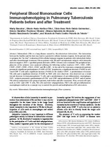
Fig. 1. Germline factor DDX4 is expressed in several blood-derived cancer cells. (a) RT-PCR results of six cell lines (KMS11, THP-1, hES, OPM-2, MM1S, and IM-9) for four gene products (PIWIL2, DDX4, GAPDH, and 36B4). Left panels, negative controls (lacking RT enzyme). Right panels, positive tests. (b) (i) Immunofluorescence results showing DDX4 localization in five cell types counterstained with tubulin and DNA. As indicated by arrows, DDX4 is localized on the mitotic spindle in cancer cells expressing DDX4. Scale bar = 5 lm. (ii) Relative signal intensity of DDX4 on the spindle of each cell line. The signal level of DDX4 was calculated by Image J and normalized to that of tubulin. The average relative value of each of the five cells undergoing mitosis is presented for each cell line. The value of IM-9 is set at 1.
chromosome replication, serving as a good DNA replication marker. The proportion of the EdU-positive cells against the total number of cells was then counted under the fluorescent microscope (Fig. 3b). The C1 and C2 cells showed decreased activities in DNA replication, yet the differences compared to control cells were relatively small. This result suggests that DDX4 may be necessary for cell proliferation but less so for S-phase entry and DNA replication. As DDX4/Vasa is also known to be involved in germ cell migration in various animals, we next tested whether DDX4
knockout affects activities of cancer cell migration by invasion assays. In C1 and C2, the number of cells migrated toward the FBS-containing well were significantly decreased, close to that of the negative control group (ORI with no FBS-containing well), whereas C3 showed relatively higher migration activities but still lower than the positive controls (Fig. 3c). These results suggest that DDX4 might be important for IM-9 cancer cell migration. Together, these results suggest that DDX4 activities function in proliferation and migration in IM-9 cancer cells.
© 2017 The Authors. Cancer Science published by John Wiley & Sons Australia, Ltd on behalf of Japanese Cancer Association.
Cancer Sci | 2017 | 4
www.wileyonlinelibrary.com/journal/cas
Original Article Schudrowitz et al.
Fig. 2. Construction of germline factor DDX4 knockout cell lines in IM-9 through clustered regularly interspaced short palindromic repeats (CRISPR)-Cas9 gene-targeting technology. (a) CRISPR guide RNAs (gRNAs) were designed within the third exon of the DDX4 gene to construct three knockout cell lines (C1, C2, and C3). (b) (i) Genomic PCR results for three CRISPR knockout cell lines (C1–C3), control cells introduced with scrambled gRNA sequence (SC), and original intact cells (ORI). (ii) Genomic PCR results detecting lentiviral vector sequence used for virus infection, showing effective introduction of CRISPR constructs in C1–C3 and SC cell lines. (c) DNA sequencing results of the third exon and its flanking region of the DDX4 gene depicted in (a). CRISPR knockout cell lines showed deletions in many of the clones sequenced, whereas controls of scrambled gRNAs showed no mutations. (d) (i) Immunoblot results showing reduced DDX4 protein expression in knockout cell lines (C1–C3) compared to control (SC). (e) Immunofluorescence results showing reduced DDX4 protein expression in knockout IM-9 cell lines (C1–C3) compared to control (SC, arrows). Numbers underneath of each image indicate the percentage of cells expressing DDX4. The number of DDX4-positive cells per the DAPI-positive cell number (a total cell population) was calculated by Image J. The total numbers of cells counted were: C1, 1222; C2, 1317; C3, 1471; and SC, 1686. Scale bar = 5 lm. F, forward; R, reverse.
DDX4 knockout downregulates expression of cell cycle regulators and oncogenes. As Vasa/DDX4 is an RNA-helicase and
known to function as a translational regulator, the cellular phenotypes described above may be due to changes in expression of DDX40 s downstream targets that are directly involved in cell replication and/or migration. To test whether DDX4 knockout in IM-9 cells changes the selected gene expression, we undertook quantitative RT-PCR. The potential downstream genes of interest included germline factors (ddx4 and piwil2) and oncogene/cell cycle factors (14-3-3r, cyclinB1, e2f1, and myc). Among those candidates, most transcripts were altered minimally (Fig. 4a) whereas the cyclinB1, e2f1, and chi3 l1 transcripts were found consistently downregulated in the DDX4-knockout cell lines, although still detectable. To test whether DDX4 knockout indeed changes protein expression of the above targets, we undertook immunoblots against each molecule of interest (Fig. 4b). As a result, we found that the positive cell cycle regulators CyclinB1 and E2F1 were downregulated, whereas a negative cell cycle regulator 14-3-3r was upregulated. As DDX4 is a translational regulator, these results suggest that CyclinB and E2F1 mRNAs could be targets of DDX4 and their protein production was downregulated in the absence of DDX4. Indeed, CyclinB is essential for M-phase entry and exit and has also been reported Cancer Sci | 2017 | 5
as one of Vasa’s targets in embryonic cells.(15) Its knockdown, therefore, may have blocked M-phase entry and indirectly resulted in spindle defects. E2F1 is a transcription factor and known to be highly active during the G1/S-phase transition, yet some of its target genes also function in G2/M phase.(28,29) Therefore, the reduction of CyclinB and E2F1 proteins that are critical for cell cycle progression might have caused cell proliferation defects in DDX4-knockout cell lines. It is also important to consider the phenotype observed here likely includes both direct and indirect effects of DDX4 knockout, because both DDX4 and some of its targets, including E2F1, likely regulate a wide range of translation and transcription in the cell. The further analysis of this process, including a broad screening of DDX40 s targets and cofactors by RNA immunoprecipitation sequencing and proteomics is thus necessary to parse out direct from indirect effects of DDX4 function. In contrast, 14-3-3r is a cell cycle repressor. It is unclear what increases its expression at this point in the DDX4-knockout. DDX4 likely targets many mRNAs for translation and one may thus speculate that DDX4 regulates a factor that suppresses 14-3-3r, and indirectly inhibits 14-3-3r protein expression. This change could also cause cell cycle delay, which is consistent with the phenotype seen in DDX4-knockout cell lines. Intriguingly, another germline factor PIWI was © 2017 The Authors. Cancer Science published by John Wiley & Sons Australia, Ltd on behalf of Japanese Cancer Association.
Original Article DDX4 functions in cancer phenotypes
www.wileyonlinelibrary.com/journal/cas
Fig. 3. Knockout of germline factor DDX4 causes defects in cell proliferation and migration. (a) Cell number analysis shows reduced counts in DDX4 knockout cell lines (C1, C2, and C3) compared to controls (scrambled [SC] and original intact cells [ORI]). This suggests that DDX4 might be important for cell proliferation. (b) (i) Immunofluorescence using 5-Ethynyl-20 -deoxyuridine (EdU) as indicator for DNA replication within knockout cell line C2 and control. Top panels shows consistent labeling of native DNA in C2 and control cells. Bottom panels show reduced DNA replication in C2 compared to the control. (ii) Graphical results of the DNA replication (EdU as indicator) in various cell lines in proportion with total cell count, normalized to SC. (c) Graphical results of invasion assay in various cell lines, normalized to control (ORI). Results indicate that DDX4 might be important for cell migration.
also upregulated in C1. As PIWI has been reported to target various transcripts in the germ line for destruction,(30) the overexpression of PIWI may have resulted in degradation of mRNAs important for cellular activities, the mechanisms of which need to be tested further in the future. Vasa has been reported to have broad targets and function as a general translational regulator in the embryo,(17) so we tested whether DDX4 knockout inhibits general protein synthesis (Fig. 4c). Each cell line was treated in methionine-depleted media for 3 h with HPG (a tagged methionine analog) that is incorporated into newly synthesized proteins and thus serves as a good tool to measure the amount of nascent protein synthesis. We found that the level of HPG incorporation was significantly decreased in DDX4 knockout cell lines. These results suggest that DDX4 might positively function in protein synthesis as it does in embryonic or germ cells(17,31,32) and help explain the phenotypic changes seen in the knockout cells. Discussion DDX4 is essential for proliferation and migration activities in somatic cancer cells. All animals tested so far appear to express
Vasa/DDX4 in their germ line at one point in their life. As its © 2017 The Authors. Cancer Science published by John Wiley & Sons Australia, Ltd on behalf of Japanese Cancer Association.
knockdown often results in infertility, Vasa/DDX4 has been considered and used as a germline marker for decades in the field.(7–13) Its function in the germ line was also concluded to be “germline-specific”, essential for functions in cell proliferation and migration of germ cells and translation of the mRNAs in the germ cells. In this study, however, we reported that DDX4 is widely expressed in several blood-derived cancer cells and contributes to cell proliferation and migration. These results directly suggest that DDX4 has functionalities outside of the germ line, yet its expression may be limited to contextspecific periods of cellular development, to rapidly dividing cells such as embryonic cells as reported previously, and in human cancer cells as reported in this study. DDX4/Vasa has also been reported to be transiently expressed during regeneration,(19) the tissues of which also undergo active cell proliferation and phenotypic changes. With the results here, we might consider that DDX4 has various downstream targets that are essential more generally for cell proliferation and migration (Fig. 5). These results are also consistent with Vasa’s activities in embryonic cells; Vasa has been reported to regulate widespread translation essential for embryonic cell division and development.(17) Although what Vasa does on the spindle has been unclear in any cell or organism at this point, in embryonic cells, DDX4/Vasa appears to Cancer Sci | 2017 | 6
www.wileyonlinelibrary.com/journal/cas
Original Article Schudrowitz et al.
Fig. 4. Knockout of germline factor DDX4 caused little change in gene expression at the transcript level yet impacted protein expression. (a) RT–quantitative PCR results for knockout cell lines (C1, C2, and C3) and two controls (scrambled [SC] and original intact cells [ORI]). DDX4 knockout showed little effect in oncogene expression at the transcript level. (b) Immunoblot results showing protein expression in DDX4 knockout cell lines (C1–C3) and control (SC). In DDX4 knockout cells, positive cell cycle regulators CCNB1 (CyclinB1) and E2F1 were downregulated and negative cell cycle regulator 14-3-3r was upregulated. Germline factor PIWI was upregulated. (c) L-homopropargylglycine (HPG) labeling assay. C2 knockout cell line and control SC are shown as representative images of HPG assay (i). Knockout cell lines C1–C3 showed reduced HPG signal, indicating nascent protein synthesis compared to the control SC, suggesting that DDX4 could have a role in positive protein regulation.
Fig. 5. Hypothetical models for the molecular pathway of germline factor DDX4 (a) and for DDX4 function in cancer cells (b).
function as a general translational regulator rather than as a regulator of specific targets. It is thus intriguing to speculate that, in cancer cells and embryonic cells alike, DDX4/Vasa may facilitate M-phase translation(33) on the spindle to assist sufficient protein production necessary for rapid cell division. This idea needs to be experimentally tested in the future by manipulating Vasa function specifically on the spindle. Through this mechanism, DDX4 could provide a molecular environment for cancer cells to maximize proliferative activities and potentially contribute in neoplastic features of cancer
cells through upregulated protein expression. The thorough screening of DDX40 s downstream targets will be essential to understand the details of DDX4 functions in cancer cell regulation. Another important question to be addressed in the future is why DDX4/Vasa becomes essential only when it is expressed in the cells. Not all cells, neither embryonic nor cancer cells, appear to have DDX4/Vasa. Yet DDX4/Vasa expression, normally or abnormally induced, seems to change the cellular dependency for translational activity to itself. Perhaps the
Cancer Sci | 2017 | 7
© 2017 The Authors. Cancer Science published by John Wiley & Sons Australia, Ltd on behalf of Japanese Cancer Association.
Original Article DDX4 functions in cancer phenotypes
www.wileyonlinelibrary.com/journal/cas
changes ensued by DDX4/Vasa expression might directly and indirectly alter the global translational landscape in the cell. We thus predict that other types of cancer cells that express the DDX4 transcript, such as germ cell cancer, breast cancer, and small-cell lung cancer (data not shown), may also depend on its activity for survival. To be noted, DDX40 s function is heavily regulated by post-transcriptional regulation. Therefore, it will be necessary to test its expression and localization at the protein level in each of these cell lines. These further exhaustive screenings as well as identification of the upstream regulator(s) of DDX4 will be essential to understand how DDX4 directly contributes to cellular activities of cancer cells. Furthermore, as DDX4 is generally not expressed in benign adult somatic cells,(14) it could serve as a potential target to detect early steps of malignancy among heterogeneous tumors and/or for a cancer therapy in the future. Our results suggest that another germline factor, PIWI, also appears to be expressed with DDX4 in these same cancer cells, and thus
may serve as important co-target for cancer therapy. The detailed functional mechanisms of DDX4 with other germline factors and its downstream targets needs to be tested further in various cancer cell types and in patient tissues.
References
18 Janic A, Mendizabal L, Llamazares S, Rossell D, Gonzalez C. Ectopic expression of germline genes drives malignant brain tumor growth in Drosophila. Science 2010; 330: 1824–7. 19 Wagner DE, Ho JJ, Reddien PW. Genetic regulators of a pluripotent adult stem cell system in planarians identified by RNAi and clonal analysis. Cell Stem Cell 2012; 10: 299–311. 20 Hashimoto H, Sudo T, Mikami Y et al. Germ cell specific protein VASA is over-expressed in epithelial ovarian cancer and disrupts DNA damageinduced G2 checkpoint. Gynecol Oncol 2008; 111: 312–9. 21 Kim KH, Kang YJ, Jo JO et al. DDX4 (DEAD box polypeptide 4) colocalizes with cancer stem cell marker CD133 in ovarian cancers. Biochem Biophys Res Commun 2014; 447: 315–22. 22 Shalem O, Sanjana NE, Hartenian E et al. Genome-scale CRISPR-Cas9 knockout screening in human cells. Science 2014; 343: 83–7. 23 Sanjana NE, Shalem O, Zhang F. Improved lentiviral vectors and genomewide libraries for CRISPR screening. Nat Methods 2014; 11: 783–4. 24 Barretina J, Caponigro G, Stransky N et al. The cancer cell line encyclopedia enables predictive modelling of anticancer drug sensitivity. Nature 2012; 492: 290. 25 Xiol J, Spinelli P, Laussmann MA et al. RNA clamping by Vasa assembles a piRNA amplifier complex on transposon transcripts. Cell 2014; 157: 1698–711. 26 Tan Y, Liu L, Liao M et al. Emerging roles for PIWI proteins in cancer. Acta Biochim Biophys Sin (Shanghai) 2015; 47: 315–24. 27 Voronina E, Lopez M, Juliano CE et al. Vasa protein expression is restricted to the small micromeres of the sea urchin, but is inducible in other lineages early in development. Develop Biol 2008; 314: 276–86. 28 Ren B, Cam H, Takahashi Y et al. E2F integrates cell cycle progression with DNA repair, replication, and G(2)/M checkpoints. Gene Dev 2002; 16: 245–56. 29 Ishida S, Huang E, Zuzan H et al. Role for E2F in control of both DNA replication and mitotic functions as revealed from DNA microarray analysis. Mol Cell Biol 2001; 21: 4684–99. 30 Zhang P, Kang JY, Gou LT et al. MIWI and piRNA-mediated cleavage of messenger RNAs in mouse testes. Cell Res 2015; 25: 193–207. 31 Carrera P, Johnstone O, Nakamura A, Casanova J, J€ackle H, Lasko P. VASA mediates translation through interaction with a Drosophila yIF2 homolog. Mol Cell 2000; 5: 181–7. 32 Liu N, Han H, Lasko P. Vasa promotes Drosophila germline stem cell differentiation by activating mei-P26 translation by directly interacting with a (U)-rich motif in its 3’ UTR. Genes Dev 2009; 23: 2742–52. 33 Gross PR. FRY BJ. Continuity of protein synthesis through cleavage metaphase. Science 1966; 153: 749–51.
1 Hanahan D, Weinberg RA. Hallmarks of cancer: the next generation. Cell 2011; 144: 646–74. 2 Yachida S, Iacobuzio-Donahue CA. Evolution and dynamics of pancreatic cancer progression. Oncogene 2013; 32: 5253–60. 3 Simpson AJ, Caballero OL, Jungbluth A, Chen YT, Old LJ. Cancer/testis antigens, gametogenesis and cancer. Nat Rev Cancer 2005; 5: 615–625. 26 4 Miles WO, Korenjak M, Griffiths LM, Dyer MA, Provero P, Dyson NJ. Post-transcriptional gene expression control by NANOS is up-regulated and functionally important in pRb-deficient cells. EMBO J 2014; 33: 2201–15. 5 Tan Y, Liu L, Liao M et al. Emerging roles for PIWI proteins in cancer. Acta Biochim Biophys Sin (Shanghai) 2015; 47: 315–24. 6 Wang Z, Liu N, Shi S, Liu S, Lin H. The Role of PIWIL4, an Argonaute Family Protein, in Breast Cancer. J Biol Chem 2016; 291: 10646–58. 7 Hay B, Jan LY, Jan YN. A protein component of Drosophila polar granules is encoded by vasa and has extensive sequence similarity to ATP-dependent helicases. Cell 1988; 55: 577–87. 8 Lasko PF, Ashburner M. The product of the Drosophila gene vasa is very similar to eukaryotic initiation factor-4A. Nature 1988; 335: 611–7. 9 Linder P. Dead-box proteins: a family affair active and passive players in RNP-remodeling. Nucleic Acids Res 2006; 34: 4168–80. 10 Raz E. The function and regulation of vasa-like genes in germ-cell development. Genome Biol 2000; 1: 1017.1. 11 Sengoku T, Nureki O, Nakamura A, Kobayashi S, Yokoyama S. Structural basis for RNA unwinding by the DEAD-box protein Drosophila Vasa. Cell 2006; 125: 287–300. 12 Gustafson EA, Wessel GM. DEAD-box helicases: post translational regulation and function. Biochem Biophys Res Commun 2010; 395: 1–6. 13 Lasko P. The DEAD-box helicase Vasa: evidence for a multiplicity of functions in RNA processes and developmental biology. Biochem Biophys Acta 2013; 1829: 810–6. 14 Poon J, Wessel GM, Yajima M. An unregulated regulator: ectopic Vasa expression and tumorigenesis. Develop Biol 2016; 415: 24–32. 15 Yajima M, Wessel GM. The DEAD-box RNA helicase Vasa functions in embryonic mitotic progression in the sea urchin. Development 2011; 138: 2217–22. 16 Schwager EE, Meng Y, Extavour CG. Vasa and piwi are required for mitotic integrity in early embryogenesis in the spider Parasteatoda tepidariorum. Dev Biol 2014; 402: 276–90. 17 Yajima M, Wessel GM. The germline factor Vasa functions broadly in somatic cells: mRNA clustering, translational regulation, and wound healing. Development 2015; 142: 1960–70.
Acknowledgments We thank Dr. Irene M. Ghobrial at Dana Farber Cancer Institute in Boston for sharing cancer cell lines. We also thank Professor Chun Geun Lee and members of the PRIMO at Brown University for generous support. This work was supported by the American Heart Association Scientist Development Grant to M.Y. (14SDG18350021), and by National Institutes of Health (2R01HD028152) and National Science Foundation (IOS-1120972) grants to G.M.W.
Disclosure Statement The authors have no conflict of interest.
Supporting Information Additional Supporting Information may be found online in the supporting information tab for this article: Table S1. Primers and guide RNA (gRNA) sequences used in this study.
© 2017 The Authors. Cancer Science published by John Wiley & Sons Australia, Ltd on behalf of Japanese Cancer Association.
Cancer Sci | 2017 | 8