(1982), Stitt et al. (1985) .... Mac Donald, F. D. and apRees, Τ. (1983b) Phytochemistry, 22, 1141. ... Stitt, Î., Cseke, C. and Buchanan, B. B. (1985) Physiol. Veg.
J. Biosci., Vol. 15, Number 2, June 1990, pp. 77–82. © Printed in India.
Enzyme activities associated with developing wheat (Triticum aestivum L.) grain amyloplasts R. MAHAJAN and RANDHIR SINGH* Department of Chemistry and Biochemistry, Haryana Agricultural University, Hisar 125 004, India MS received 19 December 1989; revised 24 April 1990 Abstract. Intact amyloplasts from endosperm of developing wheat grains have been isolated by first preparing the protoplasts and then fractionating the lysate of the protoplasts on percoll and ficoll gradients, respectively. Amyloplasts isolated as above were functional and not contaminated by cytosol or by organelles likely to be involved in carbohydrate metabolism. The enzyme distribution studies indicated that ADP-glucose pyrophosphorylase and starch synthase were confined to amyloplasts, whereas invertase, sucrose synthase, UDP-glucose pyrophosphorylase, hexokinase, phosphofructokinase-2 and fructose-2,6-P2ase were absent fro the amyloplast and mainly confined to the cytosol. Triose-P isomerase, glyceraldehyde-3-P dehydrogenase, phosphohexose isomerase, phosphoglucomutase, phosphofructokinase, aldolase, PPi-fructose-6-P-l phosphotransferase, and fructose-l,6-P2ase, though predominantly cytosolic, were also present in the amyloplast. Based on distribution of enzymes, a probable pathway for starch biosynthesis in amyloplasts of developing wheat grains has been proposed. Keywords.
Wheat; Triticum aestivum; amyloplasts; enzymes; carbohydrate metabolism.
Introduction Starch synthesis in higher plants is confined to chloroplasts in green photosynthetic tissues and amyloplasts in non-green storage tissues such as the endosperm tissue of the developing wheat grain (Keeling et al., 1988). The amyloplast consists of a starch granule and plastid stroma enclosed by a double membrane (Badenhuizen, 1969). Based on in vitro assays of enzymes presumed to be involved in starch synthesis, a biosynthetic pathway has been proposed for storage tissues including cereal grains (Singh and Mehta, 1986). Though the pathway proposed has received general acceptance, it does not take into account the intracellular compartmentation of starch synthesis within the amyloplast and the selective permeability of the plastid membranes. Hence, it could not be proved conclusively as to how translocated sucrose is converted to starch in storage tissues. The transport properties of chloroplasts have been studied in detail (Heber, 1974; Heber and Heldt, 1981). However, parallel information is completely lacking for amyloplasts. This has been mainly because of the reason that intact amyloplasts from cereal grains could not be isolated till recently (Singh, 1989). However, techniques have now been developed and successfully employed for the isolation of intact amyloplasts from cereal grains (MacDonald and apRees, 1983a, b; Echeverria et al., 1985; Entwistle et al., 1988; Santha et al., 1988). In our present attempt, we *To whom all correspondance should be addressed. Abbreviations used: BSA, bovine serum albumin; PFP, fructose-6-phosphate-1-phosphotransferase; PFK-2, fructose-6-phosphate 2-kinase; PFK, phosphofructokinase.
77
78
Mahajan and Singh
have been successful in isolating intact amyloplasts from developing wheat grains. Furthermore, we have determined the activities of the enzymes involved in carbohydrate metabolism and starch synthesis associated with cytosol and amyloplastic fractions of wheat grains. Based on the distribution of enzymes,a probable mechanism for the biosynthesis of starch in amyloplasts of developing wheat grains has been proposed. Materials and methods Chemicals All the biochemicals used in the present investigation were procured from Sigma Chemical Co., St. Louis, Missouri, USA. All other chemicals used were of high purity analytical grade obtained from BDH and/or E. Merck. Plant material Immature wheat grains were harvested from field grown wheat crop (cv. WH- 157) at day 20 after anthesis, endosperms removed and stored in liquid nitrogen for further use. Isolation of amyloplasts Amyloplasts were isolated essentially by following the methods of Echeverria et al. (1985) and Emes and England (1986) with slight modifications. Unless stated otherwise, all steps were carried out at 0–4°C. Endosperm sections (1g) were incubated for 18–20 h in 50 mM tricine-NaOH buffer (pH 7·9) containing 1 mM EDTA, 5 mM MgCl2, 0·5 Μ sucrose, 0·1% bovine serum albumin (BSA), 2% cellulase (Onozuka, R–10), 1% pectinase (Sigma), 1% polyvinylpyrrolidone, 1 mM KC1, 1 mM CaCl2 and 10 mM mercaptoethanol at 10°C. The endosperm sections were pressed gently with the help of fine brush. The homogenate was passed through a single layer of muslin cloth and underlaid with 50 mM of tricine - NaOH buffer (pH 7·9) containing 0·5 Μ sucrose, 1 mM EDTA, 5 mM MgCl2, 0·1% BSA, 1 mM KCl, 1 mM CaCl2, 10 mM mercaptoethanol and 10% percoll in the ratio of 1:1. After allowing to stand for 1 h, the homogenate was centrifuged at 4000 g for 20 min in a swing out rotor (Hitachi model) The pellet was suspended in 50 mM tricine-NaOH buffer (pH 7·9) containing 0·5 Μ sucrose, 1 mM EDTA, 1 mM MgCl2, 0·1% BSA and 10% mercaptoethanol. Amyloplasts were released by passing the above suspension through a 19 gauge syringe. The amyloplasts were then separated by layering the suspension on discontinuous ficoll gradient of 30–100% at 1×g. After setting at 1×g for3 h, the amyloplasts appearing as bands were collected. The upper layer (30–50%) was free from amyloplasts contamination and centrifuged at 15,000 g for 20 min. The pellet obtained was dissolved in the supernatant after centrifugation in a swing-out rotor at 4000 g. This was recentrifuged at 15,000 g for 30 min to get rid of nuclei and mitochondria. The supernatant obtained at this step was referred to as cytosolic fraction.
Enzymes of carbohydrate metabolism in amyloplasts
79
For getting photographs under light microscope, the amyloplasts were stained with iodine solution. The length and width of amyloplasts were measured with the help of oculometer. Enzyme assays Ficoll gradient layer (60–70%) was collected and vigorously stirred on a vortex mixer for several seconds to rupture the amyloplast membranes. The suspension was centrifuged for 5 min at 1000 g. The supernatant obtained was used directly for enzyme assays except that of invertase for which the supernatants were dialyzed overnight at 4°C against 21 of 5 mM phosphate buffer (pH 7) containing 5 mM EDTA. All assays were carried out at 30°C, in the various reaction mixtures described below. Assays were conducted in the range of concentrations of enzyme where the activity increased linearly with increase in the amount of enzyme preparation and the reaction time. The background values were routinely taken as the activities obtained with denatured enzyme (the enzymes were denatured immediately after their addition to the reaction mixtures). Triose phosphate isomerase (EC 5.3.1.1), aldolase (EC 4.1.2.13), fructose-1, 6bisphosphatase (EC 3.1.3.11), phosphohexose isomerase (EC 5.3.1.9), and phosphorglucomutase (EC 2.7.5.1) were assayed spectrophotometrically following the procedures of MacDonald and apRees (1983a). Hexokinase (EC 2.7.1.1), sucrose synthase (EC 2.4.2.13), invertase (EC 3.2.1.26), ADP-glucose pyrophosphorylase (EC 2.7.7.27), UDP-glucose pyrophosphorylase (EC 2.7.7.9) and starch synthase (EC 2.4.1.21) were assayed as described earlier (Kumar and Singh, 1984). Fructose-6-phosphate-l-phosphotransferase (PFP, EC 2.7.1.90), fructose-6-phosphate, 2-kinase (PFK-2, EC 2.7.1.105), fructose 2,6-P2ase (EC 3.1.3.46), phosphofructokinase (PFK, EC 2.7.1.11) and glyceraldehyde-3phosphate dehydrogenase (EC 1.2.1.12) were assayed as per the procedures of VanSchaftingen et al. (1982), Stitt et al. (1985), Kruger and Beevers (1985), Kelly and Latzko (1979) and Wu and Racker (1959), respectively. Results and discussion The amyloplast preparation obtained by the procedure detailed in methods was pure and not contaminated with cytosol as judged by the marker enzymes (ADPglucose pyrophosphorylase and starch synthase). The length and width of these amyloplasts ranged from 13–39 and 10–34 µm, respectively and were ovoid or round in shape. Methods involving the use of sucrose gradients employed earlier by Gaynor and Galston (1983), MacDonald and apRees (1983a) and Nishimura and Beevers (1978) for isolating plastids were not successful in the present case. Our procedure for amyloplast separation took advantage of the relatively high density of the amyloplast which allowed them to settle through a ficoll gradient. The purity of the amyloplast fraction prompted us to further study the distribution of enzymes of carbohydrate metabolism in cytosolic and amyloplastic fractions of endosperm to work out the compartmentalization of the reactions of sucrose metabolism and starch biosynthesis in developing wheat endosperm.
80
Mahajan and Singh
The enzymes listed in table 1 could be divided into 3 main groups, i.e., (i) enzymes which were present only in cytosol, (ii) enzymes which were present only in amyloplast and (iii) the enzymes present in both fractions. The enzymes of sucrose hydrolysis and further metabolism, viz., invertase, sucrose synthase, hexokinase and UDP-glucose pyrophosphorylase were absent in amyloplast and present only in cytosolic fraction, indicating these to be cytoplasmic. Similarly, PFK-2 and fructose-2,6-P2ase were also mainly cytosolic. MacDonald and apRees (1983a) and Echeverria et al. (1988) obtained similar results for invertase and sucrose synthase in soybean suspension cultures and maize endosperm. However, UDP-glucose pyrophosphorylase which was absent from amyloplasts of rice (Nakamura et al., 1989), soybean suspension cultures (MacDonald and apRees, 1983a) could be detected in the amyloplasts of maize endosperm (Echeverria et al., 1988). Hexokinase was also absent from the amyloplasts of maize endosperm. Contrary to this, ADP-glucose pyrophosphorylase and starch synthase were present only in amyloplast. Similar localization for these enzymes has been suggested for storage tissue of rice (Nakamura et al., 1989) and maize (Echeverria et al., 1988). Table 1. Enzymes of carbohydrate metabolism in the amyloplast and cytosol.
Each value is the mean of 6 replications
Phosphoglucoisomerase, phosphoglucomutase, PFK, aldolase, triose phosphate isomerase, fructose-l,6-P 2ase, glyceraldehyde-3P-dehydrogenase and PFP, though present predominantly in cytosolic fraction were also present in the amyloplast. Similar distribution for these enzymes has also been observed earlier in endosperm tissue of maize (Echeverria et al., 1988) and soybean suspension cultures (MacDonald and apRees, 1983a). The confinement of starch synthase and ADP-glucose pyrophosphorylase to the amyloplast and of UDP-glucose pyrophosphorylase to the cytosol strongly suggests the ADP-glucose, rather than UDP-glucose, is the immediate precursor of starch. The absence of invertase, sucrose synthase and UDP-glucose pyrophosphorylase from amyloplasts indicates that this organelle
Enzymes of carbohydrate metabolism in amyloplasts
81
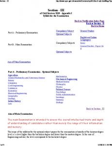
lacks any enzyme involved in sucrose metabolism. Thus sucrose cannot be considered as the substrate which could cross the amyloplast membrane but is metabolized in cytosol mainly by invertase, sucrose synthase, PFP and UDP-glucose pyrophosphorylase. Accordingly, the following pathway may be suggested for the conversion of sucrose to starch in developing wheat endosperm. The pathway is mainly comprised of 3 sections/steps. First in the cytosol, sucrose is degraded to form triose-P (DHAP via reactions catalysed by invertase/sucrose synthase, UDP-glucose pyrophosphorylase, phosphoglucomutase, phosphohexose isomerase, fructokinase, PPi-PFK (and ATP-PFK), aldolase and triosephosphate isomerase (figure 1). DHAP is then translocated to amyloplast through phosphate translocator and converted to ADP-glucose (substrate for starch) through the reactions of triose phosphate isomerase, aldolase, fructose-1, 6-P2ase, phosphorhexoseisomerase, phosphoglucomutase and ADP-glucose-pyrophosphorylase. Finally, starch is synthesized from ADP-glucose through the reactions of starch synthase and Q enzyme. If this pathway is operational, we must expect atleast two forms each of triose phosphate isomerase, aldolase, fructose–1,6-P2ase, phosphoglucomutase and phosphohexose isomerase. We have already demonstrated the presence of these forms in endosperm tissue of developing wheat grains (Anand and Singh, 1985; Sangwan and Singh, 1987a, b, 1988, 1989, 1990).
Figure 1. Proposed metabolic pathway for starch synthesis in amyloplasts of developing wheat grain. (1), Sucrose synthase; (2), UDP-glucose pyrophosphorylase; (3), hexokinase; (4), phosphoglucomutase; (5), phosphohexose isomerase; (6), PFK; (7), fructose-1, 6bisphosphatase; (8), aldolase; (9), triose phosphate isomerase; (10), phosphate translocator; (11), hexose translocator; (12), ADP-glucose pyrophosphorylase; (13), starch synthase.
Involvement of triose-P in the biosynthesis of starch in non-photosynthetic tissues has been proposed by MacDonald and apRees (1983a, b), Singh and Mehta (1986) and by Echeverria et al. (1988). These results have been supported by the observations of Ngernprasirtairi et al. (1988) who found that, in the amyloplast envelope membrane from cultured sycamore cells, the Pi translocator like protein with molecular weight of 31 kDa was immunoreactive with an antibody raised against the pea chloroplast Pi translocator. In contrast, Keeling et al. (1988) and Tyson and apRees (1988) have reported glucose-1-P to be the preferred substrate
82
Mahajan and Singh
involved in translocation and synthesis of starch in amyloplasts of developing wheat grains. However, these authors were not sure whether this was the sole source of carbon for starch synthesis in wheat amyloplasts. Accordingly, in the present case, we have shown the involvement of both triose-P and glucose- 1-P in the synthesis of starch. The uncertainity as to which carbon compound is transported across the amyloplast membrane may only be resolved by studying transport properties of the isolated translocators from functional amyloplasts as has been done with chloroplast translocators. This aspect is currently being pursued in our laboratory. Acknowledgement The senior author is thankful to the Council of Scientific and Industrial Research, New Delhi, for financial assistance in the form of a fellowship. References Anand, S. and Singh, R. (1985) Plant Physiol. (Suppl.), 77, 47. Badenhuizen, N. P. (1969) Biogenesis of starch granules in higher plants (New York: Appleton-Century Crofts) Echeverria, E., Boyer, C. D., Liu, K. C. and Shannon, J. C. (1985) Plant Physiol., 77, 513. Echeverria, E., Boyer, C. D., Thomas, P. Α., Liu, K. C. and Shannon, J. C. (1988) Plant Physiol., 86, 786. Emes, M. J. and England, S. (1986) Planta, 168, 161. Entwistle, G., Tyson, R. H. and apRees, T. (1988) Phytochemistry, 27, 993. Gaynor, J. J. and Galston, A. W. (1983) Plant Cell Physiol., 24, 411. Heber, U. (1974) Annu. Rev. Plant Physiol., 25, 393. Heber, J. and Heldt, Η. W. (1981) Annu. Rev. Plant Physiol., 32, 139. Keeling, P. L., Wood, J. R., Tyson, R. H. and Bridges, I. G. (1988) Plant Physiol., 87, 311. Kelly, G. J. and Latzko, E. (1977) Plant Physiol, 60, 290. Kruger, Ν. J. and Beevers, Η. (1985) Plant Physiol., 77, 358. Kumar, R. and Singh, R. (1984) J. Agric. Food Chem., 32, 806. Mac Donald, F. D. and apRees, Τ. (1983a) Biochem. Biophys. Acta, 755, 81. Mac Donald, F. D. and apRees, Τ. (1983b) Phytochemistry, 22, 1141. Nakamura, Υ., Yuki, Κ., Park, S. Y. and Ohya, T. (1989) Plant Cell Physiol., 30, 833. Ngernprasirtairi, J., Harinasut, P., Macherel, D., Strzalka, K., Takabe, T., Akazawa, A. and Kojima, K. (1988) Plant Physiol., 87, 371. Nishimura, M. and Beevers, Η. (1978) Plant Physiol., 62, 40. Sangwan, R. S. and Singh, R. (1987a) Indian J. Biochem. Biophys., 24, 33. Sangwan, R. S. and Singh, R. (1987b) Plant Physiol. Biochem., 25, 745. Sangwan, R. S. and Singh, R. (1988) Physiol. Plant., 73, 21. Sangwan, R. S. and Singh, R. (1989) J. Biol. Sci., 14, 47. Sangwan, R. S. and Singh, R. (1990) Indian J. Biochem. Biophys., 27, 23. Santha, I. M., Swaroop, R. and Mehta, S. L. (1988) Indian J. Biochem. Biophys., 25, 532. Singh, R. (1989) in Recent advances in plant biochemistry (eds S. L. Mehta, M. L. Lodha and P. V. Sane) (New Delhi: IC AR) p. 106. Singh, R. and Mehta, S. L. (1986) J. Sci. Ind. Res., 45, 336. Stitt, Μ., Cseke, C. and Buchanan, B. B. (1985) Physiol. Veg., 23, 819. Tyson, R. H. and apRees, Τ. (1988) Planta, 175, 33. VanSchaftingen, Ε., Lederer, Β., Batrons, R. and Hers, H. G. (1982) Eur. J. Biochem., 129, 191. Wu, R. and Racker, E. (1959) J. Biol. Chem., 234, 1029.