To my Father, George iv ...... In a study by Ojemann and Whitaker (1978), localisation of the two languages in the cerebral cortex was mapped in two bilinguals ...
http://researchspace.auckland.ac.nz
ResearchSpace@Auckland
Copyright Statement The digital copy of this thesis is protected by the Copyright Act 1994 (New Zealand). This thesis may be consulted by you, provided you comply with the provisions of the Act and the following conditions of use: • • •
Any use you make of these documents or images must be for research or private study purposes only, and you may not make them available to any other person. Authors control the copyright of their thesis. You will recognise the author's right to be identified as the author of this thesis, and due acknowledgement will be made to the author where appropriate. You will obtain the author's permission before publishing any material from their thesis.
To request permissions please use the Feedback form on our webpage. http://researchspace.auckland.ac.nz/feedback General copyright and disclaimer In addition to the above conditions, authors give their consent for the digital copy of their work to be used subject to the conditions specified on the Library Thesis Consent Form.
A behavioural and functional imaging investigation of Stroop task performance in late proficient bilinguals
Gjurgjica Badzakova-Trajkov
Department of Psychology, The University of Auckland, Auckland, New Zealand
A thesis submitted in fulfilment of the requirements for the degree of Doctor of Philosophy, The University of Auckland, 2008
Abstract In this thesis, Stroop task performance was investigated (using behavioural, electrophysiological and functional magnetic resonance imaging (fMRI) techniques) in late and proficient adult bilinguals currently living in the second language (L2) environment. Monolingual participants, matched for age and handedness, were recruited as controls. The Stroop colour-word task was considered an appropriate tool to test the general hypothesis that bilingualism might influence executive or cognitive control processes. In Study One, a dual-task paradigm was used for assessing the lateralisation of language functions (given the linguistic nature of the Stroop paradigm used here) in the bilinguals (MacedonianEnglish/M-E). Bilinguals showed a more bilateral hemispheric involvement, for both languages, compared to monolinguals. These findings also provided supporting evidence for the hypothesis of greater right-hemispheric involvement for language in bilinguals. In Study Two, two behavioural Stroop task paradigms (manual and verbal) were used in order to assess the magnitude of the Stroop effect between the groups. Bilinguals (M-E, German-English/GE) showed a trend of smaller interference scores across both languages compared to monolinguals. In Study Three, manual Stroop task performance with concurrent electroencephalograph (EEG) recording revealed that bilinguals had temporal shifts in the N400 component (of about 30-40 ms) for the interference comparison for both languages compared to monolinguals. Also, relative to monolinguals, M-E bilinguals (for both L1 and L2) and G-E bilinguals (for L2) had fewer electrodes over frontal and central sites with a significant amplitude difference in the interference comparison (i.e., a reduced interference effect). In Study Four, the neural substrates engaged during Stroop task performance were investigated using fMRI. In general, monolinguals showed greater activation in regions such as the prefrontal cortex and anterior cingulate (regions associated with good executive control). This suggested that relative to bilinguals, monolinguals require more neural resources to accomplish conflict resolution. Taken together, Stroop task performance in late and proficient bilinguals currently living in the L2 environment differed from that of monolinguals across all methods of investigation. It appears that cognitive processing changes at the executive level can be observed as a result of bilingualism. The results also provide some evidence for changes in L1 processing following late L2 acquisition, as similar results across both languages and tasks were observed for the M-E bilinguals. It is also possible
ii
that slight modifications to cerebral laterality as a result of the late learning of (and continuous exposure to) a second language could contribute to these differences in executive functioning. The language environment might therefore be a major factor in the lateralisation of language processing and executive functioning in bilinguals. These conclusions, though tentative and require further investigation, have important implications for language and executive processing in general and for theories regarding cognitive flexibility in bilinguals.
iii
To my Father, George
iv
Acknowledgements First and foremost, I would like to thank my supervisors, Dr Karen Waldie and Dr Ian Kirk, for being exceptionally supportive throughout my PhD as both supervisors and dear friends. I am extremely grateful for your constant encouragements and for making me believe in myself as a researcher. I would like to thank all the people, my colleagues, in the department who have become my friends as well, and have helped me out in any way possible. A special thanks to Branka Milivojevic for generously giving up her time and sharing her EEG and fMRI knowledge with me. On a personal level, the unwavering support of my family has guided me through this long process. To my husband Vane, thank you for being the voice of reason in the most difficult of times and for believing in me. To my beautiful daughter Anastasia, my mother Ubavka, my sister Dobrila, my mother-in-law Elica, and the rest of the family and friends, thank you for being there for me. Finally, to my late father, who has been my inspiration, I hope I made you proud. The following thesis was made possible by the University of Auckland Doctoral Scholarship and all the people who unselfishly participated in my experiments.
v
Table of Contents Abstract....................................................................................................................................... ii Acknowledgements .....................................................................................................................v Table of Contents .......................................................................................................................vi List of Tables ..............................................................................................................................ix List of Figures........................................................................................................................... xii List of Abbreviations ................................................................................................................xiv List of Publications....................................................................................................................xv List of Conference Abstracts .....................................................................................................xv Preface .........................................................................................................................................1 CHAPTER 1: Introduction ..........................................................................................................4 What is bilingualism? ..............................................................................................................4 Techniques used in earlier research.........................................................................................5 Language representation in bilinguals...................................................................................11 Language lateralisation in general.....................................................................................12 Right-hemisphere involvement in language for bilinguals ...............................................17 Left Hemisphere for Language in Bilinguals too? ............................................................27 Language switching and executive functions in bilinguals ...................................................39 Language switching hypotheses ........................................................................................40 Factors that affect language switching ability ...................................................................42 Executive functions in bilinguals ......................................................................................46 The current research ..............................................................................................................49 CHAPTER 2: General methods.................................................................................................52 Participants ............................................................................................................................52 Screening assessment ............................................................................................................53 Proficiency assessment ......................................................................................................53 Handedness........................................................................................................................55 Experimental tasks.................................................................................................................58 Dual-task............................................................................................................................58 Stroop Colour-Word Task (Verbal Version) .....................................................................59 Stroop Colour-Word Task (Manual Version One) ............................................................60 Stroop Colour-Word Task (Manual Version Two) ...........................................................61 General Testing Procedure ....................................................................................................62 Screening assessment ........................................................................................................62 Experimental tasks.............................................................................................................63 General Data Analysis...........................................................................................................63 Behavioural data ................................................................................................................63 Electrophysiological data ..................................................................................................64 fMRI data...........................................................................................................................65 CHAPTER 3: Dual-task performance in late proficient bilinguals...........................................67 Abstract..................................................................................................................................67
vi
Introduction ...........................................................................................................................68 Method...................................................................................................................................73 Participants ........................................................................................................................73 Stimuli and Procedure .......................................................................................................75 Data Analysis.....................................................................................................................76 Results ...................................................................................................................................77 Discussion..............................................................................................................................81 CHAPTER 4: Stroop colour-word task performance in late proficient bilinguals: a behavioural study ..........................................................................................................................................85 Abstract..................................................................................................................................85 Introduction ...........................................................................................................................86 Experiment 1 .........................................................................................................................90 Method...............................................................................................................................90 Results ...............................................................................................................................93 Experiment 2 .........................................................................................................................98 Method...............................................................................................................................98 Results .............................................................................................................................101 Discussion............................................................................................................................105 CHAPTER 5: Stroop colour-word task performance in late proficient bilinguals: an ERP study .................................................................................................................................................110 Abstract................................................................................................................................110 Introduction .........................................................................................................................111 Method.................................................................................................................................115 Participants ......................................................................................................................115 Stimuli .............................................................................................................................116 Procedure .........................................................................................................................116 EEG Apparatus and ERP Averaging ...............................................................................117 Results .................................................................................................................................119 Behavioural results ..........................................................................................................119 EEG results......................................................................................................................125 Discussion............................................................................................................................139 CHAPTER 6: Stroop colour-word task performance in late proficient bilinguals: an fMRI study ........................................................................................................................................145 Abstract................................................................................................................................145 Introduction .........................................................................................................................147 Method.................................................................................................................................152 Participants ......................................................................................................................152 Stimuli .............................................................................................................................153 Procedure .........................................................................................................................154 Image acquisition.............................................................................................................155 Image pre-processing and analysis ..................................................................................155 Results .................................................................................................................................157 Behavioural results ..........................................................................................................157 fMRI results.....................................................................................................................160 Discussion............................................................................................................................172 CHAPTER 7: General discussion............................................................................................180 Lateralisation of language processing in late and proficient bilinguals ..............................184 vii
The effects of bilingualism on executive functions: evidence from Stroop task performance .............................................................................................................................................186 The effect of bilingualism on L1 processing .......................................................................191 Implications, limitations, and future research .....................................................................193 Conclusions .........................................................................................................................196 Appendix A- Language Background Questionnaire ...............................................................198 Appendix B- Edinburgh Handedness Inventory......................................................................200 Appendix C- Verbal Version of the Stroop Task ....................................................................201 Appendix D- Descriptive statistics for the interference scores based on language and order for Study Two and Three ..............................................................................................................208 Appendix E- Diagram showing the response choices paradigm for the fMRI study ..............211 References ...............................................................................................................................212
viii
List of Tables
Table 1 Mean scores (standard deviation in parenthesis) for the descriptive measures (age, Edinburgh Handedness Inventory (EHI)) for each group based on the total sample. Gender balance is also presented for both groups. Quick Placement Test (QPT) scores and reading age (RA) are also presented for the bilingual groups.....................................................................................57 Table 2 Mean (standard deviation in parenthesis) for starting age of L2 acquisition, and self-reported proficiency in L1 and L2 for the bilingual participants based on the total sample. Percentage of bilinguals participants based on type of L2 learning history, language of thought, and frequency of use at time of recruitment are also presented.......................................................57 Table 3 Percentage of bilingual participants reporting specific contexts of use for L1 and L2 based on the total sample. The other category represents social outings, shopping, and/or university. .58 Table 4 Descriptive statistics for the Quick Placement Test (QPT) proficiency level and scores, starting age of L2 acquisition, age of immersion in L2 environment, time spent in L2 environment, and self-reported written/oral proficiency in L1 and L2 for the bilingual participants from Study One......................................................................................................74 Table 5 Interference data (in percentages) for both monolinguals and bilinguals across each language, tapping finger and reading condition from Study One.............................................77 Table 6 Mean median reaction times (RTs) in milliseconds and mean accuracy for each condition (congruent, congruent, incongruent) for each group in English for the manual version of the Stroop colour-word task from Study Two. Standard deviations are shown in parenthesis. .....94 Table 7 Mean median reaction times (RTs) in milliseconds and mean accuracy for each condition (congruent, control, incongruent) for each group in L1 for the manual version of the Stroop colour-word task from Study Two. Standard deviations are shown in parenthesis. .................96
ix
Table 8 Mean median reaction times (RTs) in milliseconds and mean accuracy for each condition (congruent, control, incongruent) for L1 and L2 for the bilinguals for the manual version of the Stroop colour-word task from Study Two. Standard deviations are shown in parenthesis.97 Table 9 Interference scores in percentages for monolinguals and bilinguals across language for the manual version of the Stroop-colour word task from Study Two. Standard deviations are shown in parenthesis. ................................................................................................................98 Table 10 Mean reaction times in seconds for the difference score, ratio index of interference, and interference score for each language across group for the verbal version of the Stroop colourword task from Study Two. Standard deviations are shown in parenthesis. ...........................104 Table 11 Mean median reaction times in milliseconds and mean accuracy for each group across each condition in English from Study Three. Standard deviations are shown in parenthesis.........121 Table 12 Mean median reaction times in milliseconds and mean accuracy for each group across each condition in L1 from Study Three. Standard deviations are shown in parenthesis.................123 Table 13 Mean median reaction times (RTs) in milliseconds and mean accuracy for each condition (congruent, control, semantically incongruent, response incongruent) for monolinguals and bilinguals (in both languages) from Study Four. Standard deviations are shown in parenthesis. .................................................................................................................................................159 Table 14 Brain regions showing significant differences between the Groups for the contrasts of interest in English. Cluster extent in mm3, with Talairach coordinates for the peak activation voxel, corresponding summary statistics (F- and uncorrected p-values), Brodmann area (BA) and direction of difference are also shown. More than one local maxima more than 8mm apart are shown where appropriate. Note that a single voxel extent is 45mm3. .....................................161 Table 15 Brain regions showing significant differences between the Groups for the contrasts of interest in L1. Cluster extent in mm3, with Talairach coordinates for the peak activation voxel, corresponding summary statistics (F- and uncorrected p-values), Brodmann area (BA) and direction of difference are also shown. More than one local maxima more than 8mm apart are shown where appropriate. Note that a single voxel extent is 45mm3. .....................................165 Table 16 Brain regions showing significant differences between the languages for the contrasts of interest in bilinguals. Cluster extent in mm3, with Talairach coordinates for the peak activation voxel, corresponding summary statistics (F- and uncorrected p-values), Brodmann
x
area (BA) and direction of difference are also shown. More than one local maxima more than 8mm apart are shown where appropriate. Note that a single voxel extent is 45mm3. ............169 Table 17 Interference for each language based on order in which the languages were performed across group for the manual version of the Stroop colour-word task from Study Two. Standard deviations are shown in parenthesis……………………………………………………………... . 208 Table 18 Interference for each language based on order in which the languages were performed across group for the verbal version of the Stroop colour-word task from Study Two. Standard deviations are shown in parenthesis……………………………………………………209 Table 19 Interference for each language based on order in which the languages were performed across group for the manual version of the Stroop colour-word task from Study Three. Standard deviations are shown in parenthesis…………………………………………………………….….210
xi
List of Figures Figure 1. (A) A high density geodesic sensor map displaying the 129 electrode locations (Medial Frontal-MF; Lateral Frontal-LF; Central-C; Anterior Temporal-AT; Parietal-P; Posterior Temporal-PT; Occipital-O). (B) Idealized waveform of the computer-averaged auditory event-related potential (ERP) to brief auditory sound. The ERPs cannot be detected in the raw EEG (top) and it requires averaging over many one second epochs of EEG to view them (bottom). A logarithmic scale is used to present the different ERP components (early brain stem waves I-VI), midlatency component (N0, P0, Na, Pa, Nb), vertex potentials (P1, N1, P2), and task-related endogenous components (Nd, N2, P300, and slow wave-SW; Hillyard & Kutas, 1983). .............................................................................................................9 Figure 2. A diagram of the four major brain lobes (A) and numbered Brodmann areas (Blateral view, C-medial view) (Images obtained online at www.stanford.edu/.../braintut/f_ab11lobes.gif and http://en.wikipedia.org/wiki/Image:Gray726-Brodman.png#file on 15 October 2007)............15 Figure 3. A diagram showing the classical perisylvian language areas (Broca’s and Wernicke’s areas) along with other language associated brain regions (Pinel, 2000). .............17 Figure 4. Bar graph showing interference scores (error bars represent +1SE) for English during concurrent reading for each hand separately as a function of Group from Study One..78 Figure 5. Bar graph showing interference scores (error bars represent +1SE) for L1 for each interference condition separately as a function of Group from Study One. ..............................79 Figure 6. Bar graph showing interference scores (error bars represent +1SE) during concurrent reading for the bilingual group for each hand separately as a function of Language from Study One. ...........................................................................................................................................80 Figure 7. Median reaction times (in ms) (error bars represent +1SD) for each condition in English for each group separately from a manual version of the Stroop colour-word task from Study Two. ................................................................................................................................94 Figure 8. Median reaction times (in ms) (error bars represent +1SD) for each condition in L1 for each group separately from a manual version of the Stroop colour-word task from Study Two............................................................................................................................................95 Figure 9. Median reaction times (in ms) (error bars represent +1SD) for each condition in L1 and L2 for bilinguals from a manual version of the Stroop colour-word task from Study Two. ...................................................................................................................................................97
xii
Figure 10 . Mean reaction times (in seconds) (error bars represent +1SD) for each condition in L1 and L2 for bilinguals from a verbal version of the Stroop colour-word task from Study Two..........................................................................................................................................105 Figure 11. Interference scores for each group and language from Study Three. Error bars represent +1 SE........................................................................................................................125 Figure 12. Headmap showing the electrode locations of interest. Average waveforms also presented for the conditions of interest (congruent, incongruent) for Nz, Fz*, Cz and Pz* electrode locations for each group and language separately from Study Three. The electrode labels correspond to electrodes 17, 11, 129 and 62. Labels are according to the international 10-20 system............................................................................................................................129 Figure 13. Headmap showing the seven selected areas for analysis on each side (Medial Frontal-MF; Lateral Frontal-LF; Central-C; Anterior Temporal-AT; Parietal-P; Posterior Temporal-PT; Occipital-O) from Study Three. Electrodes included in each area are also shown.......................................................................................................................................133 Figure 14. Headmaps showing the areas of significant amplitude differences between the incongruent and congruent conditions for monolinguals in English, M-E bilinguals across both languages, G-E Bilinguals across both languages, and across all groups in English and L1 from Study Three. Red indicates a significant positive difference, blue indicates a significant negative difference. .................................................................................................................134 Figure 15. (A) The GFP for the incongruent and congruent Stroop task conditions with difference wave plotted for each group and language separately; (B) The scalp topography of the difference wave at its maximum. Blue is negative potential, red is positive; (C) The scalp distribution of the significant electrodes for the incongruent-congruent comparison. The significant differences between the ERPs are seen in the marked time windows...................138 Figure 16. Significant differences between the groups in English for each Stroop task contrast of interest displayed on a cortical surface rendering and glass brain SPMs. Selected peak voxels within the clusters shown on the SPMs. Labels for shown voxels are presented in Table 14. Red numbers indicate regions of greater activation for monolinguals, whereas blue indicate greater activation for bilinguals...............................................................................................163 Figure 17. Significant differences between the groups in L1 for each Stroop task contrast of interest displayed on a cortical surface rendering and glass brain SPMs. Selected peak voxels within the clusters shown on the SPMs. Labels for shown voxels are presented in Table 15. Red numbers indicate regions of greater activation for monolinguals, whereas blue indicate greater activation for bilinguals...............................................................................................167 Figure 18. Significant differences between the languages in bilinguals for each Stroop task contrast of interest displayed on a cortical surface rendering and glass brain SPMs. Selected peak voxels within the clusters shown on the SPMs. Labels for shown voxels are presented in Table 16. Red numbers indicate regions of greater activation for L1, whereas blue indicate greater activation for L2. .........................................................................................................171 xiii
List of Abbreviations ACC – Anterior Cingulate Cortex ANOVA – Analysis of Variance BA – Brodmann Area DLPFC – Dorsolateral Prefrontal Cortex EEG – Electroencephalography EHI – Edinburgh Handedness Inventory ERP – Event Related Potential fMRI – functional Magnetic Resonance Imaging G-E – German-English L1 – First Language L2 – Second Language LVF – Left Visual Field M-E – Macedonian-English PCA – Principal Components Analysis PET – Positron Emission Tomography QPT – Quick Placement Test RT – Reaction Time RVF – Right Visual Field SPM – Statistical Parametric Mapping
xiv
List of Publications Badzakova-Trajkov, G., Kirk, I.J., & Waldie, K.E. (2008). Dual-task performance in late proficient bilinguals. Laterality, 13, 201-216. Badzakova-Trajkov, G., Milivojevic, B., Kirk, I .J., & Waldie, K.E. Stroop colour-word task performance in late proficient bilinguals: an ERP study. Submitted to Cognitive Brain Research. Badzakova-Trajkov, G., Barnett, K.J., Waldie, K.E., & Kirk, I.J. An ERP investigation of the Stroop task: The role of the cingulate in attentional allocation and conflict resolution. Submitted to Cognitive Brain Research. Badzakova-Trajkov, G., Milivojevic, B. Kirk, I.J., & Waldie, K.E. Stroop colour-word task performance in late proficient bilinguals: an fMRI study. Manuscript in preparation for Bilingualism: Language and Cognition.
List of Conference Abstracts Badzakova-Trajkov, G., Milivojevic, B., Kirk, I.J., & Waldie, K.E. (2008). The effect of bilingualism on cognitive control in the Stroop colour-word task in late proficient bilinguals: ERP study. Poster session presented at the Cognitive Neuroscience Society annual meeting, San Francisco, April 12-15. Badzakova-Trajkov, G., Waldie, K.E., & Kirk, I.J. (2005). Investigation of the Stroop Effect in Bilinguals Using EEG. Proceeds of the International Australasian Winter Conference on Brain Research, Vol 23, abstract #9.3. (http://www.awcbr.org/). Badzakova-Trajkov, G., Waldie, K.E., Corballis, M.E., & Kirk, I.J. (2004). The Stroop effect in bilinguals. Proceeds of the 31st Experimental Psychology Conference. Australian Journal of Psychology, 56 (Suppl), p104.
xv
Preface It has been well documented that humans have a unique ability for acquiring language, arguably the most complex of cognitive skills. We also have the ability to acquire and master two or more languages at a time with no apparent difficulty. Bilinguals represent an interesting study group in that they provide researchers with an opportunity to see how the brain is able to cope with two or more different language systems with their own grammar, morphology and phonology. Some of the questions that bilinguals have generated in the field are: “Does the brain use the same resources to acquire a second language like the first and, if not, what is different?”; “Are both languages stored and accessed using the same neuronal networks?”; “What sort of mechanisms are in play that enables this complex manoeuvring of the two languages while managing to keep them separate?”; What effect does bilingualism have on cognitive processes in general?” As such, research with bilinguals can provide crucial evidence regarding the universality of cognitive principles and help uncover the limits of the cognitive architecture (Kroll & De Groot, 2005). This thesis is presented as seven separate chapters. In Chapter 1, the Introduction, two current topics in the field of bilingualism will be addressed. First, the topic of how languages are represented in the bilingual brain will be discussed. Evidence from both behavioural and neuroimaging research will be evaluated. Second, the possible interaction between language switching in bilinguals and the executive functions in bilinguals, that enable the two language systems to be kept separate without interfering with each other, will be discussed. This thesis expands on existing research in the field that suggests that positive changes in executive functions can take place as a result of constant management of the two language systems. The effect of bilingualism on cognitive processing, in terms of executive functions, has not been
1
investigated comprehensively in late and proficient bilinguals. Late and proficient bilinguals currently living in their second language environment, carefully screened for proficiency and language use in both languages, were the population of interest in this thesis. Monolingual controls were matched for gender, age, and handedness. Chapter 2 presents a general overview of the research design and methods from the four studies that were conducted as part of the current research. A description of recruitment procedures, participants, tasks and stimuli, as well as electroencephalogram (EEG) and functional magnetic resonance imaging (fMRI) data collection protocols and analysis methods are included. In Chapter 3, the findings from a study employing the dual-task paradigm are presented. In a dual-task setting, participants are generally required to tap with one hand at a time whilst performing a linguistic task (e.g., reading silently). The dual-task is considered a valid and noninvasive method of assessing the structural and functional organisation of language. This study was undertaken in order to obtain a general measure of the lateralisation of language functions in the bilingual group of interest – this was deemed important because of the nature of the experimental paradigm used in the subsequent studies (e.g., as described below, the Stroop task involves language interference). Furthermore, as reviewed in the Introduction, it was of theoretical interest to determine whether the right hemisphere contributes to a greater extent in language processing in bilinguals compared to monolinguals. Chapters 4, 5 and 6 present an investigation of the Stroop colour-word task (Stroop, 1935) performance in late proficient bilinguals. The Stroop colour-word task has been utilised in clinical settings as a measure of executive functioning (Lezak, 1995). In a Stroop task, participants are generally slower to name the colour of a word when the meaning of the word is incongruent with the colour it is printed in (e.g., RED) compared to a neutral condition (e.g., 2
DOG), and/or a congruent condition (e.g., GREEN). Good performance on this task depends on a number of cognitive control processes including selective attention, mental flexibility, inhibitory control, and monitoring behaviour (MacLeod, 1991). These represent some of the cognitive domains at the executive functioning level that are thought to be possibly enhanced by bilingualism (e.g., Bialystok, 2001; Kovelman, 2006). A variety of techniques (behavioural, EEG, fMRI) were employed in order to obtain a comprehensive profile of cognitive or executive control in late and proficient bilinguals as assessed by the Stroop colour-word task. A behavioural investigation of the Stroop interference effect is presented in Chapter 4. Two experiments are included. In the first experiment, manual reaction times were investigated in late proficient bilinguals in both their languages and compared to monolinguals. In the second experiment, verbal reaction times were investigated in late proficient bilinguals in both their languages and compared to monolinguals. Chapter 5 presents an electrophysiological approach to the study of the Stroop interference effect in late proficient bilinguals, in both their languages, compared to monolinguals. Chapter 6 presents an fMRI study of the neural substrates of the Stroop interference effect in late proficient bilinguals, in both their languages, and compared to monolinguals. Finally, a general discussion of the main findings from the current research, the main implications of this research, its limitations, and suggestions for future research are presented in Chapter 7.
3
CHAPTER 1: Introduction What is bilingualism? Bilingualism is the term commonly used to represent people who have mastered two languages, whereas multilingualism represents people who have mastered more than two languages. The bilingual individual can generally function in each language according to given needs (Grosjean, 1989). Bilinguals do not form a homogenous group in terms of proficiency in each of their languages. For example, bilinguals can be balanced or equilingual (similar proficiency in both languages) or dominant in one language (more fluent and accurate in one language than the other). Bilinguals also differ in terms of the context of acquisition (age and manner) and/or context of use (frequency, purpose, modalities, sociolinguistic status; Paradis, 2004). The number of bilinguals around the world is considered to be increasing rapidly; that is, it is estimated that half of the world’s population can speak more than one language (Abutalebi, Cappa, & Perani, 2005). Bilingualism can serve both as a means for studying general cognitive and linguistic processing and as an end in itself for understanding the functional plasticity of the human brain. Two particular lines of inquiry in the field of bilingualism will be addressed in the current chapter. The first line of enquiry that will be discussed is the subject of cerebral lateralisation and localisation of language in bilinguals (relevant to Study One of the thesis) and the second line of inquiry involves language switching and the importance of executive functioning in bilinguals (Studies Two-Four). The different techniques used in earlier research to investigate the two topics of interest will be described first. The chapter will conclude with the general aims of this PhD research.
4
Techniques used in earlier research Behavioural laterality techniques Behavioural techniques such as dichotic listening procedures, tachistoscopic procedures and the dual-task procedure have found great use in the field of language lateralisation research in bilinguals. The dual-task procedure was employed in Study One of the current thesis. Under the dual-task procedure subjects are typically asked to speak or read linguistic material while simultaneously tapping the index finger as fast as possible. Rates of finger tapping during these language tasks are compared to baseline measures to assess lateralisation for language. Many researchers have regarded these behavioural techniques as lacking in value and credential (as reviewed by Zatorre, 1989). Still, they have proven widely useful due to their non-invasive nature and ease with which they are administered. Hull and Vaid (2005) have strongly argued that the behavioural laterality literature should be acknowledged for its historical and heuristic contribution in the study of brain and language. The dichotic-listening and tachistoscopic procedures will also be described here as research from these methodologies will be reviewed where relevant in this chapter. Dichoticlistening generally involves presentation of auditory linguistic stimuli monaurally (to one ear at a time) to the right and left ear. As such, it is useful in investigating receptive processing of auditory language stimuli in both hemispheres as the right ear has stronger connections to the left hemisphere and vice-versa. Tachistoscopic procedures involve brief presentations of visual language stimuli on a screen in the right and left visual fields (RVF and LVF, respectively), as information presented to the RVF is processed in the left hemisphere and vice versa. Reaction times and accuracy data are usually collected for both of these methods to assess lateralisation of function.
5
Neuroimaging methodology Neuroimaging techniques such as fMRI, positron emission tomography (PET) and (EEG) have also proven useful in bilingualism research. In the current thesis, EEG and fMRI were used in Studies Three and Four, respectively. FMRI and PET have been utilized a great deal recently because of their excellent spatial resolution. They allow us to look at the level of activation associated with each area of the brain while performing language tasks (such as listening, language recognition, reading, lexical decisions), memory tasks (such as retrieval of word lists, recalling life events), and other cognitive tasks. EEG, on the other hand, has the ability to provide excellent temporal resolution of the cognitive processes of interest. A brief description of the PET, fMRI and EEG methodology is provided next. Research from each of these approaches and their contributions will be discussed where appropriate. PET measures cerebral blood flow by using radioactive labelled water (H215O). The radioactive substance is inserted into a vein. In a short time, the substance accumulates in the brain. The substance emits abundant numbers of positrons that provide an image of the blood flow. Blood flow measurements are generally collected in a baseline condition with no task to perform and compared to measurements from an experimental condition where a specific task is being performed (Abutalebi et al., 2005). There is generally a high correlation between neural activity and increased blood flow which can provide a high spatial resolution of the brain areas associated by a given cognitive process (Hopfinger, Khoe, & Song, 2005). FMRI is a relatively new non-invasive neuroimaging technique. This technique relies on the assumption that blood flow and blood oxygenation changes are related to neural activity. The magnetic resonance (MR) signal of blood is slightly different depending on the level of oxygenation. These differential signals can be detected using an appropriate MR pulse sequence 6
as blood-oxygen-level dependent (BOLD) contrast. Higher BOLD signal intensities arise from decreases in the concentration of deoxygenated hemoglobin since the blood magnetic susceptibility more closely matches the tissue magnetic susceptibility. By collecting data in an MRI scanner with parameters sensitive to changes in magnetic susceptibility one can assess changes in BOLD contrast. These changes can be either positive or negative depending upon the relative changes in both the regional cerebral blood flow (rCBF) and oxygen consumption. Increases in rCBF that outstrip changes in oxygen consumption will lead to increased BOLD signal. Conversely, decreases in rCBF that outstrip changes in oxygen consumption will cause decreased BOLD signal intensity. FMRI has a spatial resolution of millimeters and a temporal resolution of about 3 s. However, there are some known disadvantages. For example, some structures in the brain that are located more ventrally (orbitofrontal and inferior temporal regions) may not be scanned because of interference with the magnetic field (Abutalebi et al., 2005). The interference arises from the air enclosed in adjacent structures (the middle ear and mastoid bone). Another disadvantage is the sensitivity to movements in the scanner, and speech production tasks are generally not recommended because of this. The EEG approach relies on the electromagnetic properties of brain activity. More specifically, it measures brain activity by recording electromagnetic fields generated by certain neuronal populations (Otten & Rugg, 2005). The populations of neurons must be of reasonable size, in synchrony, and in the right orientation to produce large enough electric fields that can be measured on the scalp. EEG measures the synchronized electrical activity of post-synaptic potentials from cortical pyramidal neurons. EEG data can contain a number of different oscillations or rhythmic activities. Oscillations can be observed with the naked eye or can be filtered out from the EEG data (the two types are considered to be of different nature). The extracted oscillations will be briefly mentioned here. They are categorised by their amplitude 7
and phase. Delta (0-4 Hertz or Hz) and theta (4- 8 Hz) oscillations are associated with working memory functions. Alpha range (8-12 Hz) is associated with general sensory (auditory, visual) stimulation, beta range (12-30 Hz) with motor actions, and the gamma range (30-80 Hz) with higher brain functions like perception and attention (Hermann, Grigutsch, & Busch, 2005). The most common way of examining electrical activity is by investigating event-related brain potentials (ERPs). ERPs represent small voltage changes in the electrical activity, which are specifically related to the brain’s response to an external stimulus or internal events (Coles & Rugg, 1995). The electrical activity is recorded in the order of a few milliseconds from multiple scalp locations as it changes rapidly over time (Otten & Rugg, 2005). ERPs are derived from the EEG recording by defining an epoch of the EEG that is time-locked to a stimulus. These epochs usually begin about 100-200 ms before the onset of the stimulus and end 1000 ms later. They are then averaged to produce an average ERP for that stimulus (see Figure 1).
8
A
B
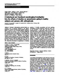
Figure 1. (A) A high density geodesic sensor map displaying the 129 electrode locations (Medial Frontal-MF; Lateral Frontal-LF; Central-C; Anterior Temporal-AT; Parietal-P; Posterior Temporal-PT; Occipital-O). (B) Idealized waveform of the computer-averaged auditory event-related potential (ERP) to brief auditory sound. The ERPs cannot be detected in the raw EEG (top) and it requires averaging over many one second epochs of EEG to view them (bottom). A logarithmic scale is used to present the different ERP components (early brain stem waves I-VI), midlatency component (N0, P0, Na, Pa, Nb), vertex potentials (P1, N1, P2), and task-related endogenous components (Nd, N2, P300, and slow wave-SW; Hillyard & Kutas, 1983).
9
ERPs are generally viewed as waveforms comprising a number of features (peaks and troughs). These features are also referred to as components and are predominantly defined by the functional process with which they are associated. That is, components are defined in terms of the cognitive function thought to be performed by the brain systems whose activity is recorded at the scalp (Coles & Rugg, 1995). Components are measured in terms of their amplitude (in μV) and latency (in ms). For example, N200 or N2 represents a negative change in amplitude (compared to a baseline where no stimulus was presented) that occurred about 200 ms following the onset of the stimulus or event of interest. ERP components can be exogenous (elicited only by the physical properties of sensory stimuli) and endogenous (depend on the interaction between the subject and the stimulus; Coles & Rugg, 1995). Endogenous components are thought to be influenced by the degree of cognitive processing and as such are the focus of study in higher order cognitive functions like language. Other measures can also be obtained from an EEG recording. These include correlations between activity in different brain regions (coherence), interhemispheric transfer of information, and topography (the distribution of wave amplitudes across the scalp; Pfefferbaum, Roth, & Ford, 1995). Some of the commonly investigated ERP components are briefly introduced next. The P50 is an early component that occurs after an auditory stimulus presentation. The N100 or N1 component is elicited by auditory stimuli and reflects early sensory and attentional processes that are associated with the ability to selectively attend to particular features in a stimulus (Michel, Thut, Morand, Khateb, Pegna, Grave de Peralta, et al., 2001). The P100 is a positive waveform and occurs after a presentation of a visual stimulus. The N200 or N2 is observed following the presentation of visual and auditory events that in some way deviate from the prevailing context (Coles & Rugg, 1995). The P300 or P3b is a positive wave that is modulated by subjective aspects of experimental stimuli, such as their salience, their task 10
relevance, and their probability (Coulson, 2004). P3a is different from the classic P300, as it appears in relation to introducing novel stimuli and is more frontally distributed rather than parietally. The N400 is a negative component that is associated mainly with linguistic phenomena like word frequency, repetition, semantic incongruence (Coulson, 2004). According to language research, the N1 is closely related to listening to speech sounds and phonetic processing, the N2 can be observed with visual presentation of words or word sequences, and the P300 is generally observed in lexical-decision tasks (e.g., word versus nonword) and sentence processing. The N400 as mentioned earlier can be easily elicited in semantically anomalous sentences (Hillyard & Picton, 1987). Finally, the P600 is another language associated component that is closely related to syntactic processing (Coulson, 2004).
Language representation in bilinguals The construct of laterality in bilinguals refers to how each of the languages that a bilingual masters is lateralised in the brain. The early research in the field of bilingualism was based on aphasia studies and suggested that either the second language in bilinguals might be represented in the right hemisphere (rather than the left) or that the right hemisphere is more actively involved in processing a second language (e.g., Vaid, 1983). This differentiation in lateralisation between the languages is to this day heavily researched and debated. Two different views have emerged in the literature regarding the question of laterality of languages in the bilingual’s brain. According to the first view, both languages occupy different areas in the same brain and the right hemisphere has a greater role in second language (L2) processing than in first language (L1) processing. In contrast, according to the second view, both languages are represented in similar areas of the brain in the left hemisphere. Evidence for each
11
of these views is presented after an introduction into the study of language lateralisation in the general population. Language lateralisation in general In this section the major components that make up language will be introduced. This is followed by a discussion of critical periods for acquiring a first and a second language. A general overview of how language is represented in the human brain will also be presented, with emphasis on the cerebral asymmetry for language. Language components The universal design of language is based on words and grammar. The word represents an arbitrary association between a sound and a meaning, and grammar represents the rules for combining vocabulary into words, phrases, and sentences in a way that is meaningful (Kandel, Schwartz, & Jessell, 2000). Grammar has three main components: morphology, syntax, and phonology. Morphology refers to the rules for combining words and affixes into larger words. Syntax consists of rules for combining words into phrases and sentences and determining relations among words. Phonology consists of rules combining sounds into a consistent pattern in the language and prosody (patterns of intonation, stress, and timing that span entire phrases and sentences). Prosody can have a pure grammatical function in distinguishing words as well as more general communicative functions such as differentiating questions from statements, supplying emphasis, indicating sarcasm, and expressing emotion (Kandel et al., 2000). Critical/sensitive period for language acquisition The critical period for language refers to the period in life during which language must be acquired if language is to develop adequately. It has been suggested that the critical period
12
for language ends before puberty (e.g., Lenneberg, 1967; Mayberrry, Lock & Kazmi, 2002). Lenneberg (1967) proposed a biologically based critical period for acquisition of first language. According to Lenneberg’s critical-period hypothesis, the critical period is linked to an initial state of equipotentiality of the cerebral hemispheres for supporting language functions, and ends with the termination of organizational plasticity following the gradual lateralisation of language functions to the left hemisphere. The two famous cases of “Victor, the Wild Boy” and “Genie” have provided great support for the critical-period hypothesis of first language acquisition. These children were found at twelve and thirteen and a half years of age, respectively, with no language skills due to social isolation. Furthermore, they were never able to achieve reasonable linguistic competence, despite a considerable effort in their rehabilitation (Hunter, 1993; Fromkin, Krashen, Curtiss, Rigler, & Rigler, 2004). The critical-period hypothesis for second-language acquisition suggests that older learners cannot achieve native-like competence in their second language. However, given that many adults are able to learn a second language proficiently, the critical-period hypothesis has limited value. There is a general consensus, however, that learning a second language in childhood is best. For example, Hakuta, Bialystok, and Wiley (2003) tested the critical-period hypothesis for second-language acquisition using 1990 US census data and found that the degree of success in second-language acquisition steadily declined throughout the life span and there was no critical age after which second-language acquisition is not possible. Other studies have also failed to show support for a critical period in second-language acquisition (e.g., Birdsong, 1992). In summary, there is no evidence of a critical period for second-language acquisition. However, it appears that the ability to acquire a second language decreases with age. According
13
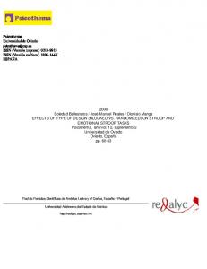
to Bialystok (2001), this decrease in ability is related to changes in cognitive strategies with aging as well as a decline in learning new skills. Cerebral asymmetry for language The left and right hemispheres are anatomically divided into four lobes: frontal; parietal; temporal; and occipital. The frontal lobes are generally associated with higher-order cognitive functions like decision making and thought, and the execution of motor movements. The parietal lobes are thought to play a role in somatosensory processing and spatial orientation. The temporal lobes are thought to play a major role in language and memory. Lastly, the occipital lobes are very important for visual processing. The different lobe regions are illustrated in Figure 2-A. Brodmann area (BA) classification (based on cytoarchitecture) is widely used in neuroimaging research and is also presented in Figure 2-B/C. Producing a sentence involves the selection of words and grammatical rules to encode ideas and intentions and then generating a set of articulatory commands to the motor system. To comprehend a sentence one must coordinate the sensory information that comes in through the auditory system or the visual system (for signing and reading) with the grammar and lexicon and then sending the interpretation to the systems underlying memory and reasoning (Kandel et al., 2000). As such, it is apparent that the relationship between brain and language is an elaborate one. Language lateralisation refers to how language is lateralised in the brain, i.e., whether it is represented in the left or the right hemisphere. Cerebral asymmetry for language refers to the general idea that the left hemisphere is more predisposed to language functions than the right. Discovery of Broca’s area and Wernicke’s area (also known as the classical perisylvian language areas) in the late 1800’s in aphasic patients provided great support for the notion of
14
hemispheric specialisation (localisation of a particular cognitive function to either the right or left hemisphere), and in particular for left hemisphere dominance for language.
Figure 2. A diagram of the four major brain lobes (A) and numbered Brodmann areas (Blateral view, C-medial view) (Images obtained online at www.stanford.edu/.../braintut/f_ab11lobes.gif and http://en.wikipedia.org/wiki/Image:Gray726-Brodman.png#file on 15 October 2007)
From numerous studies on cortical representation of language, it is now generally accepted that language is usually lateralised in the left hemisphere for about 97% of normal right-handed people and about 70% of normal left-handed people (Branch, Milner, & Rasmussen, 1964; Milner, 1975; Obler & Gjerlow, 1999). Left-hemispheric lateralisation of language has also been documented in sign language (e.g., Newman, Bavelier, Corina, Jezzard, & Neville, 2002). However, it is important to note that right hemisphere participation is also 15
evident in many aspects of linguistic competence (Baynes & Gazzaniga, 2000, Waldie & Mosley, 2000b). Studies have shown some right hemisphere ability in writing and spelling (Weekes, 1995), word recognition (Iacoboni & Zaidel, 1996), and communicative pragmatics (Brownell, Pinkus, Blum, Rehak, & Winner, 1997). Furthermore, lateralisation of language functions to the left hemisphere is not absolute, as lateralisation of language to the right hemisphere has been reported in young children after undergoing hemispherectomy (HertzPannier, Chiron, Jambaqué, Renaux-Kieffer, Van de Mootele, Delalande, et al., 2002; Ogden, 1996; Vargha-Khadem, Carr, Issacs, Brett, Adams, & Mishkin, 1997). Also, it is important to keep in mind that language requires complex patterns of information flow involving many parts of the brain (Kandel et al., 2000). The subcortical basal ganglia, the prefrontal cortex, and areas of primary visual and auditory cortex, cerebellum, as well as the perisylvian language areas and its right-hemisphere homologues are all part of the complex network that enables language use (Lieberman, 2000; Poeppel & Hickok, 2004). Although there is a preference for perisylvian language areas to regulate language, there is evidence that cortical plasticity may also allow other regions in the brain to assume this role (Elman, Bates, Johnson, Karmiloff-Smith, Parisi, & Plunkett, 1997). The classical perisylvian language areas are illustrated in Figure 3.
16
Figure 3. A diagram showing the classical perisylvian language areas (Broca’s and Wernicke’s areas) along with other language associated brain regions (Pinel, 2000).
Right-hemisphere involvement in language for bilinguals In this section, a number of hypotheses regarding L2 acquisition that suggest greater right-hemisphere involvement for language will be introduced. This is followed by a brief literature review of the studies that provide support for such hypotheses, with findings from each methodology presented separately. Hypotheses on L2 acquisition A number of factors are thought to influence the cerebral organization of each language. They can be non-linguistic (age, method of L2 acquisition such as formal or informal, proficiency) or linguistic (type of syllabic structure, systems of writing, direction of writing, orientation toward the European “logical” type of thinking; Chernigovskaia, 1992). A number of
17
hypotheses have arisen in an attempt to explain the effect of some of these factors on L2 lateralisation. In the field it is generally accepted that people who acquire their second language before the age of 6 or 7 years are considered early bilinguals, whereas if acquired after the age of 7 they are considered late bilinguals (e.g., Dehaene, Dupoux, Mehler, Cohen, Paulesu, Perani, et al., 1997; Fabbro, 2001). The younger the learner, the more similar second language learning is to the first language experience. The causes of these age effects on proficiency are highly controversial, with explanations ranging from biologically based critical periods to differences between child and adult learning contexts. It has been suggested that any cognitive or neural differences between L1 and L2 should be greater for late than early learners. For example, the age of acquisition hypothesis holds that when a second language is learned early in life, it is processed predominantly by the left hemisphere as in the first language, but when learned later in life more right hemisphere mechanisms are used (Albert & Obler, 1978; Galloway, 1982; Vaid, 1983). People who equally master all their languages are called fluent bilinguals or multilinguals. In contrast, people who master their first language to a higher degree than their second language are called nonfluent bilinguals or multilinguals. According to the stage hypothesis (Albert & Obler, 1978), proficiency modulates right-hemisphere involvement in language processing. It is suggested that the right hemisphere is more actively involved during the early stages of language acquisition. As proficiency increases, it is more likely that language will be represented and processed in a manner similar to that of the first language. A number of other hypotheses regarding second-language acquisition have also been formulated to try and account for findings that do not fit with the above mentioned hypotheses. The second language hypothesis states that the right hemisphere is more involved in bilinguals 18
for L2 compared to L1 (Genesee, 1982). The balanced bilingual hypothesis suggests that proficient bilinguals have greater right-hemisphere involvement for both languages compared to monolinguals (Galloway, 1983). The manner of L2 acquisition hypothesis applies to later L2 acquisition only and suggests greater right-hemisphere involvement for L2 acquired informally (e.g., in a natural context) than for L2 acquired in formal setting (e.g., school; Galloway & Krashen, 1980). Most of the studies reviewed below are relevant to the age of acquisition hypothesis and stage hypothesis, as age of L2 acquisition and proficiency in L2 are the most extensively studied factors with regard to language representation in the bilingual brain. Bilingual aphasia research Research in the field of bilingualism has been undertaken for a long time, creating and stirring interest in the field mostly from early reported cases of bilingual aphasia. Bilingual aphasia, or language impairment due to brain damage in bilingual people and its symptomatology or presentation, seems to have major implications about how the two languages are represented in the brain. Polyglots, or multilinguals, present an interesting problem when they become aphasic. This is more so in cases of bilinguals who have become aphasic after right-hemispheric damage (crossed aphasia). Several authors have reported greater incidence of aphasia among right-hemisphere injured bilinguals than monolinguals (e.g., Albert & Obler, 1978). This is probably the strongest evidence of right hemisphere language representation in bilinguals. This evidence has been disputed by later reports of similar prevalence of crossed aphasia in both bilinguals and monolinguals (e.g., Chary, 1986). Even so, aphasic patterns in bilinguals still raise a number of questions: Are they equally aphasic in different languages? Which of their several languages will they recover? (Benson & Ardila, 1996).
19
The “rule of Ribot” suggests that the mother tongue is the most resistant to later damage regardless of fluency, whereas Pitres (1895) claimed it is the language most familiar to the patient at the time of damage that is most likely to recover best (as cited in Pearce, 2005). Paradis (1977) comprehensively described six possible modes of language recovery in multilinguals: 1) differential: each language is impaired separately and is recovered at the same or different rate; 2) parallel: different languages are similarly impaired and restored at the same rate; 3) antagonistic: recovery of one of the languages progresses while the other(s) regress; 4) successive: one language does not show any recovery until another has been restored; 5) selective: one or more of the languages are not recovered at all; 6) mixed: two or more languages are used in some combination, i.e., unable to use one language at a time. The fact that bilingual aphasics present with a number of different recovery patterns implies that the relationship between the languages and brain areas responsible for them is a complicated one. In fact, as reviewed by Rapport, Tan, and Whittaker (1983), there are four anatomical models of the multilingual brain that are fiercely debated: (i) available space- the volume of brain damage affects multiple language use by simply limiting the total brain available for language functions; (ii) modality-performance- brain damage may differentially affect visual versus auditory modalities, thus altering the premorbid patterns of multiple language skills; (iii) lateralisation- different degrees of right- and left-hemisphere participation in the languages; and (iv) differential localisation- two or more languages occupy different loci within the language hemisphere. Evidence from Tachistoscopic studies Most evidence of right-hemisphere involvement in bilinguals comes from studies using the visual half-field paradigm. For stimuli such as words, a RVF advantage in terms of reaction times and/or accuracy is usually observed. If no such advantage is observed for bilinguals, in 20
particular for their L2, this is taken to reflect greater right hemispheric involvement in processing such linguistic stimuli. Wuillemin, Richardson, and Lynch (1994) showed that reaction times to word stimuli in the subjects’ second language varied as a function of the visual field and the age at which the subjects acquired the language. More specifically, they found that the later L2 had been acquired, the less there was a difference between reaction times to English words presented in the RVF and LVF, such that the RVF advantage usually reported for monolinguals was not found. In a similar study by Evans, Workman, Mayer, and Crowley (2002), four groups of English-Welsh bilinguals who differed in their age of acquisition and in their environment of acquisition were used. Evans et al. (2002) found that later acquired languages produced a shift in visual field advantage, i.e., they showed greater right hemispheric involvement. The authors also argued that the environment of acquisition (i.e., the language spoken in the environment) may also be an important factor in the lateralisation of language. In summary, both of these studies support the notion of greater right hemisphere processing in a later learned language. In a visual half-field study, Karapetsas and Andreou (2001) investigated visual field asymmetries for rhyme and semantic tasks in proficient and non-proficient bilinguals. They found that proficient bilinguals gave faster responses to words presented in RVF, while nonproficient bilinguals were faster in LVF. This indicated greater right hemisphere participation in the first stages of second-language acquisition. Evidence from Dichotic Listening studies Hatta and Dimond (1981) presented English-spoken digits, paired with environmental sounds, to English- and late bilingual Japanese-speakers. English speakers showed significantly greater right ear (i.e., left hemisphere) advantage when compared to the Japanese speakers. The authors suggested that one possible explanation for this finding was greater right-hemisphere 21
involvement for later acquired languages. Nachson (1986) replicated the same findings with Hebrew-English subjects. Hebrew speakers showed smaller right ear advantage to recalling English digits compared to English speakers, thus suggesting a greater right hemisphere role in processing a non-native language for late bilinguals. In a dichotic-listening study by Fabbro, Gran, and Gran (1991), hemispheric specialisation for semantic and syntactic components of language was investigated in simultaneous interpretations (translation of linguistic material was provided simultaneously with new input of linguistic material) with late bilinguals. Both students and professional interpreters performed better when L2 as a target language (translated) was sent to the left ear. Students were better at recognizing syntactic errors, while professional interpreters were better at recognizing semantic errors. Interpreters showed patterns of laterality consistent with interpreting jobs (working exclusively from L2 to L1, with L2 presented on their left ear so they can monitor their output in L1 with their right ear) whereas students did not. That is, the students’ pattern was consistent with greater right-hemisphere involvement for late bilinguals. In a study by Mägiste (1992), two tasks were used to assess hemispheric asymmetry in early proficient bilinguals. A dichotic listening task of stepwise addition (e.g., 58+2+5=?) was given to right-handed German-Swedish bilingual students (14-16 years of age). Also, the frequency and direction of conjugate lateral eye movements to verbal, spatial, and emotional questions was investigated in students aged 17-20 years. Lateral eye movements have been found to be a sensitive measure of differential hemispheric functioning with verbal material producing more rightward eyes movements (i.e., left-hemisphere processing) and spatial and emotional questions producing more leftward eye movements (i.e., right hemisphere processing; Schwartz, Davidson, & Maer, 1975). The hypothesis was that bilinguals would show more leftward eye movements on all tasks compared to monolinguals if there is more bilateral 22
hemispheric involvement. The authors found that monolingual subjects elicit mostly rightward movements when exposed to verbal questions, leftward movement to spatial questions, and leftward movement to emotional questions. The early proficient bilinguals in the study showed more bilateral hemispheric involvement compared to monolinguals. However, this is an interesting finding as it suggests less lateralisation of language functions in early and proficient bilinguals, in contrast to other studies that report similar findings with late or less proficient bilinguals. Evidence from Dual-Task studies Using the dual-task paradigm, Patkowski (2003) investigated age of acquisition as a significant contributor to laterality effects in both early and late bilinguals compared to monolinguals. Significant differences in laterality, with greater interference for the left hand under concurrent task conditions, were revealed only between the monolinguals and the late bilinguals, thus providing evidence for increased right-hemisphere involvement during speech production in L2 for late bilinguals. Green (1986) investigated tapping performance during concurrent object naming and concurrent picture-describing in groups that differed in their level of second language proficiency. All subjects were of English-speaking background and males. More left-hand interference was observed in L2 compared to L1 for subjects in the initial stages of acquiring a second language. Similar to Mägiste’s (1992) findings with dichotic listening, early proficient bilinguals in this study showed bilateral pattern for hemispheric involvement in their L1 during the concurrent tasks. The conclusion that fluency in a second language can modify how a first language is lateralised needs to be confirmed by other studies (Green, 1986). In a recent meta-analysis, Hull and Vaid (2006) combined studies that examined functional lateralisation of languages in bilinguals compared to monolinguals, from the three 23
experimental paradigms discussed so far (visual half-field presentations, dichotic-listening, and dual-task). They reported reliable left-hemisphere representation of language for monolinguals and late bilinguals. However, they reported bilateral hemispheric involvement for early proficient bilinguals. In summary, a number of behavioural studies provide support for greater righthemisphere involvement for L2 in bilinguals. Most studies discussed here argue for either age of acquisition (e.g., Patkowski, 2003; Wuillemin et al., 1994) or proficiency (e.g., Green, 1986; Karapetsas & Andreou, 2001) as the determining factors of the degree of right-hemisphere involvement in bilinguals. Surprisingly, some studies also suggest bilateral hemispheric involvement for language for early proficient bilinguals in L1 and L2 (Mägiste, 1992; Green, 1986; Hull & Vaid, 2006). Evidence from EEG studies Genesee, Hamers, Lambert, Mononen, Seitz, and Starck (1978) looked at dichotic listening during concurrent EEG with proficient adult bilinguals with different acquisition history: infant bilinguals, childhood bilinguals who became bilingual around age of 5 years, and adolescent bilinguals who became bilingual at secondary school age. Results showed shorter latency to N1 in the left hemisphere for infant and childhood bilinguals, but shorter latency in the right hemisphere for adolescent bilinguals. Overall the latency for N1 was shorter for the adolescent group compared to the other two. There were no reaction time differences. The authors argued that the results might be due to difference in strategies used by the groups, with the adolescent group relying more on right hemisphere-based gestalt-like or melodic strategy, while the early bilinguals relied more on left hemisphere-based, possibly semantic or analytic type of strategy.
24
Hahne and Friederici (2001) looked at sentence comprehension for correct, semantically incorrect, syntactically incorrect, or both semantically and syntactically incorrect sentences, in late Japanese-German bilinguals compared to native German speakers. They found that for correct sentences bilinguals elicited a greater positivity. No syntax-related ERP components were observed for the bilinguals in the syntax incorrect condition. For semantic and combined semantic and syntactic violations, bilinguals elicited late right anterior-central negativity, thus indicating greater right-hemisphere involvement. Similarly, Weber-Fox and Neville (1996) reported that with increasing age of acquisition there was reduced asymmetry in sentence type effects on the N215 and N300-500 components and absence of the 500-700 ms positive shift. According to the authors, the changes in ERP asymmetries may be associated with reduced specialisation in the left hemisphere language processing subsystems and increased righthemisphere involvement. They also found alterations in the N400 when detecting semantic anomalies when age of acquisition for L2 was 11 years or greater. Petsche, Etlinger, and Filz (1993) investigated EEG recordings by three professional interpreters. A coherence measure was used to compare different conditions. The coherence measure can indicate the degree of functional and anatomical coupling between two brain regions at any instant. Significant changes of coherence between all pairs of electrodes with respect to the averaged EEG at rest were computed for 5 frequency bands between 4 and 32 Hz. The verbal tasks of interpreting were controlled with comparable measures during mental arithmetic and listening to music. Findings showed that the temporal regions were most involved in interpreting and particularly in the uppermost beta band (24-32Hz). Coherence increases were noted in the right hemisphere while interpreting into the foreign language suggesting right-hemisphere involvement in bilinguals. More EEG research will be discussed where appropriate in later chapters. 25
Evidence from fMRI In an fMRI study by Chee, Hon, Lee and Soon (2001), both early proficient bilinguals and late less proficient bilinguals were recruited to perform a semantic judgement task. As expected, the more proficient bilinguals responded faster and displayed better accuracy. Furthermore, they found that within the left prefrontal and parietal regions the change in BOLD signal was smaller in the subject’s more proficient language. Also, the less proficient bilinguals revealed additional activation in the right inferior frontal region for L2 suggesting greater righthemisphere involvement. In an fMRI study, Dehaene et al. (1997) looked at brain activity while listening to stories in late and low proficiency bilinguals. They reported that the L1 engaged mostly areas in the left hemisphere, more specifically, in the left temporal lobe along the left superior temporal sulcus. Listening to L2 engaged both left and right temporal and frontal areas, with some subjects showing activation only in the right hemisphere. Both of these studies suggest that second language may be processed differently to first language, i.e., they provide support for the hypothesis of greater right-hemisphere involvement in L2. In summary, there is evidence from both EEG and fMRI studies that suggests greater right-hemisphere involvement in bilinguals. EEG studies suggest that right-hemisphere involvement can be reflected in ERP modulations and coherence measures. Moreover, ERP modulations are also language component specific (i.e., syntax versus semantics). FMRI studies suggest that right hemisphere activation, in particular right inferior frontal activation, might be characteristic during language processing in a less proficient language.
26
Left Hemisphere for Language in Bilinguals too? In this section, a brief literature review of studies providing support for left hemisphere representation for both languages in bilinguals is provided. A number of bilingual case studies where electrical cortical stimulation and/or WADA testing was used during language tasks are presented first. This is followed by findings from the other methodologies. Evidence from Bilingual Case studies Electrical cortical stimulation and WADA testing are important tools for language localisation prior to brain surgery. Electrical cortical stimulation involves stimulation of brain areas using electrodes during brain surgery while patients are asked to perform a language task such as object naming. WADA testing involves injection of sodium amytal to one hemisphere and testing performance of the other hemisphere on a language task such as object naming. Bilingual patients who have undergone these procedures can provide valuable information on how the two languages are represented in the brain. In a study by Ojemann and Whitaker (1978), localisation of the two languages in the cerebral cortex was mapped in two bilinguals using electrical stimulation during object naming. It was reported that the classical language areas were involved in both languages but, peripheral to this, in frontal and parietal cortex, there were additional language-specific areas. The authors also noted that for each bilingual the second language was more widely distributed in the cortex compared to their first language. They further noted that these findings provide support for the hypothesis that initially a large number of neurons are needed for naming in a second language, and increasing proficiency will result in less extensive areas being used for executing the task at hand. The findings from this study may also explain some patterns of bilingual aphasia, as the pattern will depend on whether the lesion involved is responsible for one language or both. The
27
authors also suggested that the exact sites of differential involvement may differ individually and may also depend on the characteristics of the languages concerned. In a more extensive study, Lucas II, McKhann II, and Ojemann (2004) electrically stimulated 25 bilingual patients and 17 monolingual patients during object naming. They reported distinct language-specific sites as well as shared sites that support both languages. They further reported that total cerebral areas were similar across the two languages; however, L2 specific sites were largely located in posterior temporal and parietal regions, whereas L1 specific regions were found throughout. In another cortical stimulation study that also combined WADA testing, Rapport et al. (1983) showed that multilinguals were left hemisphere dominant for all the languages, and therefore no support was found for increased participation by the right hemisphere. In summary, the cortical stimulation studies provided data most compatible with the “differential localisation” (two or more languages occupy different loci within the language hemisphere) model of cerebral localisation. Evidence from Tachistoscopic studies Endo, Shimizu, and Nakamura (1981) looked at laterality differences for processing two character nonsense Kana (Japanese phonetic symbols) word-strings, individual Kanji (nonphonetic, logographic symbols) words and individual Hangul (Korean orthographic characters that consist of a series of pronounceable symbols, therefore is closer to Kana; a word consists of one or more Hangul characters) words. Words were presented tachistoscopically in the LVF or RVF. Subjects were right-handed Japanese students with no knowledge of Hangul, and Japanese-Korean students with Hangul experience. Subjects showed a RVF advantage for recognition of Kana words, and no laterality for Kanji. However, for the Hangul stimuli there was a LVF advantage for Japanese students (note that the stimuli would appear as nonverbal 28
shapes or figures) and RVF superiority for Japanese-Korean subjects. So, for right-handed bilingual subjects, both first language (Kana) and second language (Hangul) were processed in the dominant left hemisphere. Evidence from Dichotic-Listening studies Schouten, van Dalen, and Klein (1985) found no evidence for ambilateral language organization, i.e., smaller right-ear advantage (REA) or left-ear advantage (LEA) for language oriented tasks in late proficient bilinguals. In a similar dichotic listening study with 139 EnglishHebrew bilinguals, Gordon (1980) also found that REA was the same for both languages regardless of when the second language was learned, how long it had been used, or how well it was known (based on self-rating). Morton, Smith, Diffey, and Diubaldo (1998) investigated adult dichotic processing differences in four groups based on bilingual induction age (preschool, school-age, post-pubertal and no-induction). Once again, no evidence of ambilateral language organization was found. Evidence from Dual-Task studies Furtado and Webster (1991) investigated concurrent language (reading and translating) and motor performance in bilinguals as a test of the age of acquisition hypothesis. They used early bilinguals who acquired both languages simultaneously before the age of six years, late bilinguals who acquired French first then English, late bilinguals who acquired English then French, and a monolingual group in English. Early bilinguals showed a very similar pattern of lateralised interference to both late bilingual groups, which was consistent with left-hemisphere processing. Soares (1984) also found greater right- than left-hand interference during oral tasks (talking, reciting, reading aloud) for monolinguals and bilinguals. In both studies, no differences were found between the bilinguals’ two languages or between bilinguals and monolinguals.
29
In summary, a number of behavioural studies suggest left hemisphere lateralisation for both languages in bilinguals. Also, this finding is apparent across both early and late bilinguals (e.g., Furtado & Webster, 1991) and different language proficiency levels (e.g., Gordon, 1980). Evidence from EEG Meuter, Donald, and Ardal (1987) investigated sentence comprehension during concurrent EEG in late fluent bilinguals. Results showed a larger N400 effect over left parietal sites when reading incongruent sentence endings (e.g., He had a sandwich for sleep) for both languages, suggesting similar representation in the left hemisphere for both. Ardal, Donald, Meuter, Muldrew, and Luce (1990) and, more recently, Hahne and Friederici (2001) found similar findings for reading and listening to semantically anomalous sentences in bilinguals. That is, studies reported similar patterns of brain activity for L2 and L1, and between monolinguals and bilinguals. In an interesting study, Friederici, Steinhauer, and Pfeifer (2002) trained adult participants on a miniature artificial language complete with its own vocabulary and grammatical rules, which were different to those of the subjects’ native language. Following training, ERPs were collected during grammaticality judgements of sentences from the miniature languages. The results revealed that trained participants displayed two ERP components taken to reflect early automatic and late controlled syntactic processes, just like native speakers of a language, whereas untrained participants did not. In general, automatic processes are developed through practice and require little attention, whereas controlled processes require attention in order to process less practised material (Sternberg, 1999). That is, they found no support for the notion that late acquisition of L2 dictates the recruitment of neural substrates that are different from those of monolinguals, at least for basic grammatical structures. 30
Evidence from fMRI and PET Neuroimaging techniques such as fMRI and PET, due to their precise spatial resolution, are responsible for a proliferation of studies trying to localise the areas in the brain responsible for each language in a bilingual person. In an attempt to summarise the main results from these studies, Abutalebi et al. (2005) distinguished between studies looking at language production, language comprehension, and translation. The same approach will be used in the following subsection, with studies being further differentiated based on the bilingual population used in the experiment and the factor of investigation (e.g., age of acquisition, proficiency, environmental exposure-defined as language exposure and usage in everyday life). In their review, Abutalebi et al. (2005) argued that proficiency and language exposure were the most important determinants of language representation in bilinguals and that increased proficiency in L2 leads to the engagement of the same left hemisphere neural networks subserving first languages. Language production studies The language production tasks employed in the studies reported below are very divergent (e.g., word generation, sentence generation, semantic decisions). Klein, Milner, Zatorre, Meyer, and Evans (1995) reported one of the earliest studies on language representation in bilinguals using a neuroimaging technique. In their prominent PET study with late-proficient bilinguals during a number of language production tasks (rhyme generation, synonym generation, translation), an overlap of activation was observed in similar areas (left dorsolateral cortex, left inferior temporal regions, and left superior parietal cortex), irrespective of the task requirement, for both languages. Illes, Francis, Desmond, Gabrieli, Glover, Poldrack, et al. (1999) found very similar results using fMRI with a group of late-proficient bilinguals, with main activation being observed in the left inferior frontal gyrus during a semantic judgement 31
task. Yetkin and colleagues (1996) investigated the effect of proficiency in an fMRI study during a word generation task with a group of multilinguals. Activations were observed in the inferior frontal, middle frontal and precentral gyri for all languages, with additional activations in the supplementary motor area and parietal lobe for the less fluent languages. In this study, age of acquisition information was not provided so both proficiency and age of acquisition could have contributed to the observed findings. De Bleser, Dupont, Postler, Bormans, Speelman, Mortelmans, et al. (2003) further investigated the effect of proficiency, in a PET study, by looking at cognate (words that have a similar root in both languages) vs. noncognate naming in late bilinguals who were moderately proficient in their L2. Cognate naming revealed overlapping brain regions in the left hemisphere for both languages. Noncognate naming in L2 revealed additional activation along the left inferior frontal gyrus. In summary, these studies provide evidence for proficiency being an important determinant in the cerebral representation of languages in bilinguals. Proficient bilinguals show very little difference in the neural activations during language production for both languages (e.g., Klein et al., 1995) compared to less proficient bilinguals who show some differentiation of brain activity between the languages (e.g., De Bleser et al., 2003). Kim, Relkin, Lee, and Hirsch (1997), in an fMRI study, looked at brain activation during sentence generation in early and late bilinguals of differing levels of proficiency in their L2. They found differential activation in the left inferior frontal cortex, corresponding to Broca’s area, depending on age of acquisition. For early bilinguals the observed activation for both languages was in overlapping regions, whereas for late bilinguals this was not the case. Activations in the posterior temporal regions, corresponding to Wernicke’s area, were similar across languages and groups. It is important to mention that the observed differences in activation between the late and early bilinguals might also be due to proficiency differences, due 32
to the lack of control for proficiency (proficient bilinguals in both groups is desirable). Marian, Spivey, and Hirsh (2003) have also reported that, although the same general structures are active for both languages during passive listening and viewing of words in late proficient bilinguals, differences within these general structures are present across languages and across levels of processing. In agreement with the Kim et al. (1997) findings, they reported different centres of activation for L1 and L2 within the left inferior frontal gyrus but not within the superior temporal gyrus. Chee, Tan, and Thiel (1999) looked at activations during cued word generation in L1 and L2 in early and proficient bilinguals compared to late-proficient bilinguals in an fMRI study. Activity was similar for both languages and groups, with inferior and middle frontal gyri being the main sites of generation. Based on these three studies, the effect of age of acquisition on brain activity during language production in proficient bilinguals is less clear. That is, there is support for both differential and largely overlapping activation in areas like the left inferior gyrus and superior temporal gyrus. Environmental exposure was investigated as another important factor in the cerebral representation of language in bilinguals, where environmental exposure is defined as language exposure and usage in everyday life as noted earlier (Perani, Abutalebi, Paulesu, Brambati, Scifo, Cappa, et al., 2003). In an fMRI investigation of the effect of environmental exposure on language processing in early and proficient bilinguals, Perani et al. (2003) found more extensive activation for the less exposed language compared to the more common language in the environment. The authors suggested that less exposure most likely requires the recruitment of additional neural resources to support the generation of words during the task. In fact, as suggested by Abutalebi et al. (2005), there is some support that less-automatic cognitive skills may engage supplementary neural substrates (e.g., Raichle, Fiez, Videen, MacLeod, Pardo, Fox, et al., 1994). 33
Language comprehension studies Perani, Dehaene, Grassi, Cohen, Cappa, Dupoux, et al. (1996) looked at brain activity during listening to stories in late and low proficiency bilinguals using PET. They reported greater activation when listening to stories in L1 than in L2. L1 activations were observed in the left angular gyrus, left superior and middle temporal gyri, left inferior frontal gyrus, and left temporal lobe. Right superior and middle temporal lobe and right temporal pole activations were also observed. Activations for L2 included the left and right superior and middle temporal gyri. This finding of greater activation for L1 is in contrast to studies that report greater activation for L2 (e.g., Chee et al., 2001). Price, Green, and von Studnitz (1999) used a group of late-proficient bilinguals in their PET study. Participants were required to read visually presented words. They reported an increased activation in the left temporal pole and the left and medial superior frontal cortex for L1 compared to L2. This finding is similar to that of Perani et al. (1996) reported above. As both studies used late bilinguals, it appears that the degree to which language comprehension regions are involved in each language depends on the age of L2 acquisition. In contrast, a number of researchers have argued that the proficiency of the bilingual speaker is the determining factor in the cerebral representation of languages. For example, Perani, Paulesu, Galles, Dupoux, Dehaene, Bettinardi, et al. (1998) looked at the effects of proficiency and age of acquisition on second language representation and processing in two different experiments using PET. In the first study, with late and moderate proficiency bilinguals, listening to sentences in the participants’ L1 showed engagement of the temporal lobes and temporo-parietal cortex more extensively than during L2 exposure. In the second study, the authors employed highly proficient bilinguals who were either early or late to acquire their L2. The differing amounts of activation between the languages found in the first 34
experiment were not found in either of the high proficiency groups in this study. Several brain areas (left superior and middle temporal gyrus, and left temporal pole), similar to those observed for L1 in low proficiency bilinguals, were activated by L2. According to the authors, at least for pairs of L1 and L2 languages that are similar in origin (e.g., Spanish and Catalan), attained proficiency is more important than age of acquisition as a determinant of cortical representation of L2. The fMRI study by Chee, Caplan, Soon, Sriram, Tan, Thiel, et al., (1999) supported the Perani et al. (1998) findings. They looked at sentence comprehension in early proficient bilinguals. Both languages activated the left inferior and middle frontal gyri, left superior and middle temporal gyri, left temporal pole, anterior supplementary motor areas, and bilaterally superior parietal and occipital regions. In a more elaborate study with three groups of bilinguals (early, late and low proficiency, and late and proficient bilinguals), Wartenburger, Heekeren, Abutalebi, Cappa, Villringer, and Perani (2003) looked at the neural correlates of grammatical and semantic judgements for sentences. Grammatical judgements involved determining if there is a grammatical violation in terms of disagreement of number, gender, or case (e.g., The dog run over the meadows) and semantic judgements involved in deciding if there is a semantic violation (e.g., The deer shoots the hunter). Interestingly, they found that the cortical representation of grammaticality judgements is only affected by age of acquisition, whereas the cortical representation of semantic judgements is affected by language proficiency. Differences in activation during the grammaticality judgement between the early and late proficient bilinguals were observed in the inferior frontal region. Semantic processing elicited differences in activation between the late low proficiency and late proficient bilinguals in a number of areas. Low proficiency bilinguals showed more extensive cerebral activations in Broca’s area and right middle frontal gyrus, 35
whereas the proficient bilinguals showed greater activation in the left middle frontal gyrus and right fusiform gyrus. For both late acquisition groups, increased activation was observed during the semantic task for both languages. The authors concluded that these findings indicated that both age of acquisition and proficiency affect the neural substrates of second language processing, with a differential effect on grammar and semantics. Translation and language switching Price et al. (1999) looked at translation of words from L1 to L2 and vice versa, and translation of alternating L1 to L2 and L2 to L1 in a PET study with a group of late proficient bilinguals. They referred to the latter condition as the switching condition. During translation, increased activation was observed in the anterior cingulate and bilateral subcortical structures (putamen and head of the caudate, thought to be related to control of action) with decreased activation in left extrasylvian temporal regions and temporoparietal regions (Brodmann areas (BA) 20, 21, and 39, thought to be associated with semantic processing). Case studies of bilinguals with lesions to subcortical structures, like the basal ganglia or thalamus, presenting with pathological mixing (mixing different languages within a single utterance) also suggest that subcortical structures may have an important role in language processing in bilinguals (e.g., Abutalebi, Miozzo, & Cappa, 2000; Fabbro, Skrap, & Aglioti, 2000). During the switching condition, activation was observed in the dorsal region of the left posterior inferior frontal cortex and bilateral supramarginal gyri (areas associated with phonological recoding). Hernandez, Martinez, and Kohnert (2000) used fMRI to look at early proficient bilinguals during picture naming in a single language, and picture naming in a mixed condition (this condition was taken to reflect language switching). The results revealed no differences in activation between the two languages during the naming condition in the dorsolateral prefrontal cortex, the supramarginal gyrus, the inferior frontal gyrus, and the superior temporal gyrus. 36
During the switching condition increased activation was observed only in the dorsolateral prefrontal cortex bilaterally, but more in the left hemisphere. In another fMRI study, Rodriguez-Fornells, Rotte, Heinze, Nosselt, and Münte (2002) investigated language selection in early proficient bilinguals. Subjects were instructed to respond to words in one language (pressing a button), while ignoring words in the other language as well as ignoring pseudowords. Bilinguals showed a greater activation in the left dorsolateral prefrontal cortex compared to monolinguals. The authors suggested that this region has a role in inhibiting the nontarget language, as previous research has associated this region with tasks requiring conflict resolution and selection of relevant material (e.g., MacDonald III, Cohen, Stenger, & Carter, 2000). In a PET study by Rinne, Tommola, Laine, Krause, Schmidt, Kaasinen, et al. (2000), cerebral activation patterns were analysed during simultaneous interpreting. Simultaneous interpreting involves generating the output (i.e., translation) from the input language as it is presented. Simultaneous interpreting (SI) into native language elicited left frontal activation increases, whereas SI into foreign language elicited much more extensive left-sided frontotemporal region activation increases. Overall, the results indicated involvement of lefthemispheric structures for both languages, in particular the left dorsolateral prefrontal cortex (DLPFC). Direction of translation also affected activation, which was more extensive during translation into the non-native language (often considered to be a more demanding task). SI represents a cognitively demanding task which requires temporary storage and the extraction of meaning for large chunks of the input language, while at the same time the interpreter is preparing, in the output language, the meaning of previously received chunk of language processed immediately before (Hyönä, Tommola, & Alaja, 1995). That is, differences in direction of translation may reflect differences in working memory capacity for each of the 37
languages. These studies emphasise the role of dorsolateral prefrontal regions and subcortical structures in language translation and switching in bilinguals. In summary, a number of studies provide evidence that proficiency and environmental exposure play an important role in the neural substrates activated during a language production task (left inferior frontal gyrus, middle frontal gyrus, and left posterior temporal regions). The most commonly observed areas of activation during language comprehension are the left inferior frontal gyrus, left middle and superior frontal gyrus, left angular gyrus and other left temporal regions. Based on EEG and fMRI findings, it appears that both age of acquisition and proficiency affect these neural substrates of second language processing, with a differential effect on grammar and semantics. Last, dorsolateral prefrontal regions and subcortical structures play a significant role in language translation and switching in bilinguals. The DLPFC has also been linked with semantic processing, and verbal working memory (Cabeza & Nyberg, 1997). It appears that the literature on language lateralisation is very equivocal about the question of language lateralisation in bilinguals. From the research reported above, it is clear that there is support for both differential lateralisation of language in bilinguals and for lack of such differences. Although a number of researchers have argued that language representation and processing in bilinguals is similar to that in monolinguals, this does not exclude the possibility that different neural networks within the same cortical areas independently support each of the languages (e.g., Fabbro, 1999), or that additional areas are called upon depending on proficiency and/or age of acquisition. Furthermore, a number of researchers have emphasised that other questions need to be addressed to find out how dealing with two languages on a consistent basis is different to dealing with one (e.g., Paradis, 1990; Bialystok, 2001). For example, Zatorre (1989) suggested that instead of examining the contribution of the right- and left-hemisphere, it might be of interest to examine the role of other cerebral structures and their 38
interaction with multiple linguistic systems, such as the frontal lobe. Bialystok (2001) suggested that acquiring a second language possibly affects a more general cognitive ability such as selective attention or flexibility, and it is these cognitive aspects that need to be further investigated. There is already some evidence to support this claim in bilingual children and adults (e.g., Bialystok, 2001; Bialystok & Shapero, 2005; Kovelman, 2006).
Language switching and executive functions in bilinguals Language switching and mixing is an important aspect of communication in bilingual speakers. Proficient bilingual speakers can use each of the languages independently without intrusions from the other language and then switch languages effortlessly depending on the context. Furthermore, bilinguals are also able to code-switch (introduce words from the other language into the current language of use) when the listener is also bilingual. According to a number of researchers (e.g., Bialystok, 2007; Rodriguez-Fornells, De Diego Balaguer, & Münte, 2006), language switching in bilinguals requires a cognitive control mechanism that regulates the activation of the languages. Cognitive control, in general, is required to direct thought and action in accordance with current goals and intentions (Miller &Cohen, 2001; Norman & Shallice, 1986). Cognitive control processes are very important as they filter out the irrelevant information in the environment (also called interference suppression) and have the ability to inhibit inappropriate responses or thoughts (also called response inhibition; Rodriguez-Fornells et al., 2006). It has been further argued that these cognitive control mechanisms are enhanced in bilinguals due to constant management of the two language systems (Bialystok, 2001).
39
Language switching hypotheses Although bilinguals seem to keep the two languages separate when producing words in one language, there is evidence that suggests that both languages may be active at the same time. Evidence for this comes from studies looking at a cross-language Stroop task and studies looking at lexical representations for both languages (e.g., Brysbaert, 1998; Dijkstra, 2005). The Stroop effect was first introduced to the cognitive psychology research field in 1935 (Stroop, 1935). The Stroop effect, also called the interference effect, is easily demonstrated when subjects are asked to name the colour of a printed colour word that is different to the target colour (e.g., RED). This condition leads to an increase in the number of errors and the time taken to respond compared to a condition where the colour word is consistent with the target colour (e.g., RED) or a neutral word (e.g., JUMP) is presented instead of a colour word. In bilingual versions of the Stroop task the word name can be printed in the subjects’ first language, with the response in the second language, or vice versa (cross-language Stroop task). A main finding of cross-language Stroop experiments is that Stroop interference occurs both within and between languages, but the within-language interference is stronger (e.g., Preston & Lambert, 1969). This suggests that the switch between languages is not complete, and that both processing systems remain active during language processing. In the study of lexical representations in bilinguals, researchers have made the distinction between the pre-linguistic conceptual level and the lexical level. According to models of lexical access, activation from the pre-linguistic conceptual store activates both lexical stores (e.g., Costa, Miozzo, & Caramazza, 1999). A number of studies have further shown that the activation of the non-target lexical representation activates its phonological representations as well. For example, Costa, Caramazza, and Sebastian-Galles (2000) found evidence of a cognate
40
facilitation effect, i.e., pictures that represented cognates (translations with similar phonological/orthographic form; e.g., guitarra/guitar) were named faster than non-cognates (translations with different phonological/orthographic form; e.g., pandereta/tambourine). In contrast, Marian et al. (2003) suggested that the parallel activation might only be characteristic of the early stages of language processing (e.g., phonetic), rather than lexical processing. Regardless of the level at which parallel activation takes place, a control system must be in place to regulate the production of target words even when non-target language words are interfering (Rodriguez-Fornells et al., 2006). In terms of what guides the language selection process in bilinguals, a number of researchers have discussed the idea of an activation threshold hypothesis (Luria, 1973; Grosjean, 1982; Paradis, 2000, 2004). Luria (1973) argued that naming processes are dictated by the “law of strength”, according to which every strong stimulus evokes a strong response, while every weak response evokes a weak response. Grosjean (1982) concurred with this, but referred to language switching as taking advantage of “the most available word”. He also argued that this is a very common occurrence in bilingual speech, and it happens most often when speakers are “tired, lazy or angry”, suggesting that a failure to consciously regulate the systems is at fault. According to Paradis (2000, 2004), an item is activated when there is sufficient amount of positive neural impulses at the right neural substrate. This amount is called the activation threshold. More activations of a particular item generally lead to lowering of its threshold and vice versa. Selection of one item is also thought to require the inhibition of other items by increasing their activation threshold. In his inhibitory control (IC) model, Green (1986; 1998) noted that a word must reach a certain level of activation to become available as a response. He argued that this is achieved by inhibitory processes on other possible targets, rather than an increase in activation as suggested 41
by the activation threshold hypothesis. For example, he suggested that in bilingual aphasia, a bilingual speaker calls up L2 instead of L1 not because L1 is inactive, but because there is insufficient suppression of activation in the L2 system. In summary, there is evidence to suggest that both language systems in bilinguals are active at the same time even though language output is produced from one of the languages. A number of hypotheses have been put forward to explain the language selection process. These hypotheses suggest that a language is selected based on activation threshold levels that also might be moderated by inhibitory processes on the other language. According to Meuter (2005), the language selection process is affected by a number of factors, including relative proficiency, contextual cues, and monitoring ability. Orthography of the language has also been considered as a potential factor (Preston & Lambert, 1969). These factors will be addressed next.
Factors that affect language switching ability Language switching in bilinguals is generally thought to incur a processing cost as reflected in a poorer performance by bilinguals on a number of verbal tasks (e.g., letter and semantic fluency tasks; e.g., Gollan, Montoya, & Werner, 2002). The cost of switching has been linked with a number of factors including: orthography of the language; proficiency; contextual cues; and monitoring ability (Meuter, 2005). Cross-language Stroop experiments have revealed that if stimuli are similar across languages, there would be greater interference across languages (orthographic specific hypothesis; Preston & Lambert, 1969). This assertion assumes that similar languages share more of their representation or functional systems and dissimilar languages are stored separately. This orthographic specific hypothesis postulates a greater reduction in interference in the betweenlanguage condition the greater the difference between the orthographic structures of the two
42
languages. That is, the greater the similarity between the two languages the stronger the competition for the same information processing mechanisms, and thus the smaller the reduction of Stroop interference from the within- to the between condition. So, if two languages are very similar, then the between-languages condition more closely resembles the within-language condition. Fang, Tzeng, and Alva (1981) confirmed that the ratio of between-language to within-language interference declines as language similarity decreases. However, Lee, Wee, Tzeng, and Hung (1992) failed to support the orthographic specific hypothesis. The orthographic hypothesis also predicts that a logographic script induces greater within-language interference than sound-based scripts (syllabary and alphabet; Biederman & Tsao, 1979). Biederman and Tsao (1979) found a greater interference effect for Chinese subjects in Chinese than for American subjects in English. They attributed this to potential differences in the perceptual demands of reading Chinese and English. They also concluded that the processes involved in reading Chinese are more similar to those involved in naming colours than are those involved in reading. However, Smith and Kirsner (1982) failed to replicate Biederman and Tsao’s findings for Chinese-English bilinguals. Lee and Chan (2000) also failed to show greater interference in Chinese compared to English. In terms of proficiency, it has been shown that switching from the weaker L2 to the dominant L1 is more demanding than the opposite. For example, Poulisse and Bongaerts (1994) showed that L2 production is affected by L1, with more intrusions from L1, in low proficiency bilinguals. Similarly, Meuter and Allport (1999) also showed that for low proficiency bilinguals switching costs in naming numerals were greater when switching from the L2 to L1. The authors suggested that this is due to L1 inhibition being more demanding and requiring more time to be deactivated. In proficient bilinguals the inhibition is considered to be equal, as shown in a category naming task with similar switching costs between L2 to L1 and L1 to L2 (Meuter, 43
1994). Costa (2005) further suggested that highly proficient bilinguals develop specific language-selection mechanisms that enable fluent use of both L1 and L2. In experimental settings, exogenous contextual cues that are clear and unambiguous (e.g., presentation of words prior to the target trial in the language that matches the response language for that trial) are thought to support proper language selection. The presentation of a cue is most beneficial if sufficient time is allowed between the cue and the target trial (Meuter, 2005). It has also been shown that the benefits of cueing are not as strong in a clearly dominant language (Meuter & Shallice, 2001). To establish a successful language selection process, followed by maintaining the selection, requires the use of intact monitoring skills. The need to monitor the appropriateness of speech output is most evident when errors of language choice are made (Meuter, 2005). Monitoring is thought to be controlled by the supervisory attentional system (SAS; Norman & Shallice, 1986). The SAS was introduced in order to explain the control of action and thought processes. The lowest control level is comprised of schemas (program-like entities that represent well learned actions or thought operations). These schemas are selected based on contention scheduling (a mechanism that resolves conflict independently through inhibition between activated schemas). However, contention scheduling is modulated by a supervisory system through further inhibition or activation of competing schemas (Shallice & Burgess, 1995). The SAS also bares resemblance to the Baddeley’s ‘central executive’ of the working memory model, which is a set of executive processes that manipulate the information in working memory and act upon it (Shallice & Burgess, 1995). In bilinguals, the SAS works by inhibiting all non-target language lemmas with the wrong language tag and activation of the relevant language task schemas (language task schemas control language actions such as naming a picture in L1 rather than L2; Green, 1998). 44
Imaging research has provided support for frontal lobe involvement in this monitoring strategy for bilinguals, more specifically dorsolateral prefrontal cortex (e.g., Hernandez et al., 2000; Hernandez, Dapretto, Mazziotta, & Bookheimer, 2001). Investigations of frontal lesions in bilinguals also suggest the involvement of frontal areas in the monitoring of language behaviour (Fabbro et al., 2000; Meuter, Humphreys, & Rumiati, 2002). For example, Fabbro et al. (2000) described a bilingual patient with a lesion to the left anterior cingulate and to the frontal lobe, also marginally involving the right anterior cingulate, who presented with pathological switching. Pathological switching patients alternate their languages across different utterances; utterance is a self-contained segment of speech that stands on its own and conveys its own independent meaning (Fabbro et al., 2000). In summary, orthography of the language, proficiency, contextual cues, and monitoring ability are important factors in the study of language switching. Monitoring ability in bilinguals has been closely linked to theoretical constructs like the supervisory attentional system as proposed by Norman and Shallice (1986) (Green, 1998). The importance of frontal regions, like the prefrontal cortex, in language switching has been demonstrated in bilingual patients with frontal lesions presenting with language switching pathologies. In accord with these findings, a number of researchers have proposed that the language switching mechanism is part of a more general executive attentional system (e.g., Fabbro et al., 2000; Hernandez et al., 2000). The notion of language switching in bilinguals being closely related to an executive attentional system has led researchers to explore the idea that the constant management of the two language systems has an effect on executive functions in bilinguals (e.g., Bialystok, 2001). This idea will be explored in the next section.
45
Executive functions in bilinguals Executive functions are defined here as the cognitive functions that enable control of attention, planning, categorising and inhibiting of inappropriate responses 1 (Bialystok, 2007). Executive functions generally develop later in childhood and are dependent on frontal lobe development (Bunge, Dudukovic, Thomason, Vaidya, & Gabrieli, 2002; Diamond, 2002). More on the neuroanatomy of executive functions will be presented in Chapter 6. These executive functions are used to control attention to the two language systems in bilinguals, and are therefore subject to more extensive practice (Bialystok, 2007). Price et al. (1999) similarly suggested that control mechanisms such as a central attentional or switching faculty is very important, and therefore enhanced, in bilingual processing. According to earlier research, differences between bilinguals and monolinguals can only be observed on tasks that tap into ‘control’ processes (i.e., tasks that contain a salient but irrelevant misleading cue that needs to be ignored; Bialystok, 2001; Bialystok & Shapero, 2005). Control processes are thought to include selective attention to relevant aspects of a problem, inhibition of attention to misleading information, and switching between competing mechanisms. In contrast, ‘representational’ processes include encoding problems in sufficient detail, accessing relevant knowledge, and making logical inferences about relational information (Bialystok, Craik, Klein, & Viswanathan, 2004). Research with bilingual children has shown that they develop control processes more readily than their monolingual counterparts (e.g., Bialystok & Majumder, 1998). However, the two groups progress at the same rate or slower for bilinguals in the development of representational processes (Bialystok, 2001). For example, bilingual children take longer to acquire their vocabularies in either of their languages 1
Cognitive control processes as described earlier can be seen as representing some of the processes under the set of executive functions. Researchers in the field sometimes use cognitive control and executive functions interchangeably (Miller & Cohen, 2001).
46
(Bialystok, 2001; Doyle, Champagne & Segalowitz, 1978; Pearson & Fernande, 1994), and this may even continue into adulthood as adult bilinguals were also outperformed by monolinguals on letter and semantic fluency tasks in a study by Gollan et al. (2002). Differential development of cognitive skills between bilinguals and monolinguals has also been investigated in the realm of metalinguistic ability. According to Cummins (1978), metalinguistic awareness is to look at language rather than through it to the intended meaning or, put simply, the conscious knowledge about language and its workings (Bialystok, 2001). Leopold (1961) suggested that bilingualism promoted an early separation of the word sound from the word meaning, which leads to an early awareness of the conventionality of words and the arbitrariness of language that could promote more abstract levels of thinking (Leopold, 1961). Ianco-Worrall (1972) concluded that bilingual children may reach a stage of semantic development 2-3 years earlier than monolingual children. Vygotsky (1962) suggested that a bilingual child can see the language as one particular system among many. As bilingualism induces an early separation of word and reference, it is possible that bilingual children also develop an early capacity to focus on and analyse the structural properties of language. Bialystok (1997) assessed bilingual children who could identify printed letters and their sounds but could not read, for their concepts of how print refers to language. Bilingual children understood better than monolingual children the general correspondence that printed form represents a word, i.e., they knew that written form carries the meaning and any other distracting information (e.g., pictures) are irrelevant to the meaning. According to Cummins (1979), advantages from bilingualism are specific to proficient bilinguals only (normal language competence in both languages), whereas limited bilingualism (age-appropriate skills in one of the languages) leads to cognitive and academic deficits. However, Hakuta and Diaz (1985) found advantages for both types of bilingual children. Bialystok (2001) argued that there is no 47
disadvantage for bilingual children academically as long as their proficiency in one language is adequate for the educational demands. In summary, there is evidence to suggest that bilingual children are more advanced in early language development. That is, they understand better the arbitrariness of assigning words to meaning. An important question in the study of bilingualism is whether these cognitive processing differences relate to intelligence in general. The consensus in the field is that bilingual children are no more intelligent than their monolingual peers (Bialystok, 2007). The main difference between the two groups is that bilinguals might posses an enhanced ability to control the use of their knowledge in tasks where competing or distracting information must be ignored (Bialystok, 2007). Another important question in the field is whether these effects of bilingualism on cognitive control persist into adulthood. However, very little research has been conducted on the cognitive abilities of adult bilinguals (Bialystok et al., 2004; Bialystok, Craik, & Ryan, 2006; Kovelman, 2006). Most of the current studies have employed a non-linguistic task that involves a response/attentional conflict. For example, in a Simon task that is based on stimulus-response compatibility, participants may be told to press a right response key for a blue square presented on the screen and a left response key for a red square. The stimuli are then presented on either the correct side of the screen (congruent position) or the incorrect side of the screen (incongruent position). Behavioural findings suggest a bilingual advantage, i.e., a smaller Simon effect, compared to monolinguals, suggesting an enhancement of general cognitive control processes such as response inhibition that persist into adulthood (Bialystok et al., 2004). The only two imaging studies in the field have suggested that this cognitive advantage in bilinguals is closely related to greater activation in superior and middle temporal, cingulate, and superior and inferior frontal regions (Bialystok, Craik, Grady, Chau, Ishii, Gunji, et al., 2005), as 48
well as inferior posterior regions (Kovelman, 2006) in the left hemisphere. It is obvious that further studies are necessary to reveal the full nature of these cognitive processing advantages and the neural substrates that support these.
The current research The Stroop colour-word task is considered a typical measure of executive function (Lezak, 1995). It is generally thought that the Stroop interference effect arises due to either a conflict between stimuli and responses that results in competition for the allocation of attentional resources (Phaf, Van der Heijden, & Hudson, 1990), or a conflict at the level of response selection and monitoring (Dyer, 1973). The Stroop task is considered to reflect some of the more fundamental aspects of cognitive control and goal-directed behaviour, i.e., the ability to select a weaker, task-relevant response (naming the colour) in the face of competition from an otherwise stronger and task-irrelevant response (reading the word; Miller & Cohen, 2001). In the current research, the Stroop task was used to further explore the hypothesis of positive changes in executive functions in bilinguals as a result of constant management, i.e., language switching, of the two language systems in bilinguals. The effect of bilingualism on cognitive processing, in terms of executive functions, has not been investigated in detail in late and proficient bilinguals. Late and proficient bilinguals currently living in their second language environment, carefully screened for proficiency and language use in both languages, were the subjects of interest in this thesis. The bilingual subjects were compared to monolingual controls that were matched for gender and age. The Stroop task was performed in one language at a time with no switching between languages (unlike the cross-language Stroop task, as reviewed earlier). This allows for an
49
investigation of possible changes in cognitive control within each language that a bilingual masters. Previously used measures of cognitive control in bilinguals lack this characteristic due to their non-linguistic nature. If bilinguals do possess better cognitive control, we would expect to see better conflict resolution, i.e., a smaller interference effect. A larger interference effect in bilinguals would indicate less effective cognitive control mechanisms, whereas similar interference effects between the two groups would indicate that bilinguals and monolinguals exhibit similar level of cognitive control as measured by the Stroop task. MacLeod (1991), in his review, has suggested that the left hemisphere might be more susceptible to interference than the right (as shown by Schmit and Davies, 1974). This could be due to the greater involvement of the left hemisphere in verbal processing or to left hemisphere regions, like the left parietal lobe (Posner, Walker, Friedrich, & Rafal, 1984) and left frontal lobe (Perret, 1974), being involved in disengaging attention processes during the Stroop task performance. In that sense, if bilinguals are left lateralised for language processing, we would expect a similar level of interference compared to monolinguals. In contrast, if bilinguals are more bilateral in the language processing we might observe smaller interference as the left hemisphere is not as strongly predisposed for verbal processing. In the current research, the dual-task paradigm was used to assess lateralisation of language functions in order to evaluate this possibility. The dual-task paradigm also provided the opportunity to further evaluate the hypothesis that the right hemisphere is involved to a greater degree in language processing in bilinguals. By using a variety of methodologies, i.e., behavioural, EEG and fMRI, with the Stroop task paradigm this research should provide a comprehensive account of the Stroop interference effect in late and proficient bilinguals in both their languages. Functional MRI, with its superior spatial resolution, can provide useful information about the neural substrates supporting the 50
cognitive processes of interest. EEG, with its superior temporal resolution, can provide information about the time course of neural events taking place during the different stages of cognitive processes.
51
CHAPTER 2: General methods This chapter describes the general methodology of the current research that includes four separate studies. The information is organized into five subsections: participants; screening assessment; experimental tasks; general testing procedure; and general data analysis. The participants subsection provides information on how both monolingual and bilingual participants were recruited for each of the experiments. The voluntary language background questionnaire and the computerised proficiency assessment that were used to screen potential participants are described in the screening assessment subsection. Descriptive statistics (demographic data) are provided for the participants who met all the inclusion criteria, i.e., the total sample. Each chapter includes a description of the sample of participants (all came from the total sample) that was included in the analysis specific to that study. The experimental tasks included a dual-task and three versions of the Stroop colour-word task. These tasks are introduced briefly in the experimental tasks subsection, but are described in greater detail in the relevant chapters. In the general testing procedure subsection is a description of the testing protocols used in the research. In the general data analysis subsection, statistical tests that were common to all the experiments are described.
Participants Both monolingual and bilingual participants were individuals aged 18 years old and over (no upper age limit was set). Monolingual participants were recruited using an advertising flier that was posted around the University of Auckland. Bilingual participants were recruited using one of the following methods: (1) an advertising flier that was posted around the University of Auckland; or (2) a call for participants made by the researcher at several community events.
52
Seventy adults volunteered to participate in this study (35 monolinguals, 20 MacedonianEnglish (M-E) bilinguals and 15 German-English (G-E) bilinguals). The advertising material for the monolingual participants required all volunteers to be proficient in one language only (English), right-handed, and to have no known brain damage or learning disability. Two groups of bilingual participants were targeted: Macedonian-English bilinguals, and German-English bilinguals. M-E bilinguals were used because of the relatively large Macedonian-New Zealand community in Auckland, and G-E bilinguals were used because of the large pool of foreign exchange students from Germany at the University of Auckland. Advertising material required all bilingual volunteers to be proficient speakers in both languages (Macedonian and English or German and English), right-handed, and to have no known brain damage or learning disability. All procedures for recruitment and testing were approved by the University of Auckland Human Participants Ethics Committee. Consent was obtained from all participants, after which the researcher administered the screening assessment and experimental tasks described below.
Screening assessment Proficiency assessment Potential bilingual participants were asked to fill out a language background questionnaire, which included questions regarding the age of L2 acquisition, manner of L2 acquisition, contexts of use for each language and self-reported language proficiency in both languages (see Appendix A). Filling out the questionnaire took approximately 5 minutes and participants were able to consult the researcher if uncertain. Age of L2 acquisition was checked
53
here (>6) to make sure that only late bilinguals were included in the experiments. No bilingual participants were excluded based on this criterion. Monolingual participants were not required to fill out a language background questionnaire. However, prior to performing the experimental tasks they were asked whether they were familiar, even at a basic level, with another language/s (other than English) as only pure monolingual speakers were required. Based on this criterion, 2 monolingual volunteers did not proceed with the research. Also, given that monolingual participants were purely monolingual (native speakers of English) and at university, high English proficiency was assumed. As all bilingual participants were considered late and had been formally educated in their first language for a number of years, a high level of first language proficiency was assumed. However, self-reported measures on L1 proficiency were still obtained as described above in the language background questionnaire. English, or L2 proficiency, was objectively assessed using a computerised Quick Placement Test (QPT). The QPT is a 15-20 minute adaptive computer-based test (computer selects difficulty of questions based on previous answers) and measures listening, reading, grammar and vocabulary. During a listening question, subjects read a question and a number of possible answers. They then listen to a short text and choose the best answer. For the reading task, subjects are presented with a notice, diagram, label, memo or letter containing a short text. From a number of options, they choose the phrase that most closely matches the meaning of the text in the graphic. The vocabulary questions involve subjects choosing the best word or phrase to complete a sentence (e.g., because it had not rained for several months, there was a shortage/drop/scarce/waste of water). Grammatical questions ask subjects to complete a gapped text by choosing the best word or phrase for each blank from a selection of words presented. 54
The test has been validated in 20 countries by more than 5,000 students of English. Participants are categorized into one of six levels based on their score (out of 100): 0-Beginner (0-39); 1Elementary (40-49); 2-Lower Intermediate (50-59); 3-Upper Intermediate (60-69); 4-Advanced (70-79); and 5-Very Advanced (80-100). All bilingual participants were required to be at least upper intermediate in English. Based on this criterion four bilingual volunteers were excluded from the study. The Boder Test of Reading-Spelling Patterns (Boder & Jarrico, 1982) was also used to assess reading proficiency or reading age (RA) in L2 (English). The Boder test involved a reading phase, which involves reading aloud single word lists that are standardized for age. If more than 50% is read correctly, a new list is presented. The person’s reading age is the corresponding age for the last word list with a score of more than 50%. The Spelling test involved two lists of 10 words: a known (words they read correctly) and an unknown (words they did not know how to read) word list. Each of these lists contained 5 phonetic (words that follow phonetic rules) and 5 non-phonetic (irregular) words. This test was only performed on those bilingual volunteers who went on to complete the dual-task experiment, which requires extensive reading of text passages of varying difficulty. This task is explained in greater detail later under the experimental tasks subsection. Handedness The Edinburgh Handedness Inventory (EHI; Oldfield, 1971) was used to confirm that all participants were right-handed. The inventory consisted of 10 items that question participants about their hand preference when writing, drawing, throwing, using scissors, using a toothbrush, using a knife, using a spoon, broom (upper hand), striking match (match), and opening a box/lid (see Appendix B). Participants indicate a strong preference by marking two crosses in the left
55
column or right column, depending on the direction of their preference. An equal preference is indicated by marking a cross in both the left and right columns. The laterality quotient was calculated using the equation: (R-L/R+L) x 100 (where ‘R’ equals the total number right preference responses, and ‘L’ equals the total number of left preference responses). Positive numbers indicate dextrality while negative numbers indicate sinistrality. Based on this, one bilingual participant was excluded. In the current study, all remaining participants had an EHI score between +40 and +100, indicating strong dextrality. Table 1 presents the sample size, gender balance, mean age, and EHI scores for the total sample for monolinguals and bilinguals separately. L2 proficiency scores for the bilinguals, based on the QPT, and RA, based on the Boder Test of Reading-Spelling Pattern, are also presented in Table 1. Tables 2 and 3 present data from the language background questionnaire for the bilingual groups. A number of monolingual and bilingual participants from the total sample volunteered to participate in more than one experiment over a certain time period. In the descriptive statistics table below, age at time of recruitment is used to calculate the mean ages. In the relevant chapters the mean ages for each group are adjusted to represent the age at the time of testing.
56
Table 1 Mean scores (standard deviation in parenthesis) for the descriptive measures (age, Edinburgh Handedness Inventory (EHI)) for each group based on the total sample. Gender balance is also presented for both groups. Quick Placement Test (QPT) scores and reading age (RA) are also presented for the bilingual groups. Group
Males/
Age
EHI
QPT
RA (n=14)
81.18(15.77)
79.41 (10.46)
12 (Adult RA)
Females Bilinguals M-E (n=17)
8/9
26.35 (7.29)
2 (RA=14.5) G-E (n=13)
4/9
25.85 (3.78)
91.54 (14.63)
86.85 (11.34)
Total (n=30)
12/18
26.13 (5.94)
85.67 (15.91)
82.63 (11.30)
19/14
27.45 (7.73)
89.22 (13.08)
Monolinguals Total (n=33)
Table 2 Mean (standard deviation in parenthesis) for starting age of L2 acquisition, and self-reported proficiency in L1 and L2 for the bilingual participants based on the total sample. Percentage of bilinguals participants based on type of L2 learning history, language of thought, and frequency of use at time of recruitment are also presented. Bilinguals
M-E (n=17)
G-E (n=13)
Total (n=30)
Starting age of L2 acquisition
11.41 (2.12)
10.54 (1.51)
11.03 (1.90)
6.12 (.928)
6.38 (.768)
6.23 (.858)
5.35 (.996)
5.46(.967)
5.40 (.968)
70.6%, 11.8%, 17.6%
84.6%, 7.7%, 7.7%
76.7%; 10.0%; 13.3%
38.5%, 23.1%, 38.5%
43.3%, 20.0%, 36.7%
23.1%, 30.8%, 46.2%
33.3%, 26.7%, 40.0%
Self-reported proficiency in L1 Self-reported proficiency in L2 Type of L2 learning (formal, informal, both) Language of thought (L1, L2, both)
47.1%, 17.6%, 35.3%
Frequency of use (both equally, L1 more, L2 more)
41.2%, 23.5%, 35.3%
57
Table 3 Percentage of bilingual participants reporting specific contexts of use for L1 and L2 based on the total sample. The other category represents social outings, shopping, and/or university. L1
L2
Home
Work
Other
Home
Work
Other
100%
0%
41.2%
17.6%
88.2%
70.6%
69.2%
38.5%
69.2%
61.5%
92.3%
100%
86.7%
16.7%
53.3%
36.7%
90.0%
83.3%
Bilinguals MacedonianEnglish (n=17) GermanEnglish (n=13) Total (n=30)
Experimental tasks A Dual-task and three versions of the Stroop colour-word task (two manual versions and one verbal) were used in this research project. The tasks are briefly described below. A full and detailed description for each task is presented in the relevant chapters. Dual-task In brief, the task consisted of speeded right- and left-finger tapping alone, right- and leftfinger tapping during reading aloud of narrative passages, and right- and left-finger tapping during silent reading of narrative passages (two single task baseline conditions and four tapping and reading conditions). Bilingual participants were required to undertake a reading assessment in English (Boder & Jarrico, 1982) as a measure of their reading ability in order to make sure the reading level of the passage presented was not too difficult for them and interfere with the tapping process more than expected. Therefore, the passages presented in English were appropriate for their reading ability. A reading inventory was not available in L1, and the participants were asked to glance at printed versions of the passages in L1, and pick the one they 58
felt most comfortable reading. The task was programmed in Microsoft Visual Basic 6.0. The entire passage was presented one at a time and displayed on a computer screen for 14 s. Responses were recorded by using the space bar as the tapping apparatus. Tapping rate data was collected across all conditions. The conditions were presented in random order by the program. This task was available in Macedonian and English. The task lasted for about 5 minutes. Bilingual participants performed the task twice (once in each language), and the order was counterbalanced. Stroop Colour-Word Task (Verbal Version) The stimuli for this experiment were printed on three separate cards (21.5 x 14 cm), each containing six rows of four items (Helvetica, 28 point font) spaced 1 cm apart, one for each condition represented in the test. The 24 stimuli on each card were printed in four different colours (red, blue, green, yellow). The baseline condition consisted of dots, in the word or control condition common words (when, hard, and, over) were presented in lower-case letters, and in the colour or incongruent condition colour words were printed in lower case in incongruent colours (see Appendix C). This version of the Stroop colour-word task is known as the Victoria Stroop test (Regard, 1981). The word condition and colour condition were reproduced for the Macedonian and German versions. The three cards were always presented in the same sequence: baseline, control, incongruent. Participants were required to name the colour aloud as quickly as possible for each of the stimuli. They were timed using a stopwatch. The task did not take more than 3-4 minutes to complete. Language order was counterbalanced for the bilingual participants. Reaction time data and accuracy were recorded.
59
Stroop Colour-Word Task (Manual Version One) In this task colour words were randomly presented on a computer screen in either a congruent condition (where the ink colour was compatible with the word e.g., RED) or an incongruent condition (where the ink colour was incompatible with the word e.g., BLUE). In addition, non-colour words were presented as a control/neutral condition (e.g., DOG). Stimulus presentation was controlled using E-Prime v1.1 (Psychology Software Tools, Pittsburgh, PA, USA). Participants were required to name the colour the word was presented in using four colour-coded keys on the keyboard (“v”-red, “b”-blue, “n”-green, and “m”-yellow). A practice block was included prior to the experimental block to ensure good performance (practice accuracy for all participants was at least 80%). This task was used in two separate studies (Chapter 4 and Chapter 5). In the first experiment the task was available in Macedonian and English. In the second experiment the task was available in Macedonian, German, and English. In the second experiment, where EEG recording was used concurrently, the number of trials was also increased in order to improve the averaging process. For the first experiment, the practice block consisted of 36 practice trials, and the experimental block consisted of 24 control, 24 congruent, and 24 incongruent trials. For the second experiment, the practice block consisted of 40 trials, and each of the two experimental blocks consisted of 52 control, 52 congruent, and 44 incongruent trials. Reaction time data and accuracy were recorded. The first Stroop experiment was completed in about 8 minutes and the second one in about 14 minutes. The continuous electroencephalography recording (250 Hz sampling rate; 0.1-100 Hz analogue bandpass) was done using a 128-channel Ag/AgCl electrode net (Electrical Geodesics Inc., Eugene, OR, USA). Electroencephalography recording was acquired using a common
60
vertex (Cz) reference and later re-referenced to the average reference off-line. More detail regarding the EEG methodology used in the current research is provided in Chapter 5. Stroop Colour-Word Task (Manual Version Two) This task was used with concurrent fMRI (as described in Chapter 6). A blocked experimental design was chosen for the study. As in previous versions, the colour words were either congruent, incongruent, or control/neutral. Stimuli in the incongruent condition were further divided into semantically incongruent and response incongruent. Semantically incongruent stimuli contained semantic conflict only as the response key was the same for both the target colour and the colour word. Response incongruent stimuli contained both semantic conflict and response conflict as the response key for the target colour was different from the colour word. Participants were required to name the colour the word was presented in using a mouse as the response apparatus. The stimuli were presented in the fMRI scanner through a projector. Red and blue target colours were mapped onto the left mouse button whereas green and yellow were mapped onto the right mouse button (see Appendix E). A practice block containing 18 stimuli in each condition was administered outside the scanner to ensure good performance in the scanner. This task was available in Macedonian and English, and language order for the bilinguals was counterbalanced. Stimulus presentation was controlled using E-Prime v1.1 (Psychology Software Tools, Pittsburgh, PA, USA). Data was collected over two runs. In each run a total of eight experimental blocks (18 stimuli each) were included, with each condition being repeated. A total of 72 control stimuli, 72 congruent stimuli, 72 semantically incongruent stimuli, and 72 response incongruent stimuli were included. This task lasted for about 16 minutes. Reaction time data and accuracy were recorded. Images were acquired using a 1.5T
61
Siemens Avanto scanner (Erlangen, Germany). Image acquisition parameters are presented in the relevant chapter.
General Testing Procedure Monolingual and bilingual participants were recruited for each of the four studies. Some monolingual and bilingual participants volunteered for more than one experiment. During a testing session, the following general testing procedure took place: (1) participants were provided with participant information sheets regarding the experiment/s; (2) a consent form was signed; (3) the handedness inventory was completed; and (4) participants performed the experimental task/s. The bilingual participants were also asked to complete the language background questionnaire, the QPT, and The Boder Test of Reading-Spelling Pattern prior to undertaking the experimental tasks. Screening assessment The screening assessment took place in a testing room at the Psychology Department, University of Auckland. Instructions for the consent form, the EHI, and the language background questionnaire were provided on the forms. However, the experimenter was available to clarify these instructions and answer any queries the participant may have had regarding how to complete the forms. The reading test was administered by the experimenter following the general rules for administration of this test. The QPT assessment took place in the same room on a computer on a separate computer desk. The QPT program included detailed instructions for the participant; therefore, the experimenter was not required to instruct the participant. However, the experimenter remained available in case the participant had any queries or there were
62
computer problems. Scores were automatically generated by the program at the completion of the test. Experimental tasks Following the screening assessment, participants were tested on the experimental tasks. The Dual-task, the Verbal, and Manual Version One of the Stroop colour-word task were administered in the same testing room as the screening assessment. The Manual Version One of the Stroop colour-word task was also administered at the EEG lab in the psychology department at the University of Auckland. The Manual Version Two of the Stroop colour-word task was administered at the MRI Centre for Advanced Research at the University of Auckland. Bilingual participants performed each of the experimental tasks in both of their languages and language order was counterbalanced, as mentioned earlier. Each of these tasks are briefly described above, and then in greater detail in the respective chapters.
General Data Analysis Behavioural and imaging (EEG and fMRI) data were analysed separately. General statistical analyses that were performed on the behavioural data across all experiments are described below. The general statistical approach for the EEG and fMRI data is also briefly introduced below. A detailed description of the data analysis methods is presented in the relevant chapters. Behavioural data For each study, the data analysis procedure is discussed in greater depth in the respective chapters. All analyses were performed using the Statistical Package for the Social Sciences (SPSS) version 12-14.0. Significance level of .05 was used for all tests. Bonferroni correction 63
was applied to all post-hoc tests. Greenhouse-Geisser correction was used for violations of the sphericity assumption. The behavioural data (reaction time (RT) and accuracy) was generally subjected to three separate analyses. In the first analysis the groups were compared in their performance in English. In the second analysis the groups were compared in their performance in their respective L1. In the third analysis the bilinguals’ performance was compared in their L1 versus their L2. Split-plot Analysis of Variances (ANOVAs) with condition as a within-subjects factor and group as a between-subjects factor was employed for the first two analyses. A two-way repeated measures ANOVA with condition and language as within-subjects factors was employed for the third analysis. This was extended to a split-plot ANOVA with group as a between-subjects factor when more than one bilingual group was assessed. Electrophysiological data EEG data from the Stroop colour-word task (Manual Version One) was segmented into appropriate epochs consisting of a pre-stimulus baseline and a post-stimulus period. Automatic eye-movement correction was made on all segments using the method of Jervis, Nichols, Allen, Hudson, and Johnson (1985). The data was then filtered using the bi-directional filter (Alarcon, Guy, & Binnie, 2000) with a high-band pass of 0.1 and a low pass of 30. EEG data was analysed using three different approaches. First, EEG data from selected electrodes was investigated. Second, EEG data from selected brain areas was investigated. Third, Principal Components Analysis (PCA)-derived Bonferroni method as described by Hamm, Johnson, and Kirk (2002) was employed. This analysis is described in detail in Chapter 5.
64
fMRI data Functional data from the Stroop colour-word task (Manual Version Two) was subjected to image processing and analysis using the SPM5 software (Welcome Department of Imaging Neuroscience, London, UK; http THE://www.fil.ion.ucl.ac.uk). The data was first subjected to pre-processing steps (realignment, normalisation, smoothing). These are explained in more detail in Chapter 6. Functional data was analysed in a two-step procedure, a within-subjects GLM timeseries regression, and random-effects factorial ANOVA with the resulting parameter estimates as the dependent measure. For each monolingual participant four contrast images were obtained (one for each condition: control, congruent, semantically incongruent, and response incongruent), whereas for each bilingual participant eight contrast images were obtained (four for each language). These contrast images were imported into one of three factorial-design ANOVAs for further analysis. Two 4x2 factorial-design ANOVAs with Condition (control, congruent, semantically incongruent, response incongruent) as a within-subjects factor and Group (monolinguals, bilinguals) as a between-subjects factor, one for English and one for L1, were evaluated. A 4x2 factorial-design ANOVA with Condition and Language (L1, L2) as within-subjects factors was evaluated for the bilingual group only. For each of the factorial ANOVAs only planned comparisons were performed, using an F-test, as assessing all possible main effects, interaction effects and simple effects tests would inflate the type I error rate and would make interpretations very difficult. Planned comparisons were also considered a suitable approach for addressing the aim of this study, which was to investigate potential differences in the Stroop effect between the groups (monolinguals vs.
65
bilinguals) and/or languages (L1 vs. L2). The first comparison looked at the response incongruent condition versus the congruent condition as this is the same contrast as in the EEG study. The existing paradigm allowed for investigating three additional contrasts that possibly reflect different aspects of the Stroop effect. The first additional comparison looked at the effect of competing information (response incongruent, semantically incongruent, and congruent versus neutral), the second additional comparison looked at the effect of semantic conflict (semantically incongruent versus congruent), and the third additional comparison looked at the effect of response conflict (response incongruent versus semantically incongruent). Uncorrected threshold of p.10), the reported analyses combined the data from males and females. The interference data (see Table 5) was subsequently subjected to four separate analyses: (1) baseline tapping performance by group; (2) concurrent English passages by group (L2 for bilinguals); (3) concurrent L1 passages (English for monolinguals) by group; and (4) L1 passages versus L2 passages for bilinguals only.
76
Results Baseline tapping data were subjected to a Tapping Finger (Right, Left) × Group (Monolingual, Bilingual) ANOVA with Tapping Finger as the repeated measure. As expected, the Tapping Finger main effect was significant (F (1, 28) =20.5, p