University of Cambridge, Downing Street, Cambridge CB2 3EJ, U.K., t Gonville and Caius College, Cambridge CBz iTA, U.K.. SUMMARY. This review attempts ...
J. exp. Siol. (1979), 8i, 217-279 With 27 figures frinted in Great Britain
217
A COMPARATIVE SURVEY OF THE FUNCTION, MECHANISM AND CONTROL OF CELLULAR OSCILLATORS BY M. J. BERRIDGE* AND P. E. RAPPf * A.R.C. Unit of Invertebrate Chemistry and Physiology, Department of Zoology, University of Cambridge, Downing Street, Cambridge CB2 3EJ, U.K., t Gonville and Caius College, Cambridge CBz iTA, U.K.
SUMMARY
This review attempts to survey in a uniform manner the available evidence concerning the generation and behaviour of several well-investigated cellular oscillators. Members of two broad classifications are contrasted: (i) cytoplasmic oscillations, where the periodic phenomena is generated by an instability in a metabolic pathway and (ii) membrane oscillators in which a membrane potential rhythm is generated at the membrane. Interactions between the cytoplasmic and membrane compartments are considered and the effects of these interactions on oscillatory behaviour is discussed. Because of their biological importance and the greater body of experimental results, particular attention is directed to a study of membrane potential oscillations. These systems can be approximately classified in two groups: (i) systems in which a periodic potential results from oscillatory changes in permeability and (ii) systems in which potential oscillations result from the periodic activity of an electrogenic pump. The examples considered include the glycolytic oscillator, oscillations in vein contraction in the slime mould Physarum polycephalum, rhythmic aggregation in Dictyostelium discoideum, neural oscillators, the periodic potential in Purkinje fibres and the sino-atrial node and rhythmic behaviour in smooth muscle. Questions considered include the generation of periodic activity, the modulation of the oscillation by drugs and other metabolic and membrane effectors and the question of the functional role of these oscillations.
INTRODUCTION
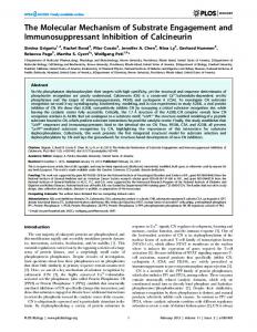
Even a cursory examination of periodic phenomena in biological systems reveals that almost every organism examined displays some form of rhythmic activity. This conclusion is supported by the preceeding papers in this volume and by Table 1 in this chapter. Even within a single organism almost every major organ system can, under appropriate circumstances, generate sustained oscillations. For example, in man, functions associated with respiration, digestion, movement, circulation and nervous activity can oscillate. Biological oscillations cover a very broad frequency spectrum as summarized in Fig. 1. The periods of oscillation can range from a fraction of a second to several hours, and in some cases, periods of a year have been reported. Jt is important at the outset to distinguish high-frequency oscillations (with frequenfcies ranging from seconds to minutes) from circadian rhythms or those of longer
2l8
M. J. BERRIDGE AND P. E. RAPP 1 msec
I s
1 min
Ih
I day
1 month I year
I
A I
Organism Complex regulations Smooth muscle
Circulation Peristalsis Respiration Heart E.E.G. Ciliated ephithelium Nervous action
I
10" 3
10" 2
10"'
10°
10' 102 103 Period duration (s)
10"
10s
106
107
108 sec
Fig. i. Periodic activity in man. Ranges in period are indicated by horizontal bars. Predominant periods are indicated by triangular peaks. (Taken from Golenhofen (1970) as modified from Hildebrandt (1967).)
Table 1. A summary of the nature and the propos* Tissue or cell type
Nature of oscillation
Dictyostelium discoideum
Periodic release of cAMP and contractility Shuttle-streaming
Physarum
polycephalum
Acetabularia
Periodic action potentials
Macrophages
Membrane potential hyperpolarizations Membrane potential hyperpolarizations Bursts of action potentials
L-cells Aplysia burster cells /?-cells Anterior pituitary
Smooth muscle Sino-atrial node
Proposed function Aggregation and differentiation Distribution of materials and chemotaxis May provide positional information during regeneration Contractility and possibly chemotaxis Contractility
Release of neurohormone Release of insulin Release of hormone Slow wave potential changes Pacemaker activity for myogenic rhythm Cardiac contraction Action potentials
Bursts of action potentials Action potentials
duration. Since the cellular basis for the latter is still a matter for considerable speculation (Hastings & Schweiger, 1976; Jacklet, 1978) we restrict our attention in this review volume to the high-frequency oscillations produced by specific cell types and for which both physiological and biochemical explanations are beginning to appear. The wide range and functional importance of such spontaneous activity is summarized in Table 1. In some cases the endogenous rhythm regulates the activity of the cell generating the rhythm (e.g. the myogenic rhythm in smooth muscle) or it provides a driving signal to control the activity of neighbouring cells (e.g. the cardiac pacemaker). In addition to such pacemaker functions, oscillatory activity is apparent in a number of secretory cells (Table 1). Such oscillatory phenomena could also play some role in the spatial organization of development as occurs during regeneration in, Acetabularia (Novak & Bentrup, 1972). The production and detection of periodic
Mechanism of cellular oscillators
219
cyclic AMP signals are important during differentiation in the slime mould Dictyostelium discoideum. The expression of a surface glycoprotein involved in cell adhesion may also depend upon pulses of cyclic AMP (see Gerisch et al., p. 45). However, there are examples where the spontaneous activity has no obvious function and may simply reflect the dynamic nature of cellular control mechanisms. As an understanding of the mechanisms responsible for driving such rhythmical activity begins to appear, it is of interest to consider whether or not there is a common mechanism underlying all such oscillatory behaviour. In this article we attempt to summarize some of the major points which emerged during the course of this meeting on Cellular Oscillators. Much of the information appears in a more detailed form in the preceding articles to which frequent reference will be made. Details of other oscillating systems, which were not specifically dealt with during the meeting, will also be described. A major problem in trying to unravel the mechanisms responsible for oscillatory activity is to detect the basic instability responsible for generating the rhythm. Since a wide range of cellular processes will be entrained to the basic oscillator, it is always difficult to isolate those processes directly responsible for oscillatory activity. One way of trying to understand the basic oscillator is to identify the input and output properties of the oscillatory system (Fig. 2). A characteristic feature of most oscillatory cells is that the periodicity of the rhythm can be altered by a variety of external signals. A detailed analysis of the mode of action of such signals may help to detect some of the processes involved in generating the oscillation. In the heart, for example, the ability of adrenaline to accelerate pacemaker activity in the sino-atrial node and in Purkinje fibres is apparently mediated by cyclic AMP, which acts by modifying some of the key processes of the membrane oscillator. By uncovering the nature of this cyclic AMPsensitive process, it may be possible to identify a key component of the oscillator. A knowledge of the output signal can also provide important clues about the nature of the oscillator. A simple example to illustrate this point is a typical myogenic system such as smooth muscle where rhythmical contractions are presumably driven by oscillations in the intracellular level of calcium. Therefore, the oscillator must have a component which is connected in some way with the mechanisms responsible for generating such calcium signals. Identification of the nature of these input and output signals can thus provide valuable insights into the cellular mechanisms responsible for generating oscillatory activity. There appear to be two distinct kinds of cellular oscillators, those based in the surface membrane and cytoplasmic oscillators originating from inside the cell (Fig. 2). As pointed out by Tsien (p. 209), these two oscillators are not mutually exclusive and it is likely that in some cells they may co-exist and even interact with each other. Such interactions are all the more likely when the two oscillators possess some common component or intermediate as shown in Fig. 2. One component which is common to many membrane and cytoplasmic oscillators is calcium. Calcium occupies such a central position because its intracellular level is determined by processes located both in the surface membrane as well as within the cell. It is not too surprising, therefore, to find that this important second messenger features prominently in many cellular oscillators. The membrane or cytoplasmic oscillatory mechanisms might be developed to a ^reater or lesser extent depending on the functional role of the cell in question. For
220
M. J. BERRIDGE AND P. E. RAPP Input for frequency modulation
Output
Output
Fig. 2. The location and relationships of cellular oscillators. A membrane oscillator composed of a variable number of components (a-d) is responsible for generating a rhythmical output usually in the form of fluctuations in membrane potential. A chemical output may also be generated by various cytoplasmic oscillators (d-g). The two oscillators might be linked to each other by sharing a common component (d). The frequency of these oscillators can be adjusted by a variety of input signals which interact with specific components of the oscillators.
example, the glycolytic oscillator, which is most commonly studied in yeast cell extracts, is a biochemical process occurring in the cytoplasm. In contrast the rhythmical trains of action potentials generated by neural or cardiac pacemaker cells originate from processes mainly restricted to the membrane. However, the organization of the cell as a functional unit is such that membrane and cytoplasmic oscillatory mechanisms can never be entirely independent of each other. Even in those cells where a membrane oscillator predominates, interactions between membrane events and internal biochemical processes are an important aspect of most oscillatory systems and are often an essential feature of many of the mechanisms responsible for frequency modulation. The main properties of some cytoplasmic and membrane oscillators will be described first before considering how they may function in a variety of specific examples.
CYTOPLASMIC OSCILLATORS
Theoretical work (Savageau, 1976) suggests that most metabolic control systems will respond to a concentration displacement by a rapid monotonic return to a steady state. There are, however, well-established examples of metabolic control networks whose steady state is dynamically unstable (Tyson & Othmer, 1978; Goldbeter & Caplan, 1976). Control circuits of this type would be characterized by sustained oscillations in the concentrations of the participating metabolites. Metabolic oscilla* tions have been investigated experimentally both in vivo and in vitro. Perhaps the
Mechanism of cellular oscillators
221
most interesting aspects of these metabolic oscillations is that they display frequencies within the second to minute range which thus makes them possible candidates to drive some of the cellular oscillations which will be described later. The glycolytic oscillator
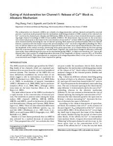
Glycolysis has provided a classical system for studying metabolic oscillations (see Hess, p. 7). Most of the observations have been performed on cell-free extracts of yeast (Boiteux & Hess, 1974), skeletal muscle (Tornheim & Lowenstein, 1974, 1975), cardiac muscle (Frenkel, 1968) and Ehrlich ascites tumour cells (Ibsen & Schiller, 1967, 1971). However, the phenomenon may not be confined to cell extracts because oscillations have been recorded from intact yeast cells (Chance et al. 1973). The possibility that the glycolytic pathway might oscillate within intact cells has important implications and it is not too surprising, therefore, to find that the glycolytic oscillator has been invoked to explain several cellular oscillators. For example, Sachsenmaier & Hansen (1973) have proposed that such a metabolic oscillator might drive rhythmical contractile activity in Physarum. Components of the glycolytic oscillator may also play a role in inducing the slow potential waves in /?-cells (Matthews & O'Connor, p. 75) and in molluscan burster cells (Chaplain, see p. 113). The main features of the glycolytic oscillator are summarized in Fig. 3. Phosphofructokinase (PFK) is the key enzyme whose activity is sensitive to allosteric control by various components and products of the overall glycolytic pathway. In particular, the enzyme is very sensitive to adenine nucleotides in that it is inhibited by ATP but activated by ADP and AMP. The enzyme is also activated by its substrate fructose-6phosphate (F6P). When the various glycolytic intermediates in the pathway are measured at different times, they are found to oscillate (Fig. 4). Some intermediates oscillate in phase, whereas others oscillate as much as 1800 out of phase (Fig. 3^, 4). By analysing such phase relationships it is possible to construct the sequence of events which occur during an oscillatory cycle. The two extremes in the activity of PFK are shown in Fig. 3(6, c). When PFK has been active it builds up the level of fructose-1,6bisphosphate (FDP), which provides substrate for the rest of the cycle leading to an increase of ATP which then rises towards a peak at the expense of ADP and AMP (Fig. 3d, 4). However, as ADP, AMP and F6P fall, the activation of PFK declines and is further inhibited by the increase in ATP. As PFK switches off, the production of FDP declines as does ATP thus leading to an accumulation of ADP and AMP. Additionally, a decline in the activity of PFK will result in a build up of its substrate F6P. As these intermediates accumulate they once again switch the enzyme back to an active state (Fig. 3 c) and the cycle will repeat itself. The existence of a single control point at PFK seems to be sufficient to explain oscillatory activity in extracts from skeletal muscle and beef heart (Frenkel, 1968; Tornheim & Lowestein, 1975). However, the phase relationships in yeast cells are much more complex and indicate that an additional control point exists at pyruvate kinase (see Hess, p. 10). One of the interesting features of this glycolytic oscillator is its sensitivity to the rate of substrate entry into the pathway. For example, increasing the concentration of glucose over a fairly wide range increases the frequency of the oscillation (Boiteux & Hess, 1974) and fiis feature may help in trying to assess the possible significance of the glycolytic Oscillator in various cells. While the existence of glycolytic oscillations has been demonstrated in several 8
EXB 8l
222
M. J. BERRIDGE AND P. E. RAPP Glucose
(a)
Fig. 3. The glycolytic oscillator, (a) A summary of the glycolytic pathway (thick arrows) together with the NAD and adenine nucleotide cycles (thin arrows). The dotted lines represent the allosteric control of phosphofructokinase (PFK) by ATP, ADP, AMP and fructose-6phosphate (F6P). The circles indicate those enzymes which seem to be important for oscillatory activity: GDH-glyceraldehyde dehydrogenase; PGK-phosphoglycerate kinase; PK-pyruvate kinase. (ft) and (c) Changes in the concentration of key intermediates at two points during the oscillatory cycle. Large-face lettering of metabolites is used to indicate high concentrations while smaller lettering indicates a comparatively low concentration. In one part of the cycle (b) PFK is being switched from an active to an inactive state by ATP whereas later in the cycle enzyme activity is switched back to the active state as ADP, AMP and F6P begin to accumulate (c). (d) As PFK is switched back and forth between its two activity states, the glycolytic intermediates oscillate with some components 180° out of phase. Fig. 4 gives actual measurements of these oscillations in extracts of skeletal muscle.
Mechanism of cellular oscillators
223
500
/JM
jUM.
350 1
20
'
40
60
Time (min) Time (min) Fig. 4. Oscillations in the concentrations (expressed as /iM) of various glycolytic components in extract of skeletal muscle. (Taken from Tornheim & Lowenstein, 1974.)
cell-free extracts, there is little evidence on whether or not they exist in intact cells apart from the study on yeast cells mentioned earlier. It is unlikely that such oscillations can exist in cells which have active aerobic respiration where a continuous supply of ATP would tend to suppress glycolytic oscillations by damping out the oscillations in the adenine nucleotide cycle. On the other hand, oscillations in glycolysis might induce oscillations in mitochondrial metabolism through a periodic input of pyruvate. The other problem to consider is the way in which the glycolytic oscillator might be linked to various effector systems. Any fluctuation in the level of ATP would certainly have repercussions for a number of processes. Protons may represent another important output from the oscillator because each time a molecule of F6P is converted to FDP a hydrogen ion is released (Fig. 3 a). Chaplain (see p. 113) has proposed that fluctuations in the level of ATP and hydrogen ions might be important in regulating ion permeability in burster neurones. In insulin-secreting /?-cells there appears to be an interesting relationship between glycolysis and membrane permeability (Dean, Matthews & Sakamoto, 1975). The enzymes glyceraldehyde-3-phosphate dehydro|^ase (GDH) and phosphoglycerate kinase (PGK) seem to be particularly important 8-2
224
M. J. BERRIDGE AND P. E. RAPP
in that the flux of metabolites through these enzymes somehow alters potassiufl conductance leading to membrane depolarization (see Matthews & O'Connor, p. 757. In addition to effecting ionic permeabilities, variations in intracellular pH could alter contractile activity as will be described presently (p. 244). It is clear from this brief survey that oscillations in the glycolytic pathway are potentially capable of generating a variety of output signals which could drive a range of oscillatory phenomena. However, an unequivocal demonstration of the existence of glycolytic oscillations in intact cells other than yeast cells has not appeared. Attempts to monitor fluctuations in the NAD/NADH ratio in Physarum were unsuccessful due to changes in cell geometry which occurred during each contraction (Sachsenmaier & Hansen, 1973). More sophisticated techniques for monitoring intracellular metabolism will have to be devised in order to assess the contribution of the glycolytic oscillator to other cellular oscillations. Mitochondrial oscillations
Isolated mitochondria display a range of oscillatory activity (Fig. 5) (Boiteux & Hess, 1974; Goldbeter & Caplan, 1976). The oscillations in NADH fluorescence are particularly significant because spectrophotometric analysis of intact slime mould (see Fig. 17 on p. 246) and smooth muscle (see Connor, p. 164) have revealed similar fluctuations in the redox state of NAD and cytochrome b respectively. Since most of the NAD and cytochrome b is concentrated in mitochondria, it is reasonable to speculate that they may also oscillate within the intact cell. Mitochondrial respiration is sensitive to both external and internal controls which makes it difficult to assess whether the oscillations originate from within or are driven from outside by a periodic input such as pyruvate originating from glycolytic oscillations as described in the previous section. The release of calcium from mitochondria may also be regulated by the level of phosphoenolpyruvate (PEP) (Roos, Crompton & Carafoli, 1978), which suggests another possible way in which mitochondrial function might be entrained to glycolytic oscillations. According to Mitchell's chemiosmotic theory, oxidative phosphorylation is driven by a proton gradient established when hydrogen ions are extruded from the matrix as electrons pass down the electron transport chain (Carafoli & Crompton, 1976). This proton gradient can then be harnessed either to ATP production or it can be used to drive the uptake of calcium. In liver mitochondria, the uptake of calcium takes precedence over ATP formation which emphasizes the proposed role of these organelles as calcium buffers (Rossi & Lehninger, 1964). If the intracellular level of calcium rises above its normal 'operational' level, the mitochondria seem to be capable of switching from ATP production to calcium transport. If the proton gradient is being used to drive calcium entry, the level of mitochondrial ATP will fall which will have repercussions on the mitochondria because the ADP/ATP ratio seems to be one of the important control factors for oscillatory activity. It is quite conceivable, therefore, that oscillations in the intracellular level of calcium due to some other mechanism might induce oscillatory activity in mitochondrial respiration. If the mitochondria are actively engaged in calcium accumulation this could result in oscillatory changes in intracellular pH. When calcium was injected into neurones of the snail Helix asper^B there was a transient decrease in intracellular pH as the calcium was exchanged f3f
Mechanism of cellular oscillators NADH fluorescence H* uptake
K+uptake
Swelling
lA AA \\JA\/A\/AV A 1 \ / V /Viv V
/ /
A
/
M min)H \ /\
j
r\
225
Redu ction
M-equival. g protein
r
,0,
oLS.
L
Fig. 5. Oscillation of various properties of isolated mitochondria. (Taken from Boiteux & Hess, 1974.)
protons across the mitochondrial membrane (Meech & Thomas, 1977). The pH returns to normal presumably as hydrogen ions were extruded from the cell. These studies on mitochondria suggest that they could play an important role not only in contributing to oscillations in internal calcium but they may also be responsible for establishing oscillations in intracellular pH. Cell suspensions of the slime mould Dictyostelium discoideum display oscillations in hydrogen release which lag slightly behind the peaks in light scattering (Malchow, Nanjundiah & Gerisch, 1978). The precise source of the hydrogen ions which are being extruded from the cell has not been established, but if they reflect the existence of an intracellular pH oscillation, this could have some interesting implications for contractile systems, especially those in non-muscle cells as described later (p. 244). Cyclic nucleotide-calcium interactions
Second messengers such as the cyclic nucleotides and calcium have been implicated as control elements in a number of oscillatory systems (Durham, 1974; Goldbeter & Caplan, 1976; Rapp & Berridge, 1977). They could both function in the generation of oscillatory activity in addition to playing an important role in modulating the frequency of cellular oscillators. These second messengers, especially calcium, also play an important role in linking internal metabolic events to changes in membrane properties. An important reason for proposing that second messengers might generate oscillatory activity stems from the fact that cyclic nucleotides and calcium interact with each other through a variety of positive and negative feedback loops (Berridge, 1975; Rapp & Berridge, 1977). The ubiquitous calcium binding protein called calmodulin (calcium dependent regulator or modulator protein) mediates many of the actions of calcium within cells including some of the interactions with cyclic nucleotides. For example, when cells are activated there is usually an increase in the intracellular level of calcium which begins to bind to calmodulin to form a complex which can activate a variety of cellular processes including the hydrolysis of cyclic AMP and cyclic GMP (Fig. 6). The calcium-calmodulin complex (CaM) can activate both phosphodiesterase and adenylate cyclase (Wang, 1977). During the action of CaM there is a preferential ^tivation of the cyclic GMP phosphodiesterase. In the presence of a fixed concentrafTon of calmodulin, the adenylate cyclase from rat brain displays a biphasic response to
226
M. J. BERRIDGE AND P. E. RAPP Si
Signal
Membrane phosphorylalion
AC
'.
Cyclic AMP
PDE O
5'-AMP
ATP
Bnal
Ca1* pump
ADP
Calmodulin
Ca-Calmodulin complex
Cyclic GMP
PDE
5-GMP
Fig. 6. The role of calmodulin in cyclic nucleotide—calcium interactions. Cyclic AMP, cyclic GMP and calcium form a triumvarate of second messengers which are all linked together through a variety of interactions. Many of the effects of calcium on the cyclic nucleotides are mediated through a specific receptor protein calmodulin.Guanylate cyclase exists in both a soluble (GC.) and membrane-bound (GCm) form. For simplicity, the effects of the cyclic nucleotides on calcium homeostasis have been omitted. AC, adenylate cyclase; PDE, phosphodiesterase.
calcium (Wolff, Brostom & Brostom, 1977). At low concentrations, calcium is stimulatory but becomes inhibitory at high levels of calcium (Fig. 6). Studies on intact cells have also indicated that high levels of calcium may inhibit adenylate cyclase (Butcher, 1975; Campbell & Siddle, 1976). Calmodulin thus occupies a pivotal position between the cyclic nucleotides and calcium. Another important function of calcium which seems to require the participation of calmodulin is membrane phosphorylation. Schulman & Greengard (1978) and Greengard (1978) have shown that the calcium-calmodulin complex seems to be responsible for phosphorylating specific proteins of synaptosomal membranes. The functions of these proteins have not been established. Perhaps the ability of calcium to increase potassium conductance, which is such a key feature of many cellular oscillators, might be mediated through such a phosphorylation reaction. Another possible outcome of such phosphorylation might be the activation of calcium extrusion. The calcium-calmodulin complex activates the surface calcium pump of red blood cells (Gopinath & Vincenzi, 1977; Jarrett & Penniston, 1977). If such a feedback mechanism is widespread in other cell types it could also be important in oscillatory systems because any increase in the level of calcium will automatically activate the surface pumps which extrude calcium (Fig. 6). In the case of cyclic GMP there is considerable indirect evidence to suggest that aj increase in the intracellular level of calcium may be responsible for activating guanj* late cyclase (Schultz et al. 1973; De Rubertis & Craven, 1976; Ohga & Daley, 1977).
Mechanism of cellular oscillators
227
The activation of guanylate cyclase is complicated by the existence of soluble and paniculate or membrane-bound forms of the enzyme (Fig. 6, GC m and GCg), which may be affected differently by calcium (Mittal & Murad, 1977). Cyclic GMP has proved somewhat of an enigma because, despite the fact that its concentration rises significantly during the activation of many different cells, its precise function is still unknown. It has been suggested that cyclic GMP might act in smooth muscle by inhibiting the entry of calcium (Schultz, Schultz & Schultz, 1977). Interactions between cyclic GMP and calcium may also be important in photoreceptors (Lipton, Rasmussen & Dowling, 1977). This ability of cyclic nucleotides to modulate the level of calcium has been studied more extensively in the case of cyclic AMP. There are numerous reports in the literature to suggest that some of the affects of cyclic AMP may depend on its ability to modulate the movement of calcium across both surface and internal membranes (Fig. 6) (Berridge, 1975; Rasmussen, Jensen & Goodman, 1976; Rasmussen & Goodman, 1977; Putney, Weiss, Leslie and Marier, 1977; Fitzpatrick and Szentivanyi, 1977). The biochemical basis of many of these feedback interactions operating between cyclic AMP and calcium have yet to be determined so it is difficult to construct precise control loops. However, on the basis of available evidence it is possible to organize some of these second messenger interactions in the form of classical feedback control loops which could generate oscillations under appropriate conditions (Durham, 1974; Rapp & Berridge, 1977). Feedback interactions involving cyclic AMP feature significantly in many of the models designed to account for cyclic AMP oscillations generated by the slime mould Dictyostelium discoideum (see p. 248). Apart from the possible direct contribution of these various second messengers to the generation of oscillations, the way in which they interact with each other may also be an important component of some of the mechanisms responsible for modulating the frequency of oscijlatory activity. It will be evident from the following sections that an important featurt of rhythmical activity in many cells is an oscillation in the intracellular level of calcium. Since both cyclic AMP and cyclic GMP seem to be capable of adjusting the level of calcium, it is not too surprising to find that such second messenger interactions have been implicated in modulating the frequency of various cellular oscillators (see subsequent sections for details). Calcium-induced calcium release
Another possible mechanism for inducing oscillations in the intracellular level of calcium stems from the observation that calcium might be capable of inducing a regenerative release of calcium from internal reservoirs. This phenomena of calciuminduced calcium release was uncovered in studies on skinned muscle fibres where calcium was able to induce a regenerative release of calcium from the sarcoplasmic reticulum (Endo, Tanaka & Ogawa, 1970; Fabiato & Fabiato, 1975). A similar phenomena occurs in medaka eggs where a local increase of calcium at the point of fertilization spreads as a wave towards the opposite pole (Gilkey et al. 1978). Of considerable interest in the current context was the observation that under appropriate conditions this release of calcium from the sarcoplasmic reticulum of cardiac muscle occurred spontaneously (Fig. 7). The fact that the frequency of these oscillations accelerated as *he level of calcium was increased is particularly interesting because raising the level of calcium seems to accelerate a number of cellular oscillators. If such a rhythmical
228
M. J. BERRIDGE AND P. E. RAPP
0
J
"
A pCa
7-65
\
7-50
X
B pCa
7-65
|
7-40
l_L_L_i c pCa
7j65
4
pCa
J_M55
J
7-0
650
f
5-50
10s Fig. 7. Cyclical contractions of a skinned cardiac cell. Note how the amplitude and the frequency of the oscillations increased as the external concentration of calcium was increased. (Taken from Fabiato & Fabiato, 1975.)
release of calcium occurs in intact cells it could be responsible for driving various oscillatory mechanisms. Such calcium-induced calcium release has already been incorporated into models to explain oscillatory behaviour in Physarum (WohlfarthBottermann, see p. 23), Purkinje fibres (Tsien et ah, see p. 211), in macrophages and L-cells (Nelson & Henkart, see p. 57) and in sympathetic ganglion cells (Kuba & Nishi, 1976; see p. 242). Membrane oscillators
A large number of oscillatory processes take place in the membrane and are characterized by regular fluctuations in membrane potential. Such potential oscillations can develop from regular fluctuations in either ionic permeability or in ion pumping mechanisms. Examples of the latter include intestinal smooth muscle and various fungal and algal cells. In smooth muscle, the slow waves are generated by regular fluctuations of an electrogenic sodium pump (Connor, Prosser & Weems, 1974; see also Connor, p. 153, for details). At present there is no clear indication as to why the pump should oscillate. Since the slow waves are sensitive to agents which alter metabolism, Connor, Kreulen & Prosser (1976) have postulated that the 'rhythmic pace^ maker may be located in the metabolic paths which make ATP available to the pump
Mechanism of cellular oscillators
229
A RL2a: spontaneous -180 —
B
UM300a: spontaneous
-130
-no0
1
0
1
-50
1 -100 -150
0
l
1
2 1
3 1
I
1
l
1 l l l l k 1 I I I \\W\\\ l^DIIHDiHIUlli\WWW\I 1/1/1/1/V1/1/
IN A -\
11111111 n it
I
r(h)
-50 -100 -150
Fig. 8. Spontaneous membrane potentials produced by Neurospora crassa (A and B) and by Acetabularia crenulata (C). (A and B taken from Slayman, Long & Gradmann, 1976; C taken from Gradmann, 1976.)
or in a feedback pathway between ion pump and metabolic cycle'. Some evidence for the idea that pump activity is closely linked with metabolism was derived from the fact that the level of NADH seems to oscillate in phase with the slow waves (Connor et al. 1976). A close correlation between metabolism and pump activity has also been observed in fungal and algal cells. In Neurospora crassa the mycelium can display several different types of oscillatory activity. Some of the higher-frequency spontaneous oscillations are shown in Fig. 8 A and B. These potential oscillations in Neurospora seem to originate from the periodic activity of an electrogenic hydrogen pump (Gradmann & Slayman, 1975; Slayman, Long & Gradmann, 1976). Although the pump seems to be linked to metabolism, the source of oscillatory activity has not been established. Gradmann & Slayman (1975) seemed to have ruled out oscillations in ATP but they raise the possibility that cyclic AMP may play a role. Some evidence for the latter has come irom the observation that the phosphodiesterase inhibitor caffeine can markedly
230
M. J. BERRIDGE AND P. E. RAPP
enhance oscillatory frequency. The so-called 'metabolic' action potentials in the algal cell Acetabularia (Fig. 8C) are driven by periodic changes in an electrogenic chloride pump (Novak & Bentrup, 1972). Gradmann (1976) considers that these action potentials are driven by a metabolic event which could be a fluctuation in ATP which declines during depolarization but increases during repolarization. As mentioned earlier, these oscillations in membrane potential could provide positional information during regeneration (Novak & Bentrup, 1972). The role of electrical fields in the morphogenesis of Acetabularia is described by Goodwin & Pateromichelakis (1979). In all the examples of membrane oscillators which are driven by periodic pump activity there appears to be a close dependence on metabolism which is not too surprising since the pump requires a constant input of energy. However, in none of the cases described so far is there a clear indication of whether or not the potential oscillations are actually driven by fluctuations in energy metabolism or how they might be coupled to metabolism except in the case of Acetabularia where ATP may be an intermediary. Metabolism seems to be of less importance in the second main group of oscillators which are driven by periodic fluctuations in ionic permeability. Despite the fact that the nature of the ionic channels responsible for oscillatory activity varies considerably between different cell types, certain generalizations are beginning to emerge. For example, the depolarizing and hyperpolarizing phases of most of the oscillators seems to depend on an interplay between at least two separate channels. The membrane is depolarized by an inward flow of current carried either by sodium or calcium. This depolarizing phase then gives way to a hyperpolarizing phase due to the onset of an outward current usually carried by potassium. An interplay between fluctuations in an inwardly directed flow of calcium and an outward flow of potassium is a characteristic feature of many membrane oscillators (Table 2). The decay of this outward potassium current usually exposes the inward current mechanisms which once again depolarize the membrane thus completing the cycle. In order for such oscillators to operate over an extended time span the ion gradients necessary for theflowof current through these various channels must be maintained through active ion pumps. Except in the examples described earlier, voltage changes due to pump activity do not figure significantly in those membrane oscillators where potential fluctuations are the result of changes in ion permeability. In some of the membrane oscillators, the inward and outward current mechanisms are related to each other through calcium. During depolarization, calcium enters from outside or is released from internal reservoirs leading to a build up of internal calcium which then interacts with the outward current mechanism by switching on a calcium-dependent potassium conductance. The ability of calcium to switch on potassium conductance is widespread (Meech, 1978; Putney, 1979) and is particularly important in most of the membrane oscillators described in the following sections (Table 2). Since calcium is an integral component of these membrane oscillators, it is clear that such oscillators will be susceptible to any process which effects calcium homeostasis. For example, the ability of cyclic nucleotides to modulate the frequency of heart cell aggregates (Goshima, 1976) may thus depend on their ability to influence the intracellular level of calcium as described in the preceding section. Cellular metabolism, may also be important in regulating oscillatory activity because it provides the energy
Mechanism of cellular oscillators
231
Table 2. A summary of those membrane oscillators where potential fluctuations have been attributed to alterations between an inward depolarizing current (usually carried by calcium and/'or sodium) and an outward hyperpolarizing current carried by potassium Tissue
Inward current
Outward current
References
Anterior pituitary
Ca"+
K+
Kidokoro (1975); Poulsen & Williams (1976); Taraskevich & Douglas
/S-cell
Ca8+
Adrenocortical cells Molluscan burster cells L cells Macrophages Sino-atrial node
Ca 8+ Ca 8 + /Na + ? ? Ca 1+ /Na+
Cardiac Purkinje fibres
Ca 8+ /Na+
Sympathetic ganglion cells
Na+ or Ca a+
K+(Ca8+-dependent) Matthews & O'Connor (see p. 75); Atwater et al. (1979) K+ Matthews & Saffran (1973) K+(Ca2+-dependent) Meech (see p. 93) K+* Nelson & Henkert (see p. 49) K+* K+(ip is Ca 2+ Brown, Noble & DiFrancesco dependent) (see p. 175) K+(g Kl and g Ka Isenberg (1977) are Ca 2+ -dependent) K+» Kuba & Nishi (1976)
• It has been proposed that these potassium currents are activated by calcium but direct evidence is lacking.
necessary to extrude calcium following each depolarizing phase. If metabolism oscillates by one of the mechanisms discussed earlier, then it is easy to see how oscillations in the supply of energy might be translated into oscillations in calcium and hence membrane potential. Such a mechanism may be found in L cells (see Nelson & Henkart, p. 49), where oscillations in intracellular calcium are thought to arise through a process of calcium-induced calcium release from the endoplasmic reticulum. This calcium oscillation may then feedback onto the membrane to produce the characteristic oscillations in transmembrane potential (see fig. 2 on p. 220). Calcium is thus intimately connected with both cytoplasmic and membrane oscillators and will feature significantly in the following descriptions of a variety of cellular oscillators. OSCILLATIONS IN SECRETORY CELLS
A wide variety of secretory cells display oscillations which are mainly restricted to regular fluctuations in membrane potential. In many cases, these membrane potential oscillations seem to be a reflection of the intracellular events connected with the role of calcium in stimulus-secretion coupling. For example, in /?-cells and in the molluscan burster cell, the membrane oscillations are intimately connected with the mechanisms responsible for calcium entry whereas in other secretory cells the membrane oscillations seem to be a consequence of changes in calcium concentration, perhaps resulting from a cytoplasmic oscillator. P-cells
Insulin secreting ytf-cells provide an excellent example of how oscillations can arise through the relationship between membrane events and underlying biochemical pathways. Both calcium and cyclic AMP have been implicated in the control of insulin secretion (Fig. 9). The main action of glucose is to increase the intracellular level of
M. J. BERRIDGE AND P. E. RAPP
232
Glucose
Glucagon
rv
Ca 2 *'
Insulin
r\
AC
ATP
G6P
Y
'••[•'].•-'
Pyruvate Cyclic AMP
1
o
Calcium' store,
Fig. 9. A summary of the control mechanisms responsible for insulin secretion from pancreatic /?-cells. AC, Adenylate cyclase.
calcium which is then responsible for triggering the release of insulin by exocytosis (Fig. 9). In order to stimulate the entry of calcium, glucose must be metabolized by the glycolytic pathway (Dean et al. 1975). Studies with glycolytic intermediates and inhibitors seem to indicate that the enzymes glyceraldehyde-3-phosphate dehydrogenase (GDH) and phosphoglycerate kinase (PGK) (see Fig. 3 in Matthews & O'Connor, p. 78) are of critical importance in linking glycolysis to the membrane events responsible for calcium entry. As metabolites pass through these enzymes there is a decrease in potassium conductance (Fig. 9). The addition of glucose to /?-cells causes a marked decrease in the flux of radioactive potassium from prelabelled cells (Henquin, 1978). Electrophysiological experiments have revealed that this decrease in potassium conductance results in the expected increase in resistance and membrane depolarization (Dean et al. 1975; Atwater, Ribalet & Rojas, 1978). However, as the membrane depolarizes with increasing levels of glucose it becomes unstable and the potential begins to oscillate. At certain glucose concentrations the potential displays slow waves with bursts of action potentials on the crests of the waves (see Fig. 2 from Matthews & O'Connor, p. 77). The nature and frequency of these oscillations are surprisingly similar to the bursting pattern of certain molluscan neurones. In particular, each burst is preceded by a slow depolarization very reminiscent of the pacemaker depolarization seen in these other oscillatory systems. In order to explain these potential oscillations, Matthews & O'Connor (p. 75) have put forward a detailed membrane model whose basic features are very similar to the membrane models which have been proposed for burster cells (Meech, p. 93) and for various pacemaker cells in the heart (Noble et al., p. 175, Tsien et al., p. 205). As in these other membrane oscillations (Table 2), fluctuations in potassium permeability due to the presence of
Mechanism of cellular oscillators
233
glucose are responsible for the pacemaker depolarization which initiates each burst as the potential reaches the threshold for the voltage-dependent calcium channels responsible for spike activity. The ionic mechanisms responsible for repolarization have not been established. Matthews and O'Connor consider that these calcium channels may close due to a build up of calcium near the membrane which switches off further entry. They also consider that there may be a separate potassium conductance mechanism responsible for terminating the fast spikes. As spike activity continues during the crest of the wave there presumably is a gradual increase in the intracellular level of calcium which will trigger the release of insulin. The accumulation of intracellular calcium during the burst serves another important function in that it is probably responsible for once again switching on the potassium conductance which terminates the burst as the membrane hyperpolarizes below the threshold for spike activity (Atwater et al. 1979). Presumably the subsequent removal of calcium will cause these potassium channels to close thus initiating the depolarization to switch on the next burst. In the absence of voltage-clamp information it is difficult to decide whether the glucose-sensitive potassium conductance is synonymous with this proposed calcium-dependent potassium conductance. Alternatively, there might be separate potassium conductances and glucose may act to inhibit a pacemaker potassium conductance which causes the membrane to enter a potential domain where the slow wave oscillations can be established. A related phenomenon has been observed in cardiac atrial cells which can be induced to display pacemaker activity by applying a steady current to slightly depolarize the membrane (Brown & Noble, 1969). An important consequence of the yS-cell oscillations is that they must be accompanied by oscillations in the intracellular level of calcium which must rise periodically during each burst (Atwater et al. 1979). As this increase in calcium during the course of a burst is thought to be responsible for repolarization, the subsequent depolarization will depend on how fast this calcium is removed. Since the intracellular level of calcium seems to be sensitive to cyclic AMP (Charles et al. 1975; Sehlin, 1976), it is conceivable that the latter might have some role to play in these oscillations. Charles et al. (1975) have speculated that cyclic AMP may provide a positive-feed forward signal for secretion. Glucose was found to raise the level of cyclic AMP which, in turn, may act by releasing internal calcium (Fig. 9). Interactions between cyclic AMP and calcium may thus have some role to play in the intricate control mechanisms which generate oscillatory activity and regulate the release of insulin. In addition to responding to glucose, the cell is also sensitive to glucagon and the neurotransmitter acetylcholine. Gagerman et al. (1978) have shown that like glucagon, acetylcholine seems to act by potentiating the action of glucose. Acetylcholine can also modulate the bursting pattern and may provide another tool for trying to analyse the relationship between oscillatory activity and insulin secretion. Calliphora salivary glands The salivary glands of the blowfly Calliphora erytkrocephala are long tubular organs which function to secrete a watery saliva containing amylase (Berridge & Prince, 1972; Hansen Bay, 1978). The secretion of enzyme and fluid is regulated by 5-hydroxytryptamine (5-HT). A detailed analysis of fluid secretion has revealed that both cyclic AMP and calcium function as intracellular intermediaries during the action of 5-HT
M. J. BERRIDGE AND P. E. RAPP
234
10 mV
40
30
S. 20
Fig. io. Oscillations in transepithelial potential recorded across the salivary gland of the blowfly Calliphora erythrocephala. The regular downward deflexions are the result of passing constant current pulses across the gland to record changes in resistance which are represented as changes in potential on the lower diagram. Note that the negative peaks of the oscillation correspond with marked decreases in resistance.
(Prince, Berridge & Rasmussen, 1972; Prince & Berridge, 1973). Electrophysiological measurements indicate that the lumen is normally 15-20 mV positive with respect to the bathing medium. During the action of a maximal dose of 5-HT, there is a rapid depolarization of the apical membrane and the luminal potential falls close to zero. This apical membrane has a potassium pump which extrudes potassium into the lumen and is responsible for the positive luminal potential, whose magnitude depends on the ease with which chloride can neutralise this charge movement. At rest, the chloride permeability is low and residual pump activity can develop sufficient charge to account for the large positive resting potential. Despite a large increase in pump activity during the action of 5-HT, the apical membrane depolarizes due to a large increase in chloride conductance which effectively short-circuits the pump causing the potential to fall close to zero. The increase in chloride conductance is triggered by calcium (Prince &
Mechanism of cellular oscillators
235
Effect of S-HT 2 X 10"'M
(6)
Effect of CaJ* 10 HIM
I 5 mV 1 min
(e)
Effect of IBMX I m « IBMX
= 2-5 X
10"'M
5-HT
1 mM
Fig. 11. The effect of various treatments designed to alter the intracellular levels of cyclic AMP and calcium on the frequency of transepithelial potential oscillations across the salivary gland of Calliphora. (a) The effect of varying the concentration of 5-HT between 2 and 5 x icr" M. (6) The effect of lowering the levels of calcium from 10 to o-i mM. During the course of this experiment the gland was perfused with 25 x io" 0 M 5-HT. (c) The effect of 1 mM isobutylmethylxanthine (IBMX) on the frequency produced by a gland during continuous treatment with 25 x 10"' M 5-HT.
Berridge, 1973; Berridge, Lindley & Prince, 1975). The second messenger responsible for activating the potassium pump has not been established as current evidence indicates it could either be calcium, cyclic AMP or both. Some of the uncertainty arises from the fact that there appears to be an interaction between these two second messengers whereby cyclic AMP seems to stimulate the release of internal calcium (Prince et al. 1972) in much the same way as just described for the /?-cell. The increase in pump activity observed during the action of cyclic AMP could thus arise either from a direct action of this cyclic nucleotide or indirectly due to a release of internal calcium. Such second messenger interactions might form the basis of oscillations in transepithelial potential which occur under certain conditions. If the concentration of 5-HT is varied over the normal dose-response curve from a threshold level (IO~ 9 M) up to a concentration which stimulates fluid secretion
236
M. J. BERRIDGE AND P. E. RAPP 8
maximally (icr M), the transepithelial potential is found to oscillate (Fig. 10). These oscillations in transepithelial potential occur in phase with oscillations in resistance measured by passing constant current pulses across the gland (Fig. 10). Since a decrease in resistance is attributable to the calcium-dependent change in chloride conductance, it is reasonable to assume that these potential oscillations in Calliphora reflect an underlying oscillation in the intracellular level of calcium. Rapp & Berridge (1977) have proposed that such calcium oscillations might be generated through feedback interactions operating between cyclic AMP and calcium. However, the precise nature of the oscillator remains to be determined. Some evidence for the participation of cyclic AMP and calcium has been obtained by studying the effect of varying the level of these two second messengers on the frequency of the potential oscillations. All treatments which are expected to raise the level of either cyclic AMP or calcium accelerate the oscillator. 5-HT acts to increase the level of both cyclic AMP and calcium. At low 5-HT concentrations (2 x io~9 M), the oscillations have a low frequency which accelerates considerably as the level of 5-HT is increased to 5 x io~9 M (Fig. 11 a). Recent experiments have shown that 5-HT increases the entry of calcium across the plasma membrane through a coupling process which involves the hydrolysis of phosphatidylinositol (Berridge & Fain, 1979; Fain & Berridge, 1979). The amount of phosphatidylinositol hydrolysed increases as the dose of 5-HT rises above io~9 M and is closely correlated with a parallel increase in the rate of calcium entry. The observation that 5-HT accelerates the oscillator (Fig. 11 a) may thus depend on this ability to increase the rate at which calcium enters the cell. Some evidence for the importance of calcium can be obtained by varying the level of external calcium. At low calcium concentrations there is a significant decrease in frequency (Fig. 11 b). Since 5-HT also seems to act by raising the level of cyclic AMP (Berridge, 1970; Prince et al. 1972), the latter may also play a role in regulating the oscillator. The phosphodiesterase inhibitor isobutyl methylxanthine (IBMX), which probably acts to increase the intracellular level of cyclic AMP, was also found to accelerate the oscillator (Fig. 11 c). Note that all the treatments in Fig. 11 which resulted in increases in frequency were always associated with decreases in amplitude. The fact that the frequency of the oscillator can be adjusted by treatments designed to alter the intracellular levels of either calcium or cyclic AMP seems to implicate these two second messengers as important components of the oscillator. Nerve cells
A variety of nerve cells seem to be capable of generating rhythmical activity of various kinds. In many cases, a rhythmical output from the nervous pathway is the result of a complicated series of interactions within a neural network. However, there are also clear indications that individual nerve cells are capable of regular pacemaker activity but this intrinsic rhythm is often modulated by synaptic input. For example, the neurosecretory cells of a fly display an irregular pattern of neuronal activity which is converted to a regular one if synaptic input is blocked by high magnesium solutions (Bruce & Wilkens, 1976). Similar pacemaker activity occurs in locust motoneurones (Woollacott & Hoyle, 1977). Woollacott & Hoyle (1977) have observed that the frequency of these motoneurones altered during learning and they propose that such changes in pacemaker frequency might be the physiological basis underlying learning.
Mechanism of cellular oscillators
237
Ca2+/Na*
Ca 2 + /Na +
K+
00 „
(iv)' &
Fig. 12. A summary of the ionic mechanisms responsible for bursting activity in molluscan neurones. The diagram indicates the state of the ionic channels responsible for the various inward and outward currents which produce the changes in membrane potential (Em) during the course of a typical burst cycle. The thickness of the arrows provides an approximate indication of how much current is Mowing through each channel at different stages during the burst. The changes in the intracellular level of calcium (based on measurements made by Stinnakre & Tauc, 1973, and Thomas & Gorman, 1977) are illustrated below the potential trace. The build-up of intracellular calcium helps to terminate the burst by switching on the Ca 2+ dependent potassium channel as shown by the dashed lines. The four conductances depicted in the diagram are: (i) Ca 2 + /Na + channels responsible for the rapid upstroke of the action potential (i'in.f»,t). (ii) Ca a + /Na + channels responsible! for the slow regenerative inward current (iiQ-tiow) which underlies pacemaker activity, (iii) One of the potassium channels responsible for repolarization during the action potentials, (iv) Ca 2+ -dependent potassium channel which contributes to pacemaker activity. See text for further details of the stages (a)—(/).
Chaplain (see p. 124) has also pointed out that microneurones which might be responsible for memory storage in vertebrates also display spontaneous activity often in the form of slow waves. The existence of such intrinsic excitability may be an essential feature of the nervous system and it becomes of considerable interest to understand the cellular basis of this rhythmicity. Most of our information on the ionic basis of neuronal rhythmicity has come from studies of molluscan pacemaker neurones. Molluscan pacemaker neurones
A number of neurones within molluscan ganglia generate slow membrane potential oscillations which trigger bursts of action potentials on the crests of waves. When these neurones are isolated from their synaptic input, this endogenous bursting rhythm is remarkably constant and will persist for many hours with a constant frequency. Under normal conditions, however, the frequency of this rhythm becomes •regular as it is modulated by synaptic input. As in many other oscillating systems, The way in which the rhythm is modulated can provide valuable clues as to the nature of the oscillator.
238
M. J. BERRIDGE AND P. E. RAPP
A number of authors have proposed a membrane model to explain bursting activity (Junge & Stevens, 1973; Wilson & Wachtel, 1974; Barker & Gainer, 1975; Smith, Barker & Gainer, 1975; Eckert & Lux, 1976; Johnston, 1976). A detailed description of this membrane model and the evidence for the ionic components responsible for bursting is presented by Meech (see p. 93). An intriguing feature of this model is its basic similarity to the membrane models put forward to account for cardiac pacemaker activity (see subsequent sections). The potential oscillations in bursting neurones depend on cyclic variations between an inward current (which depolarizes the membrane) and an outward current (which hyperpolarizes the membrane). Voltage-clamp experiments have revealed that these fluctuating inward and outward currents can be separated into at least five separate components (see Meech, p. 93). There are two mixed inward currents carried by either sodium or calcium and at least three outward potassium currents. The fluctuations in membrane potential are due solely to changes in ionic permeability with little or no contribution from the active pump mechanisms. The latter, however, play a crucial role in maintaining the ionic gradients responsible for driving current through the various channels as they open and close during the course of a burst cycle. The properties of the channels which carry these currents and their contribution to the burst are summarized in Fig. 12(0-/): (a) Before dealing with the channels directly responsible for the pacemaker wave it is convenient to describe the currents responsible for the action potentials which occur on the crests of the wave. As the potential depolarizes due to the development of the slow inward current (Fig. 12, ii) it reaches threshold for the initiation of spike activity. The upstroke of the spike results from the opening of a voltage-dependent channel (^inrast)tnat c a n carry both sodium and calcium (Fig. 12, i). (b) The membrane repolarizes after each action potential due to rapid inactivation of *Infast together with the activation of a delayed voltagedependent outward potassium current (Fig. 12, iii). However, the membrane does not hyperpolarize completely and the potential rapidly returns back towards the threshold for the next action potential where the sequence of events just described in (a) and (b) are repeated. The voltage-dependent potassium channel (Fig. 12, iii) gradually inactivates during the burst and the process of repolarization is probably taken over by the calcium-dependent potassium current (Fig. 12, iv), which slowly develops as the calcium concentration begins to rise. The reason why the membrane remains depolarized during the burst is because of the existence of the slow inward current (zmsiow) (Fig. 12, ii) mainly carried by calcium (Eckert & Lux, 1976), which persists during prolonged depolarization. As we shall see later, the gradual development of iin slow provides the background current which depolarizes the membrane as the potassium current wanes during the pacemaker wave. (c) In the middle or the burst (i.e. at the crest of the slow wave) the inward current carried by * )ns i ow just balances the developing outward potassium current. This potassium current is calcium-dependent (Meech, 1976, 1978; Thomas & Gorman, 1977) and its onset depends upon the gradual build up of calcium during the burst (see Fig. 12). Some calcium enters continuously through the tln slow channels which remain open throughout the burst but most of the calcium enters intermittently during each burst. Since the width of each action potential increases during the burst (see fig. 1 A in Meech's
Mechanism of cellular oscillators
239
article on page 95) the amount of calcium entering will be larger during the final spikes of the burst. This increase in the intracellular level of calcium during a burst has been measured directly using aequorin (Stinnakre & Tauc, 1973) and arsenazo III (Thomas & Gorman, 1977; see Fig. 4 of Meech's article on page 98). (d) This calcium-dependent potassium conductance continues to develop until it reaches the point where it hyperpolarizes the membrane and the potential falls below the threshold for action potentials thus terminating the burst. This developing potassium current coinciding with an inactivation of the slow inward current causing the 'distinctive "sweep" which drives the cell towards the bottom of the cycle' (Wilson & Wachtel, 1974). (e) At the point of maximum hyperpolarization, there is a large outward current through these calcium-dependent potassium channels which are now fully open. (/) The membrane does not remain hyperpolarized because the cell begins to lower the calcium concentration by active transport mechanisms which will thus reduce this calcium-dependent potassium conductance to initiate the so-called pacemaker depolarization. In addition to the decrease in potassium conductance, an increase in the slow regenerative inward current (z"in slow) contributes to the pacemaker depolarization. This inward current which inactivates slowly is particularly important because it is responsible for the negative slope region which is present in the steady state currentvoltage (I-V) curve (Wilson & Wachtel, 1974; Smith et al. 1975). The I-V curve is N-shaped (Fig. 13) with the region of negative slope spanning the normal range of the pacemaker potential (i.e. — 60 to — 30 mV). Therefore, as the potential starts to depolarize the inward current begins to develop in a regenerative manner which causes the depolarization to accelerate towards the threshold for the initiation of action potentials (Wilson & Wachtel, 1974). This slow inward current inactivates slowly and is thus responsible for keeping the membrane depolarized during the burst and is only overcome when the build up of internal calcium switches on the potassium current which completes the cycle by hyperpolarizing the membrane. The pacemaker wave is thus driven by two main currents (Fig. 12, ii and iv). The regenerative inward current provides the basic membrane instability in the pacemaker range as it drives the membrane depolarization. At the end of the burst the inward current is terminated by the calcium-dependent outward potassium current mechanism switching on once again to hyperpolarize the membrane thus inactivating the inward current. The decay of this potassium current once again unveils this regenerative slow inward current which then takes over and inexorably drives the membrane towards the threshold for the next burst. These two competing currents are the basic components of the membrane oscillator and thus represent likely control points for modulating the bursting pattern. Indeed, Levitan et al. (see page 135 for further details) and Wilson and Wachtel (1978) have some evidence to indicate that during synaptic modulation of bursting activity in certain cells neurotransmitters may act on these same ionic conductances responsible for mediating oscillatory activity. In particular, the neurotransmitters responsible for the prolonged inhibitory postsynaptic potential (IPSP), which can last for several seconds or longer, seem to act switching off the slow inward current (Fig. 12, ii) which underlies bursting activity, ffnee this inward current is responsible for the negative resistance region of the I-V
M. J. BERRIDGE AND P. E. RAPP B
mV, -100 -80 -60 -40./-20
10 0
nA
-10 -20 -30
Fig. 13. Current-voltage curves from burster cells in Aplysia. (A) Cell L3 before (•) and during the action of acetylcholine (O), which produces long-lasting inhibition. (B) Cell RI6 before (•) and during the action of dopamine (O), which also produces prolonged inhibition. (Taken from Wilson & Wachtel, Copyright 1978 by the American Association for the Advancement of Science.)
curve, it was found that this region of the curve was lost during application of acetylcholine to neurone L3 or dopamine to R15 of Aplysia (Fig. 13) (Wilson & Wachtel, 1978). The sensitivity of the potassium channel to calcium suggests that fluctuations in the latter could influence the timing of the burst cycle and thus constitutes an important interface between the membrane oscillator and cellular metabolism. So far the oscillator has been described in terms of membrane events which raises the obvious question of whether the oscillator is entirely confined to the membrane or whether it is driven in some way by an underlying internal metabolic oscillator. The completeness of the membrane model seems to argue against the necessity of having a metabolic oscillator. However, it is still possible that the changes in inward and outward currents which underly pacemaker activity might be regulated by fluctuations in some key metabolic intermediates. Chaplain (see p. 113) has suggested that a glycolytic oscillator may be closely linked to the membrane oscillator. When added to burster cells, a range of key glycolytic intermediates have a profound effect on the bursting pattern (Chaplain, 1976). Some of the most active intermediates such as fructose-6-phosphate (F6P) activate phosphofructokinase (PFK) whereas others (citrate, 3-phosphoglycerate) activate fructose-1,6-bisphosphatase (FDPase). These two enzymes, especially PFK, are responsible for initiating oscillations in glycolysis as described earlier (see page 221). Chaplain considers that PFK and FDPase may be linked together in the form of a substrate cycle which generates periodic fluctuations in the level of hydrogen ions and ATP in the immediate vicinity of the membrane. One of the problems is to establish directly whether such a glycolytic oscillator exists and how it might be linked to membrane events. Chaplain has proposed that the main link between this metabolic oscillator and the membrane is mediated through hydrogen ions altering the slow inward current. However, there is experimental evidence to indicate that variations of pH are more likely to alter potassium conductance (Meech, see p. 107). Clearly, further evidence is necessary to test this interesting suggestion that bursting activity might be linked in some way to glycolysis. There seems to be general agreement that the control of calcium homeostasis represents a key feature not only in the establishment but also in the regulatior^ oscillatory activity (Eckert & Lux, 1976; Meech, p. 93). As noted earlier, the build ilp1
Mechanism of cellular oscillators
241
of intracellular calcium during a burst is derived from calcium entering mainly through \he I'iniut channels which open phasically during each action potential. However, there are clear indications that the existence of action potentials are not essential for maintaining the pacemaker wave. If action potentials are blocked using tetrodotoxin (TTX) and calcium-free media, the pacemaker wave persists even though there are no action potentials on the wave crests (Junge & Stephens, 1973; Strumwasser, 1974; Barker & Gainer, 1975). Under such conditions the slow wave can persist for many hours (see fig. 14 in Meech's contribution to this volume). So-called calcium-free media apparently have enough calcium to maintain the basic rhythm because if the medium also contains the chelator EGTA to reduce calcium to very low levels, then the slow wave is abolished. Junge & Stephens (1973) have also suggested that some calcium might be released from internal reservoirs during the depolarizing phase and this may also help to maintain the rhythm when the external calcium concentration is low. The possible intervention of internal calcium in oscillatory activity is discussed more fully in the next section on sympathetic ganglion cells. The other second messengers, cyclic AMP and cyclic GMP, appear to mediate some of the hormonal effects on burster cells (see Levitan et al., on p. 144, for details). Increasing the intracellular level of cyclic nucleotides either by direct addition of various derivatives or by using phosphodiesterase inhibitors seems to slow down the overall pacemaker wave by greatly prolonging the depolarizing phase (see Fig. 13 of Levitan et al. on page 146). This means that there is an enormous increase in the number of action potentials during each burst. Therefore, cyclic nucleotides seem to act by preventing repolarization. Since a build up of calcium seems to be important in switching on the calcium signal responsible for repolarization, it would appear that cyclic nucleotides may act by somehow preventing the calcium level from rising. There is some indirect evidence for such a mechanism in that calcium antagonists such as D600 and lanthanum which prevent calcium entry produce long-lasting bursts (Barker & Gainer, 1975) which are very similar to those seen during the action of cyclic nucleotides. It is not clear which of the cyclic nucleotides might be mediating these effects. When the neurones are treated with the non-metabolizable GTP analogue Gpp(NH)p there is a specific increase in the level of cyclic AMP resulting in a long-lasting hyperpolarization in contrast to the prolonged depolarization produced when both cyclic AMP and cyclic GMP act together (Levitan et al., on p. 149). Such observations raise the possible existence of complicated interactions operating between these second messengers including calcium. Rapp & Berridge (1977) have speculated on the possible role of second messenger interactions in oscillatory phenomena. Some of these cyclic nucleotide effects might be mediated by altering calcium homeostasis. Sympathetic ganglion cells
Calcium also seems to play an important role in generating the rhythmical hyperpolarizations observed in sympathetic ganglion cells of the bullfrog during the action of caffeine (Kuba & Nishi, 1976). Caffeine, which is known to be a strong stimulant in the central nervous system, causes the ganglion cells to slowly depolarize which then aives rise to regular hyperpolarizing responses (Fig. 14). These rhythmical hyperPblarizations seen in sympathetic ganglion cells bear a remarkable resemblance to the
242
M. J. BERRIDGE AND P. E. RAPP 7-2
->-^->-»-->^^-T--^>--W-^->--V->-->--V--
1-8 0-9 045 0 1 min
Fig. 14. The effect of varying the external calcium concentration on the frequency of t spontaneous hyperpolarizations produced by sympathetic ganglion cells of the bullfrog duri treatment with 3 mM caffeine. The figures refer to calcium concentration in mM. (Taken frc Kuba & Nishi, 1976.)
spontaneous hyperpolarizing responses which have been described in macrophages (Gallin et al. 1975; see also Nelson & Henkart on p. 49). As in macrophages, the spontaneous hyperpolarizations in the ganglion cells results from a large decrease in resistance due to an increase in potassium permeability. Kuba & Nishi (1976) have proposed that these fluctuations in potassium conductance are driven by periodic changes in the intracellular level of calcium. As the calcium level is lowered the frequency declines and disappears in the absence of external calcium (Fig. 14). It is not clear why caffeine should induce this rhythmical activity. Since the addition of dibutyryl cyclic AMP had no effect on the rhythm it would appear that caffeine is not acting as a phosphodiesterase inhibitor to raise the intracellular level of cyclic AMP. Kuba & Nishi (1976) favour the view that caffeine somehow liberates calcium either from the membrane or from some intracellular reservoir. The rhythmicity is thought to arise from the periodic release of internal calcium through a calciuminduced calcium release mechanism resembling that described for the sarcoplasmic reticulum of skinned muscle fibres (see p. 227). It is of some interest, therefore, to find that the endoplasmic reticulum of squid giant axons is capable of sequestering calcium in much the same way as does the sarcoplasmic reticulum of muscle cells (Henkart, Reese & Brinley, 1978). The junctions between the plasma membrane and endoplasmic reticulum are also remarkably similar to the corresponding triad junctions in skeletal muscle (Henkart, Landis & Reese, 1976). Anterior pituitary and adrenal cortical cells
Spontaneous activity has been recorded in a number of other secretory cells under a variety of experimental conditons. In some cases the significance of such oscillations are not immediately apparent since they appear only under non-physiological conditions. For example, if adrenocortical cells are exposed to adrenocorticotrophic hormone (ACTH) in a potassium-free solution, they begin to produce spontaneous action potentials with periods ranging from 2 to 12 s (Matthews & Saffran, 1973). The ionic basis of these action potentials was not studied in detail. They were not blocked by tetrodotoxin which suggests that the depolarizing phase may be driven by calcium. Similar fluctuations, which have been described in various cells of the anterior pituitary, may be of physiological significance since they occur under normal conditions and their firing rate can also be modulated by the hypophysiotropic hormonsi (Kidokoro, 1975; Poulsen & Williams, 1976; Taraskevich & Douglas, 1977, 1978). In ovariectomized rats, there is a marked hypertrophy of the gonadotropic cells
Mechanism of cellular oscillators
243
J - 5 0 mV
0-2 nA
10s Fig. 15. Spontaneous oscillations in the membrane potential of the anterior pituitary are shown in trace (a). Note that the rhythm accelerates when the membrane was depolarized but slows down when hyperpolarized. The currents necessary to alter membrane potential are illustrated in trace (6). (Taken from Poulsen & Williams, 1976.)
which also display spontaneous hyperpolarizations (Poulsen & Williams, 1976) resembling those described earlier in sympathetic ganglion cells (Fig. 15). The frequency of these hyperpolarizing responses is increased if the membrane is depolarized by injecting current (Fig. 15) or by raising the extracellular concentration of potassium. Other pituitary cells, particularly those involved in the secretion of growth hormone and prolactin, produce spontaneous calcium action potentials (Kidokoro, 1975; Taraskevich & Douglas, 1977, 1978). The frequency of these action potentials was also increased by passing a depolarizing current and in certain cells the frequency was also accelerated by the hypophysiotropic thyrotropin releasing hormone (Taraskevich & Douglas, 1977). On the other hand, dopamine and noradrenaline were found to decrease the spontaneous firing of the prolactin-secreting cells of fish (Taraskevich & Douglas, 1978). The rhythmical activity found in the prolactin-secreting cells is particularly interesting because the release of hormone from these cells seems to occur spontaneously. A clonal cell line isolated from the anterior pituitary of rats secretes both growth hormone and prolactin continuously (Kidokoro, 1975). This continuous release of hormone seems to be triggered by these spontaneous calcium-dependent action potentials. The mechanisms responsible for this spontaneous electrical activity have not been established. Apart from a direct involvement of calcium in stimulus-secretion coupling, there are numerous reports that both cyclic AMP and cyclic GMP may also play some role in initiating secretion (Berridge, 1975). Interactions between this triumvarate of second messengers could play some role either in initiating spontaneous activity or in mediating the modulatory action of the catecholamines or the hypophysiotropic hormones. Some support for the latter comes from the observation that dopamine, which inhibits spontaneous firing in prolactin-secreting cells, has been found to inhibit adenylate cyclase activity (DeCamilli, Macconi & Spada, 1979). OSCILLATIONS IN CONTRACTILE CELLS
Some of the best-known cellular oscillators are found in contractile systems. Rhythmical contractile activity occurs in single cells (e.g. the slime moulds Dictyojtelium and Physarum, macrophages, L-cells) or in cellular aggregates (smooth muscle and heart). All these oscillators have one feature in common, they all seem to be
244
M. J. BERRIDGE AND P. E. RAPP
driven by rhythmical fluctuations in the intracellular level of calcium which is responsible for triggering the interaction between actin and myosin. Even the non-^ muscle cells such as Dictyostelium, Physarum and various tissue culture cells seem to employ actomyosin which implies that periodic contractile activity is likely to be driven by regular fluctuations in the intracellular level of calcium. However, the actomyosin system in these non-muscle cells shows some important differences from that in muscle cells in that it appears to be much more labile. In particular, the subunits of actin may constantly associate and dissociate during contractile activity. If this cyclic transformation between globular (G) and filamentous (F) actin is important in oscillating cells, it suggests another form of control which might be mediated through hydrogen ions. Studies on the acrosome reaction of echinoderm spermatozoa have revealed that this G to F transformation of actin might be regulated by intracellular pH since the extrusion of hydrogen ions was essential for polymerization (Tilney et al. 1978). In most of these contractile cells, however, most attention has been focused on how the oscillations in intracellular calcium are generated. Dictyostelium discoideum The life-cycle of the slime mould Dictyostelium discoideum has been described in detail by Bonner (1967). In an excellent review, Newell (1977&) has surveyed recent biochemical studies of this organism. The life-cycle has three distinct periods: a unicellular growth phase, an aggregation phase and a multicellular phase (Fig. 16). Unicellular amoeboid cells feed on bacteria and divide by binary fission. If the supply of bacteria is exhausted, the amoebae aggregate to form a multicellular stage. Starvation is the direct stimulus for the transition from the unicellular stage to the aggregative stage (Rickenberg et al. 1975; Marin, 1976). The response to deprivation of bacteria is not immediately obsovable as amoebae first pass through a preaggregative interphase of several hours. During aggregation 200-100000 cells converge on a centre from distances as far as 20 mm. Of present interest is the observation that cellular movement to centres during aggregation is not continuous but rather periodic; individual cells move rhythmically. Aggregation can produce whorls and streams of cells, or, if the cell density is high enough, concentric rings or spirals of cells (Arndt, 1937; Bonner, 1944; Shaffer, 1957; Gerisch, 1968). The period of rhythmic cell movement is not constant during aggregation. At the start of aggregation the period is approximately 10 min. It decreases rapidly to 5 min and then decreases slowly to 2-5 min (Gerish, 1965; Durston, 1974 a, b). The definitive demonstration of periodic signalling during aggregation of Dictyostelium discoideum came with the identification of cyclic AMP as the naturally occurring chemotactic agent and with the demonstration of autonomous oscillations in intracellular and extracellular cyclic AMP in aggregating colonies (Konijn et al. 1967; Gerisch & Wick, 1975). Th« ability of cells to spontaneously produce and secrete a periodic pulse of cyclic AMP (autonomous signalling competence) begins to appear in a small fraction of cells after about 8 hours of starvation. In order to avoid the development of small aggregates, there appears to be a control mechanism which restricts the development of autonomous signalling to a small number of pacemakes cells (Raman et al. 1976). Movement towards a pacemaker cell is continuous for 1
Mechanism of cellular oscillators
245
Spore germination (X 1000) Amoebal growth and division
•s
I
—Starvation— Pulsatile" signalling (X10)
Chemotaxis
Relay
Streaming-
Slug migration
Culmination
Morphogenetic development phase Fig. 16. A summary of the life-history of the slime mould, Dictyostelium discoideum. The oscillatory activity described in this article concerns the pulsatile signalling system which operates early in the aggregation phase. (Taken from Newell, 1977a.)
period of about 100 s. During that time the cell moves about 20 /«m (Alcantara & Monk, 1974). Gerisch & Wick (1975) have directly measured cyclic AMP pulses in both the intracellular and extracellular compartments. Intracellular cyclic AMP varies between 3 and 20 /iM and extracellular cyclic AMP varies between o and 10 /tM. The periodic rise in intracellular cyclic AMP indicates that the oscillation in external nucleotide is due to an increase in net synthesis followed by a release of cyclic AMP. The latter may occur through exocytosis (Maeda & Gerisch, 1977). Further, the periodic rise in internal cyclic AMP could be due to rhythmic activation of adenylate cyclase, rhythmic inhibition of phosphodiesterase or periodic regulation of both enzymes. The direct measurement of a marked oscillation in adenylate cyclase activity (Roos, Scheidegger & Gerisch, 1977; Klein, Brachet & Darmon, 1977) suggests that ihe ultimate source of the observed oscillation is periodic de novo synthesis of cyclic AMP.
246
M. J. BERRIDGE AND P. E. RAPP
Light scattering" Cytochrome 6
Adenylate cyclase and intracellular cyclic AMP concentration
Intracellular cyclic GMP concentration
PH
25 20 10 15 Time (min) Fig. 17. The phase relationship of various components of the oscillator in Dictyostelium discoideum. The curves have been normalized for the purpose of comparison ((a) and (6) redrawn from Gerisch & Hess, 1974; (c) and (d) redrawn from Gerisch et al. 1977; (e) redrawn from Malchow, Nanjundiah & Gerisch, 1978.)
Cyclic AMP oscillations seem to fulfil an important function in the control of developmental events. There is evidence that cyclic AMP pulses (but not a continuous cyclic AMP signal) may be required for cell differentiation from the growth to aggregation competent stage (see Gerisch et al., p. 43). Darmon et al. (1975) found that aggregateless mutants subjected to periodic cyclic AMP pulses are able to complete development but did not respond to a continuous supply of cyclic AMP. Oscillations in the levels of cyclic AMP may thus be important not only for aggregation but also for initiating the developmental programme. During aggregation there are both spatial and temporal differences between the individual cells within the aggregating centre, which greatly complicates attempts to analyse the oscillating system biochemically. In well-stirred cell suspensions, however, all the cells undergo periodic activity simultaneously and thus provide an opportunity for measuring a number of biochemical parameters (Fig. 17). The peaks in the light-scattering trace are produced by a decreased absorption due to changes iri
Mechanism of cellular oscillators
247
cell shape and agglutination (Gerisch & Hess, 1974). Since calcium is almost certainly involved in the reactions necessary for cell movement, it is reasonable to assume that these light-scattering changes in Dictyostelium are probably associated with periodic changes in the intracellular level of calcium. Mockrin & Spudich (1976) found that actomyosin from Dictyostelium was sensitive to calcium. Not only is calcium important in generating mechanical force but it may also be important in directional control during chemotaxis. Nuccitelli, Poo & Jaffe (1977) found that a calcium current was related to the direction of movement in the giant amoeba Chaos chaos. Oscillations in intracellular calcium thus appear to be one important output signal from the oscillator. Another important output from the oscillator is a periodic release of cyclic AMP. In phase with the changes in light scattering there is a marked change in the activity of adenylate cyclase (Klein et al. 1977; Roos et al. 1977), resulting in large fluctuations in the intracellular level of cyclic AMP some of which is released to the medium as a chemotactic signal (Gerisch et al. 1977; see also Gerisch et al, p. 33). In addition, there are also cyclic GMP oscillations whose peaks slightly preceed those for cyclic AMP (Fig. 17) (Wurster et al. 1977). These second messenger oscillations are accompanied by oscillations in the redox state of cytochrome b (Hess & Gerisch, 1974) and also in the rate at which hydrogen ions are extruded to produce fluctuations in the pH of the medium (Malchow, Nanjundiah & Gerisch, 1978). With so many oscillating components, it is difficult to assess their relationships either to each other or to the basic oscillator. One way of trying to determine their direct involvement in generating periodic activity is to find out whether they can interfere with the oscillator. On the basis of such studies it is clear that cyclic AMP is a key intermediate because it can induce marked phase shifts in light scattering and hydrogen-ion extrusion when added at specific points during the oscillatory cycle (Hess & Gerisch, 1974; Malchow, Nanjundiah & Gerisch, 1978; see also Gerisch et al, p. 41). Therefore, cyclic AMP must be of central importance because not only can it alter the oscillator as an input signal, but it is also one of the major output signals from the oscillator. Further evidence in favour of a central role for cyclic AMP comes from studying the changes which occur during the chemotactic response to this cyclic nucleotide. As the amoebae become aggregation competent, they develop surface receptors capable of detecting levels of cyclic AMP as low as io~8 M (Mato et al. 1975; Gerisch & Malchow, 1976). When cyclic AMP is added to amoebae, it induces most of the changes shown in Fig. 17. Cyclic AMP causes a decrease in light scattering and when cyclic AMP is applied to individual amoebae they contract and protrude pseudopods in the direction of the source (Gerisch & Malchow, 1976). In addition to inducing these changes in motility, cyclic AMP also stimulates both adenylate and guanylate cyclases leading to an increase in the intracellular level of both cyclic AMP and cyclic GMP (Wurster et al. 1977; Mato et al. 1977). Cyclic AMP is also capable of stimulating a release of protons (Malchow, Nanjundiah, Wurster, Eckstein & Gerisch, 1978). Since all these events seem to be linked to cyclic AMP during chemotaxis, it is reasonable to suppose that similar relationships may exist during the generation of endogenous oscillations of cyclic AMP. A characteristic feature of this complex excitatory response to the addition of cyclic
248
M. J. BERRIDGE AND P. E. RAPP
AMP is its transient nature. For example, when cyclic AMP is applied to Dictyostelium the level of cyclic GMP rises rapidly but soon returns to its basal value despite the continuous presence of external cyclic AMP (see Gerisch et ai, p. 36). Receptor binding studies clearly reveal that this apparent desensitization is not due to a change in receptor affinity but seems to result from a complex series of intracellular events. The nature of these intracellular events are probably intimately connected with the mechanisms responsible for generating oscillatory activity. A number of models have been proposed to explain how the cells of Dictyostelium produce an endogenous oscillation of cyclic AMP through a periodic activation of adenylate cyclase. In one model put forward by Cohen (1977) the activation of adenylate cyclase is thought to be under allosteric control through an unidentified variable which somehow reflects energy metabolism. The nature of this variable is not defined although various possibilities are put forward such as the ATP/AMP ratio or the level of cytoplasmic calcium (Cohen, 1977). The fact that the activity of adenylate cyclase is linked to metabolism is certainly consistent with the fact that there are oscillations in the redox state of cytochrome b (Fig. 17). The possible contribution of a glycolytic oscillator can apparently be ruled out by the fact that Geller & Brenner (1978) were unable to detect oscillations in ATP, GTP, several amino acids, isocitrate, a-ketoglutarate or in the glycolytic intermediates glucose-1-phosphate and glucose-6phosphate. However, they note that their assay would not detect low amplitude oscillations. Further reason for excluding the possible involvement of the glycolytic oscillator stems from the fact that the enzyme PFK in Dictyostelium is not an allosteric enzyme. Goldbeter (1974, 1975) published a more specific mathematical model of the autonomous oscillation in cyclic AMP that depends on the biochemical results of Rossomondo & Sussman (1972, 1973). Their experiments indicated that 5'-AMP activated adenylate cyclase and that cyclic AMP activated ATP pyrophosphohydrolase. The activation curves of both enzymes were sigmoidal. This produces the cross-activation control circuit shown in Fig. 18 A. Goldbeter's mathematical realization of this scheme contains three variables: ATP, cyclic AMP and 5'AMP. The enzymes are assumed to be allosteric dimers that obey Monod-Wyman-Changeaux concerted transition kinetics. For appropriate parameters, the corresponding system of three first order nonlinear differential equations possess an attracting periodic solution. Dynamically this model is very similar to Goldbeter's previous model for glycolytic oscillations (Goldbeter & Lefever, 1972). The feedback activation of ATP pyrophosphohydrolase is not essential for producing oscillations. A reduced system that does not contain that enzyme still admits periodic solutions. Several difficulties can be identified with the cross-activation model depicted in Fig. 18 A. ATP is a variable and according to the equations must oscillate. Experiments by Geller & Brenner (1978) failed to detect oscillations in ATP, but, as they note, their experimental error is in excess of the ATP oscillation required by Goldbeter's theory. Roos et al. (1977) also found that ATP does not oscillate (above their experimental uncertainty) and with direct reference to Goldbeter (1975) state: 'the findings that the total cellular ATP concentration does not oscillate except within the limits of experimental error, poses certain restrictions on an allosteric model of adenylate cyclasq oscillations in which substrate oscillations are essential'. It is difficult to immediately
Mechanism of cellular oscillators
249
nal
ft
V
i i t V
Cyclic AMP
v
Cyclic , GMP
1
1
Fig. 18. A summary of some models which have been proposed to account for the periodic activation of adenylate cyclase (AC). All diagrams have been redrawn to facilitate comparison. (A) A cross-activation model (adapted from Goldbeter, 1975). (B) Goldbeter-Segel model (redrawn from Goldbeter & Segel, 1977). (C) The possible involvement of cyclic GMP in the activation of adenylate cyclase (modified from Fig. 14 in Gerisch et al. 1977). ATPase, ATP pyrophosphohydrolase; PDE, phosphodiesterase; R, receptor; GC, guanylate cyclase.
see how this conclusion can be supported by the experimental results reported in this paper. The amplitude of the cyclic AMP oscillation is on the order of icr 5 M, while the uncertainty in the ATP concentration appears to be about io~* M. Thus it would seem that the amplitude of the cyclic AMP oscillation is an order of magnitude less than the experimental uncertainty in substrate concentration. Goldbeter & Segel (1977) have also pointed out that the periodic variation in ATP need not exceed a range of ±10% around the mean and such small changes may be difficult to detect experimentally. Roos et al. (1977) and Goldbeter & Segel (1977) have noted that the existence of an ATP compartment for adenylate cyclase would eliminate the
250
M. J. BERRIDGE AND P. E. RAPP
requirement for oscillation in bulk cellular ATP. Experiments on brain adenylate cyclase have given some support for this possibility (Reporter, 1972; Skolnick & Daly, 1975; Lindl, Heinl-Sawaya & Cramer, 1975). Though the requirement for oscillations in ATP is not, on the basis of experimental evidence now available, a critical failure of the Goldbeter model, other problems remain. Newell (19776) has noted that the concentrations required for activation of adenylate cyclase by 5'-AMP and ATP pyrophosphohydrolase by cyclic AMP are much greater than the normal concentrations of these compounds. According to Rossomondo & Sussman (1973), half maximal activation of ATP pyrophosphohydrolase requires a cyclic AMP concentration of about 16 mM. Roos et al. (1977) report a maximum intracellular concentration of 20 //.M. The feedback activation of adenylate cyclase by 5'-AMP (Fig. 18A), which was reported by Rossomondo & Sussman (1972, 1973), is essential to the Goldbeter model. Other investigators have been unable to reproduce this observation. Roos & Gerisch (1976) found that adenylate cyclase activity was not significantly changed by 1 mM 5'-AMP. Klein (1976) reported that 5'-AMP actually inhibits the enzyme. Unless the present contradiction can be resolved in favour of the Rossomondo and Sussman result, the Goldbeter model must be regarded as unconfirmed. The original Goldbeter model could not account for the control of oscillations by extracellular cyclic AMP or for the relay of signals (Hess, Goldbeter & Lefever, 1978). This problem is explored in a subsequent model (Goldbeter & Segel, 1977; Goldbeter, Erneux & Segel, 1978). This model is also a three variable positive feedback loop. The variables are ATP, internal cyclic AMP and external cyclic AMP (Fig. 18 B). The activation of adenylate cyclase by external cyclic AMP (Roos & Gerisch, 1976) provides the destabilizing control that can produce oscillations. Again it is assumed that adenylate cyclase is an allosteric dimer of the Monod-Wyman-Changeux type and again this model is dynamically equivalent to the Goldbeter model of the glycolytic oscillator and to the previous Goldbeter Dictyostelium model for the reduced system lacking ATP pyrophosphohydrolase. For correct choices of parameters, the differential equations have a periodic solution. For a small domain of the subcritical parameter space, the equations can display relay behaviour in response to pulses of external cyclic AMP. By a careful choice of parameters it is possible to produce an impressive degree of agreement between the model's predictions and published experimental results. The greatest discrepancy is in the delay between intracellular and extracellular cyclic AMP peaks. The model's predictions are an order of magnitude too small. However, as Goldbeter & Segel (1977) note, it is probably true that the 'assumed linear transport of cyclic AMP across the membrane is an oversimplification'. It is almost certainly possible to increase the time delay significantly by revising the model to include a more sophisticated treatment of cyclic AMP release, without seriously altering the other more successful aspects of the model. One of the weaknesses of the models described so far is that they fail to take into account the oscillations in cyclic GMP described earlier. An oscillation in adenylate cyclase activity might be a secondary event which is driven by oscillations in guanylate cyclase activity. If this is the case, the models described above have incorrectly identified the critical enzyme. A model which attempts to relate the activation of both guanylate and adenylate cyclase has been proposed by Gerisch et al. (1977) (Fig. 18C).
Mechanism of cellular oscillators
251
A rise in the level of cyclic GMP is responsible for activating adenylate cyclase to produce an increase in the level of cyclic AMP which always lags slightly behind the cyclic GMP pulse. The control loop is completed by cyclic AMP feeding back to inactive guanylate cyclase. The model can also account for the excitatory response mentioned earlier in that the surface cyclic AMP receptor (R) will respond to cyclic AMP by stimulating guanylate cyclase which produced a pulse of cyclic GMP. Just how these cyclic nucleotides might regulate each other's activity has not been established but Gerisch et al. (1977) suggest that phosphorylation-dephosphorylation mechanisms might be responsible. Another weakness of all these models is that they fail to include calcium which seems to be essential for motility. It was argued earlier that the oscillatory change in light scattering which reflects a change in cell shape probably implies that an intracellular calcium oscillation is an integral component of the oscillator. In an attempt to incorporate this aspect, Rapp & Berridge (1977) speculated that interactions between cyclic AMP and calcium might play some role in the Dictyostelium oscillator. The precise nature of such interactions have not been established. As in Physarum (see next section), the oscillation in light scattering and the chemotactic response are relatively insensitive to removal of external calcium (Gerisch et al. 1975). In the presence of EGTA, the cyclic GMP and cyclic AMP peaks continue unchanged (see Gerisch et al., on p. 41). However, when calcium was readmitted these cyclic nucleotides stopped oscillating and remained at basal levels suggesting that excess calcium may suppress the activity of adenylate and guanylate cyclase. A number of studies have already investigated the effect of calcium on adenylate cyclase. Most available evidence suggests that calcium, at very high concentrations, inhibits adenylate cyclase. Klein & Brachet (1975) found that imM EDTA produces an over 40-fold stimulation of cellular cyclic AMP. Presumably this effect is due to calcium as Klein (1976) subsequently reported that while 1 mM EDTA and EGTA had no effect on adenylate cyclase, the addition of 1 mM calcium totally inhibited the enzyme. Mason, Rasmussen & diBella (1971) found that the rate of cyclic AMP production at io~° M external calcium was greater than the rate observed at io~3 M. Roos et al. (1977) found that 8-5 mM calcium inhibited adenylate cyclase in both its activated and non-activated state. In cell homogenates, 3 mM EGTA did not prevent the decay of the activated state and when EGTA was added to a homogenate with basal adenylate cyclase activity, the enzyme was not activated. They argue that this indicates that the periodic activation of adenylate cyclase in their preparation is probably not due to fluctuations in the level of ionic calcium. In any calcium-sensitive system caution must be exercised in interpreting results using calcium concentrations in the millimolar range which is far greater than the normally encountered physiological concentration. Further, it is possible that while high concentrations of calcium inhibit adenylate cyclase, the ion could be an activator at lower concentrations. This has been found in preparations of brain adenylate cyclase where calcium, in conjunction with the calcium dependent regulator protein, activates the enzyme (Wolff, Brostrom & Brostrom, 1977). At higher calcium concentrations the available calcium sites on the calcium dependent regulator are saturated and the remaining calcium seems to inhibit adenylate cyclase. If adenylate cyclase purification procedures remove the CDR protein, calcium will appear to be an inhibitor at all
252
M. J. BERRIDGE AND P. E. RAPP
concentrations; indeed it was the apparent loss of activity during purification that ledj to the discovery of the protein. The calcium-dependent regulator protein has not yef been reported in Dictyostelium but growing evidence from other organisms and tissues suggests that it may well be ubiquitous. It has been reported in the related myxomycete Physarum (Kuznicki, Kuznicki & Drabikowski, 1978). Though calcium almost certainly can effect the levels of cyclic AMP, the effect that cyclic AMP might have on calcium concentrations has not been fully established. Chi & Francis (1971) reported that externally applied cyclic AMP caused an increase in calcium efflux from cells. They used large concentrations of cyclic AMP (IO~ 4 M) and got no efflux response for concentrations less than io~5 M. In contrast, Konijn et al. (1967) got chemotactic responses with cyclic AMP concentrations less than io~6 M. Chi and Francis suggested that the insensitivity of their preparation could be explained if calcium release was sensitive to cyclic AMP only at brief intervals. Durham has been unable to reproduce their results (personal communication and unpublished results reported in Ludlow & Durham, 1977). He has speculated that the Chi and Francis result might be an artifact due to a transient oxygenation effect analogous to those reported in calcium efflux in Physarum (Ludlow & Durham, 1977). Wick, Malchow & Gerisch (1978) were also unable to reproduce the Chi and Francis results. They have suggested that the difference might be due to the concentration of cyclic AMP (Wick et al. use pulses of io~6 M cyclic AMP) and due to the sampling procedure. It is evident from this discussion that the interrelationships between the cyclic nucleotides and calcium are very confusing. More detailed experimental work must be undertaken before the role of calcium in theoscillatory system of Dictyostelium is fully understood. The existence of a number of different models for the oscillator in D. discoideum clearly indicates that we are some way from understanding how this oscillator works. Since experimental observations have pin-pointed adenylate cyclase as a key component, this enzyme features prominently in all the models and it seems that future studies must focus attention on the way in which this enzyme is regulated. The first objective must be to establish whether the enzyme is activated through an allosteric interaction with 5'-AMP as proposed in the Goldbeter model or whether it is modified covalently through a cyclic GMP-dependent phosphorylation event as proposed by Gerish et al. (1977). Physarum polycephalum Physarum is a unicellular myxomycete consisting of a multi-nuclear mass of cytoplasm at the leading edge and a posterior region that is differentiated into a vein network. The veins are composed of an ectoplasmic peripheral tube surrounding an endoplasmic core (see page 16, Fig. 1). Plasmodia show a characteristic endoplasmic streaming within the veins. Material flows down hydrostatic pressure gradients (Kamiya, 1959). Different pressures in different parts of the plasmodia are generated by oscillating contractile units. The periodic change inflowdirection is termed shuttle streaming and produces rhythmic radial volume changes in the veins. Typically periods are in the 1-3 min range. There is no single seat of contractile activity; the entire vein network participates in generating the local hydrostatic gradients. SmalL isolated sections of plasmodia or even droplets of endoplasm will continue to contract
Mechanism of cellular oscillators
253
shythmically indicating that both the driving oscillator and the contractile machinery 5re distributed throughout the plasmodia. In addition to shuttle streaming of endoplasm in the veins, the whole plasmodium is capable of slow net movements in response to chemotactic agents such as glucose, galactose and mannose (Durham & Ridgway, 1976) but are repelled by cycloheximide, sodium iodoacetate and potassium cyanide (Durham & Ridgway, 1976; Kincaid & Mansour, 1978 a, b). The local oscillations are intimately connected with chemotaxis. Time-lapse cine"filmsof Physarum show waves of alternate contraction and relaxation sweeping like peristalsis over the surface (Stewart, 1964). The relation between oscillations and chemotaxis is also indicated by results analysing the relation between chemotactic properties and their effects on the frequency of oscillatory activity (Durham & Ridgway, 1976). These results will be discussed in greater detail presently. One of the principal attractions of Physarum as a preparation for investigating rhythmic phenomena is the wide range of experimental techniques that can be employed in the examination of this behaviour. In addition to the microcinematographic techniques already mentioned (Stewart, 1964; Rhea, 1966; Grebecki & Cieslawska, 1978), oscillations can be measured by photodiodes which record the periodic changes in the transmission of light through small areas of a plasmodium which result from changes in thickness or sideways movement (Durham & Ridgway, 1976; Ludlow & Durham, 1977). Tensiometric measurements (Kamiya, 1970; WohlfarthBottermann, 1957a) have been especially important in these studies. By an extension of previous procedures, Wohlfarth-Bottermann (19776, and see page 17) can measure both longitudinal and radial contraction oscillations. Laser interferometry (Baranowski, 1976) and infra-red techniques (Samans, Gotz von Olenhusen & WohlfarthBottermann, 1978) have also been employed. Unlike many of the oscillating systems considered in this paper, electrophysiological techniques have received a limited application in Physarum investigations (Kishimoto, 1958a, b; Rhea, 1966). It is usually found that conventional microelectrodes are quickly sealed off. The results accumulated by the above techniques indicate that the contractile process has at least three components: longitudinal contractions, radial contractions and the cytoplasmic flow itself. The fact that under certain conditions of measurement these components appear to be out of phase suggested that there might be separate oscillators. However, the recent more sophisticated forms of monitoring these separate components clearly demonstrated that the oscillations are in phase and that they are driven by a single oscillator (Wohlfarth-Bottermann, see p. 17). By analysing the contractile rhythm in 30 different regions of the same plasmodium, Grebecki and Cieslawska (1978) found a remarkable degree of synchrony and concluded that 'the plasmodium of Physarum represents an imperfectly synchronized monorhythmic contractile system'. A reasonable first step in determining the source of the oscillation and the mechanism of its coordination over distance is to identify and characterize the contractile units. A review of motility in Physarum and related systems has been published by Komnick, Stocken & Wohlfarth-Bottermann (1973). There is a longitudinal system of actomyosin fibrils within the periphery of the strand and a circular system surroundBig the endoplasmic channel (Nakajima & Allen, 1965; Wohlfarth-Bottermann, 1965, 19756). The presence of F-actin has been confirmed by decoration with heavy „
EXB 8l
254
M. J. BERRIDGE AND P. E. RAPP
meromyosin (Wohlfarth-Bottermann, 1977). With a view to ultimately determining thd dynamics of the oscillation, several investigators performed experiments designed tu determine the degree of similarity between the cytoplasmic actomyosin system of Physarum and the well understood systems in muscle. Plasmodial F-actin is a double helix. One period contains 13 G-actin units (Hatano, Totsuka & Oosawa, 1967). The same structure and periodicity is observed in muscle F-actins. The amino acid composition of plasmodial and muscle actins are similar (Hatano & Oosowa, 1966a) and the same is true of the composition of plasmodial and muscle myosins (Hatano & Ohnuma, 1970). The case for similar dynamic properties was made even stronger by a series of functional substitution experiments. Hatano and his colleagues (Hatano et al. 1967) demonstrated that plasmodial and muscle G-actins can copolymerize. Plasmodial actin forms an actomyosin complex with muscle myosin (Hatano & Oosawa, 19666; Adelman & Taylor, 19696) and plasmodial myosin can form an actomyosin complex with muscle actin (Hatano & Tazawa, 1968). The structural similarities between plasmodial actomyosin and its counterpart in muscle systems immediately suggests an important role for calcium. This has been established by a series of experiments. Ohta (1958) provided the first evidence for a calcium control by demonstrating that the generation of motive force decreases in response to the application or injection of calcium chelating agents. Protoplasmic movement is controlled by ionic calcium. The threshold is in the concentration range of io~7 to io~6 M (Hatano, 1970). These values are comparable to those of muscle actomyosins. The ATPase of crude extracts of plasmodial actomyosin is activated by calcium (Nakajima, i960; Adelman & Taylor, 1969a, b; Nachmias & Asch, 1974). However, calcium sensitivity is lost in more highly purified actomyosin (Hatano & Tazawa, 1968). This sensitivity is restored by a plasmodial activating factor that is analogous to muscle tropomyosin (Tanaka & Hatano, 1972). Thus it is seen that the contractile unit in Physarum is completely analogous to the contractile unit of muscle. Motive force is generated by a calcium sensitive actomyosin system. This conclusion directed attention to a search for a calcium storage system analogous to the sarcoplasmic reticulum. Calcium ions are sequestered into microsomes isolated from Physarum (Kato & Tonomura, 1977). The cytoplasm contains calcium pumping vacuoles able to accumulate calcium ions from concentrations below io~6 M (Braatz & Komnick, 1970). This vacuolar calcium has been identified by histochemical methods and energy dispersive X-ray analysis (Braatz & Komnick, 1973). Ettienne (1972), using an ammonium oxalate precipitation method, has demonstrated the presence of calcium in vacuoles of plasmodia. However, he reported that vacuolar calcium is present in the ectoplasm and not in the endoplasm. This observation was not reproduced by Braatz (1975), who found a homogeneous distribution of both precipitate locations. Further, Braatz found that in contracted veins, precipitate is largely in the cytoplasm (75 % of the granules) while in a relaxed sample precipitate is predominantly in vacuoles (65% of the granules). The treatment of contracted cytoplasm by benzamide, a relaxing agent, produced a distribution characteristic of relaxed strands (62% of the precipitation sites were in vacuoles). Similarly, the treatment of relaxed cytoplasm by the contracting agent polylysine produced samples with 75% of the granules in the cytoplasm. However, it should bfl noted that while ammonium oxalate precipitates calcium, it does so only at high calciurfi
Mechanism of cellular oscillators
255
1-
10
20
30
Time (min) Fig. 19. The effect of 1 mM glucose (given at arrow) on the motile activity (continuous line) and efflux of "Ca a+ (dots) from the slime mould Physarum. Immediately after the glucose is administered the organism passes through a state of ' shock' after which the contractile rhythm is greatly increased. (Taken from Ludlow & Durham, 1977.)
concentrations. It is possible that physiologically significant levels of calcium are undetected by this method. Even taking this into account, the Braatz results seem to suggest that there is a significant shift between the cytoplasmic and vacuolar compartments during the contraction-relaxation cycle. A valuable confirmation of this picture would be an experiment that monitors cytoplasmic calcium continuously. Ridgway & Durham (1976) loaded Physarum with the calcium specific photoprotein aequorin and found an oscillation in photo-output that had a well-defined phase relationship with the contraction oscillation. However, Wohlfarth-Bottermann (see p. 23) notes that the oscillation in light output might well be due to rhythmic changes in the volume of the sample area due to vein contraction. Accordingly, Braatz's result must be regarded as the best available evidence for a periodic redistribution of calcium. While the preceding evidence helps elucidate the contraction mechanism, it does not immediately identify the source of the driving oscillation. This question must now be considered. The well-documented oscillations in the glycolytic pathway with periods comparable with those of shuttle streaming suggest that an oscillation in cellular energy production could drive the periodic contractions. Physarum is attracted to a range of chemoattractants including sugars and amino acids. Carlile (1970) noted that the chemoattractive properties of sugars closely parallel their ability to support growth. On the other hand Physarum will also respond to sugars such as 3-0-methyl-Dglucose and 2-deoxy-D-glucose which certainly do not support growth (Kincaid & Mansour, 1978 a, b). The way in which these chemoattractants act is a challenging problem with parallels in several other cells such as macrophages (see Nelson & Henkart, p. 49) and Dictyostelium discoideum (see Gerisch et ah, p. 33) which are also oscillating systems. It remains to be seen, however, whether or not the oscillatory phenomena displayed by these different cells is linked in any way with their ability to respond to chemoattractants. Chemotactic agents certainly exert a marked effect on the Physarum oscillator. Ueda, Gotz von Olenhusen & Wohlfarth-Bottermann (1978) 9-2
256
M. J. BERRIDGE AND P. E. RAPP
JJJ ,1J_L._ Fig. 20. The effect of anaerobic conditions on the contractile activity of Physarum measured by changes in light absorption. (Taken from Sachsenmaier & Hansen, 1973.)
found that attractants can decrease the amplitude of the oscillator. Durham & Ridgway (1976) and Ludlow & Durham (1977) found that attractants accelerate the cycle (Fig. 19). The effect of these sugars on the frequency is similar to the glycolytic oscillator where, in cell-free extracts of yeast, the period of oscillation in NADH fluorescence depends on the rate at which substrate enters the glycolytic pathway (Hess, Boiteux & Kriiger, 1969). An oscillation in reduced pyridine nucleotide level in Physarum has been reported by Daniel (1970). The possibility that the contraction rhythm is driven by a glycolytic oscillator was considered by Sachsenmaier & Hansen (1973), but they found that experiments designed to monitor NADH levels were complicated due to the distortion of optical measurements caused by rhythmical contractions. The evidence of oxygen sensitivity, which would be an important experimental parameter in a glycolytic system, is conflicting. Sachsenmaier & Hansen (1973) found that the removal of oxygen slows the frequency of the oscillation and slightly increases the amplitude (Fig. 20). Kamiya (1959) found no decrease in amplitude in anaerobic conditions. However, Wohlfarth-Bottermann (see p. 21) observed a decline in amplitude in anaerobic cultures but no change in frequency. Also, he noted that while iodoacetate reduced contractile amplitude a complete cessation of oscillations was obtained only if iodoacetate was accompanied by anaerobic conditions. Wohlfarth-Bottermann concludes that 'variations in frequency were not significant irrespective of whether aerobic or anaerobic cell respiration was available. This seems not to be in favour of an assumption that there is a metabolic oscillator which functions to create regular oscillations via the level of ATP (see page 23). However, should this indeed prove to be the case, a successful resolution of the Physarum oscillator will have to include an explanation of how chemoattractants can effect the oscillator's frequency. This question will be reconsidered after examining two other possible sources of periodicity. While the analogy with muscle contractile units argued above suggests that calcium is the most likely candidate for regulating contractile activity, it is possible that fluctuations in hydrogen ion concentration may be important. Isenberg & WohlfarthBottermann (1976) noted that actin i.iay be transformed between F and G actin during each contraction-relaxation cycle. However, on the basis of a detailed structural analysis Nagai, Yoshimoto & Kamiya (1978) argue against the idea of a cyclic G-Hl transformation. Instead they propose that the F actin aggregates in different patterns'
Mechanism of cellular oscillators
257
during the contraction-relaxation cycle. If a G-F transformation does exist, then it •night be driven by a pH oscillation as described earlier. While pH oscillations have been observed in cell suspensions of the related myxomycete Dictyostelium discoideum (Malchow, Nanjundiah & Gerisch, 1978), there is some evidence arguing against the existence of a pH oscillation in Physarum (see Wohlfarth-Bottermann, p. 29). On balance, therefore, it seems that the contraction of actomyosin in Physarum is most likely to be driven by calcium. Some direct evidence for the participation of calcium was derived from experiments where endoplasm was replaced with artificial media containing different concentrations of calcium (Ueda & Gotz von Olenhusen, 1978). At very low calcium concentrations (1 x io~7 M) there were no contractions, but oscillations appeared as the concentration was raised above 2x io~7 M. Very high concentrations inhibited oscillations so there appears to be a narrow concentration range within which normal oscillations are apparent. In contrast to this sensitivity to variations in internal calcium, the Physarum oscillations are exceptionally insensitive to the concentration of external calcium. Hatano (1970) found that cytoplasmic streaming did not respond to changes in external calcium unless the preparation was first treated with caffeine. Ludlow & Durham (1977) found that while calcium in excess of io~3 M was a chemotactic repellent, the organism is chemotactically insensitive to calcium concentrations in the io~8 to io~3 M range. The insensitivity to external calcium suggests that the contraction-relaxation cycle is driven by a cyclic flow of calcium between the internal vacuoles mentioned earlier and the cytoplasm. As in muscle, caffeine can stimulate small plasmodial fragments to contract in the absence of external calcium which has led Matthews (1977) to propose that oscillations are driven by the rhythmical uptake and release of calcium from an internal store. If we pursue this analogy further, it is reasonable to speculate that the activity of this internal membrane system is controlled by electrical activity in the surface membrane. For example, the membrane might have an oscillator resembling that in the sino-atrial node of the heart or in smooth muscle which is responsible for triggering the release of internal calcium as it passes through each cycle. Typically, these systems are characterized by a sensitivity to external calcium, and by a transmembrane calcium current. However, it has just been argued that the Physarum oscillations are insensitive to calcium. Further evidence against a membrane calcium current was obtained by measuring the efflux of 45Ca2+ from prelabelled cells (Ludlow & Durham, 1977). Despite a change in oscillator frequency there was no change in calcium efflux in response to the addition of glucose (Fig, 19). Any large increase in the entry of cold calcium would have been expected to reduce this efflux. The normal variation across samples was 10%. A 200% increase in efflux was produced by EDTA which did not, however, affect frequency. This elegant and straightforward procedure would seem to indicate that there is no membrane calcium current in Physarum. Recent additional evidence against a calcium current has been published by Wohlfarth-Bottermann and his colleagues (Wohlfarth-Bottermann & Gotz von Olenhusen, 1977; Wohlfarth-Bottermann, see p. 26). An influx of calcium may not be necessary for an oscillatory release of calcium from intracellular membrane stores since this (flight be mediated by a mechanism resembling that described earlier for the isolated sarcoplasmic reticulum of cardiac muscle (see p. 227). A similar oscillatory release of
258
M. J. BERRIDGE AND P. E. R A P P
internal calcium has also been proposed by Nelson & Henkart (p. 49) to explain1 oscillations in membrane potential of macrophages and L-cells. Before completely eliminating the possibility of a surface calcium current it should be remembered that there are deep invaginations in the surface of the plasmalemma which in turn are covered by the slime layer. It could be possible that calcium moving back and forth between the cell and the slime covered invaginations would not be flushed out by the perfusing solution. Further, the amount of calcium cycling across the plasmalemma may be a very small triggering concentration and only a fraction of the amount of calcium necessary to cause a contraction. This is the case for the cardiac muscle cell where the amount of calcium entering during each action potential is far too small to induce a contraction. However, it is arguable that a triggering role for a small amount of calcium is immediately more likely in the case of the cardiac muscle cell since the internal calcium is sequestered into a highly interconnected reticular structure which has a well-defined geometrical relationship with the invaginations of the sarcolemma. A small amount of calcium in the cisternae could plausibly serve as the trigger for the release of sequestered calcium. In Physarum, calcium is presumably stored in widely displaced autonomous vacuoles. In such an amorphous system (recall Braatz's results on the homogeneous distribution of precipitated ammonium oxalate; Braatz, 1975), the triggering capacity of a small calcium current across the cell membrane is perhaps less obvious. However, since we know so little about the membrane events associated with oscillatory contractions in Physarum, it is reasonable to propose that there may be alternative membrane events responsible for triggering the release of internal calcium (Wohlfarth-Bottermann, see p. 29). The accumulated evidence concerning the source of the oscillation would seem to suggest the following conclusions: (i) During a contraction-relaxation cycle calcium shifts between the cytoplasm and the calcium concentrating vacuoles. (ii) The oscillation is not driven by a glycolytic oscillator. (iii) The contraction rhythm is not driven by an oscillatory proton pump. (iv) The periodic release of sequestered calcium is apparently not triggered by a transmembrane calcium current. All four conclusions must be regarded as provisional and subject to revision in the light of subsequent experiments. It is possible that an oscillatory instability in the cyclic nucleotide-calcium interactions could produce the driving signal for the oscillation. This possibility has been considered in connexion with another myxomycete, Dictyostelium.
The preceding discussion has included a description of the observed oscillatory behaviour in Physarum and has considered possible mechanisms for generating this oscillation. A final question is: what functional purpose is fulfilled by the oscillations. The first advantage of shuttle streaming is found in the distribution of material through the organism. While this might be a minor matter in most unicellular organisms it should be recalled that cells of Physarum can, in admittedly extreme conditions, be a metre in extent (Wohlfarth-Bottermann, see page 16). Certainly cell sizes in the centimetre range are not atypical. The responsiveness of frequency to chemotactic agents (in particular the increase in
Mechanism of cellular oscillators
259
frequency affected by attractants) offers another clue to understanding the functional role of the oscillator. Slow net movement proceeds in the direction defined by the highest frequency. Evidently the frequency of individual contractile units is determined independently by local chemical conditions. Imposed on this is a loose coupling that provides an imperfect degree of synchronization. The automatic spatial averaging of the frequencies of local contractile units is thus produced by this loose coupling and immediately provides a very accurate determination of the optimal direction of movement. Durham & Ridgway (1976) give the following summary: 'if any point senses food, it increases its frequency of alternation and thus draws the rest of the organism towards itself, superimposing a purposeful motion on the random element'. Cardiac pacemakers
Each heart beat is triggered by an action potential which is generated within specialized cells located in the sino-atrial node and spreads through the atria and then to the ventricles via the Bundle of His. While the sino-atrial node is the primary pacemaker whose rhythm dominates the heart, the Purkinje fibres are also spontaneously active but as their rhythm is somewhat slower (Fig. 21) they are normally driven by signals spreading from the sino-atrial node. These spontaneously active cells in the heart are of particular interest because so much is known about the ionic mechanisms responsible for pacemaker activity (Brown et al., p. 175; Tsien et ai, p. 205; Katzung, 1978). The oscillator resides primarily in the membrane but as Tsien points out (see p. 210) there is evidence for an internal oscillator in Purkinje fibres especially during the action of cardiac glycosides. The membrane oscillator is also susceptible to various biochemical agents especially during the action of positive and negative chronotropic drugs. The sino-atrial node is richly innervated by both sympathetic and parasympathetic fibres. Adrenaline accelerates the rhythms of both the sino-atrial and Purkinje fibres (Fig. 21) while acetylcholine slows the rate down. Since adrenaline seems to act through cyclic AMP in both the sino-atrial node and in Purkinje fibres, it is clear that there are important interactions operating between the membrane oscillator and underlying biochemical events. The way in which these neurotransmitters modulate frequency has thus provided important insights into the key components of the membrane oscillator. Both the sino-atrial cells and Purkinje fibres are clearly modified contractile cells since they both contain a few myofibrils stretching from one side of the cell to the other (Fig. 21). These myofibrils are still functional since contractions can be recorded in phase with the action potentials which are generated by these pacemaker cells. The sino-atrial node is made up of small fusiform cells which display rather few junctional complexes with neighbouring cells. Unlike neighbouring atrial cells, these nodal cells lack transverse tubules and the sarcoplasmic reticulum is much reduced and tends to be closely associated with the flat surface membrane. The cytoplasm contains numerous mitochondria. Purkinje fibres are much larger and of variable shape often interdigitating with each other leading to extensive areas where the membranes of adjoining cells are closely apposed to form long intercellular clefts (Fig. 21). The potassium concentration within these narrow clefts may vary with cell activity and this g|>uld explain the increase in maximum diastolic potential observed during the action of adrenaline (Cohen, Eisner & Noble, 1978). Cell junctions are positioned at frequent
260
M. J. BERRIDGE AND P. E. RAPP -5Jim Gap junction
mV Control
Myofibril Sarcoplasmic reticulum Mitochondrion
Sarcoplasmic reticulum
• /
Adrenaline
nfl
Mitochondrion Glycogen Myofibril Intercellular cleft Fig. 21. T h e structure and electrical activity of the sino-atrial node (top) and Purkinje fibres (bottom) of the mammalian heart. T h e arrows on the electrical traces indicate the change in slope of the pacemaker depolarization which occurs during the action of adrenaline, which causes a marked increase in frequency. Note that adrenaline also increases the height of the action potentials as well as the maximum negative potential at the end of the repolarizing phase.
intervals and many of these are gap junctions which allow current to flow between neighbouring cells and thus enables these Purkinje fibres to rapidly conduct impulses throughout the ventricles. As in the sino-atrial node, there is a reduced sarcoplasmic reticulum much of which is closely associated with the plasma membrane which also lacks transverse tubules. This sarcoplasmic reticulum may be involved in the oscillatory release of calcium observed in Purkinje fibres during poisoning with cardiac glycosides (see Tsien, p. 211). The ionic basis of the membrane oscillator in the sino-atrial node and Purkinje fibres is sufficiently different that it is necessary to consider them separately. This difference also extends to the way in which adrenaline and acetylcholine alter pacemaker frequency (Brown et al. 1975; Tsien & Siegelbaum, 1978). Sino-atrial node The sino-atrial node is the primary pacemaker in the heart. It is characterized by a relatively low membrane potential which does not extend below — 60 mV. Like other membrane potential oscillators, the potential is unstable and after each action potential it gradually declines. This pacemaker depolarization triggers the next action potential when it reaches a threshold of approximately — 40 mV (Figs. 21, 22). The ioni?
Mechanism of cellular oscillators
26l
fime-dependent Time-independent
Fig. 22. Summary of the major ionic events responsible for pacemaker'activity in the sinoatrial node. The thickness of the arrows gives a rough indication of the amount of current flowing through the various channels at different stages during the cycle. The currents depicted in the diagram are: «o»a+/N«+ ~ a n inward current mainly carried by calcium responsible for the upstroke of the action potential; i'p, an outward potassium current responsible for repolarization and pacemaker activity; iKj - an outward background potassium current; » Na + b -an inward background sodium current which helps to depolarize the membrane as i, declines during the pacemaker depolarization (d). For simplicity, the current 1} has not been included since its properties have not been clearly defined. (For further details consult points a-d in the text.)
mechanisms responsible for pacemaker activity are described in detail by Brown, DiFrancesco & Noble (see p. 175) and will thus be summarized rather briefly with reference to Fig. 22. The membrane potential at any point in time is determined by the amount of current which is flowing across the membrane. As for many of the other membrane oscillators (Table 2), sodium and calcium carry current inwards and thus depolarize the membrane whereas potassium flows outwards causing hyperpolarization. In order to understand how the potential oscillates, it is necessary to understand the properties of the channels which regulate how these ions enter or leave the cell. All these channels are voltage-dependent in that they are opened when the membrane is depolarized beyond a critical threshold and closed when the membrane repolarizes. In addition, some of these channels are also time-dependent in that they do not respond instantaneously to a change in voltage but they open and close with a characteristic time constant. It is the time-dependent potassium current (ipaoemaker which is abbreviated to iv) which is crucial for pacemaker activity in the sino-atrial node. Those channels which are time-independent contribute to the so-called background currents which play an important role during pacemaker depolarization. The best way to understand Hhe contribution of these channels to pacemaker activity is to follow their activity "during the course of an action potential (Fig. 22a-d):
262
M. J. BERRIDGE AND P. E. RAPP
(a) The upstroke of the action potential is caused by the opening of a voltage- and time-dependent channel which carries inward current (*ca2+/Na+)It is insensitive to tetrodotoxin and thus differs from the fast sodium channel found in other excitable cells including the Purkinje fibre. This inward current carried by sodium and calcium ions causes a rapid depolarization to approximately + 10 mV (Fig. 22a). (b) Repolarization is caused by an inactivation of this inward current together with the switching on of the time-dependent potassium current (ip). In the sinus venosus of frog, which is very similar to the mammalian sinoatrial node, this current can be separated into two components itmt and zsloW (for details see Brown et al., on p. 182). The outflow of potassium through these ip channels and through the time-independent i Kl channels is responsible for repolarizing the membrane. (c) The potential reaches its minimal level at the end of the repolarizing phase because the *' channels are fully open. (d) The rate at which the ip channels close determines the slope of the subsequent pacemaker depolarization. The gradual depolarization is assisted by the time-independent background current carried by sodium ions (»Na+h )» which thus helps the membrane to depolarize towards the threshold for the next action potential. The primary cause of the pacemaker depolarization is thus the time-dependent decay of the ip potassium current. It now remains to consider how acetylcholine and adrenaline act to alter the frequency of this membrane oscillator. There is considerable evidence to implicate cyclic AMP in the positive chronotropic action of adrenaline (Yamazaki, Fujiwara & Toda, 1974; Tunganowski et al. 1977). In contrast, the possible role of cyclic GMP in mediating the negative chronotropic effect of acetylcholine is still in doubt. The sino-atrial node certainly possesses an active cyclic 3',5'-nucleotide phosphodiesterase capable of hydrolysing both cyclic nucleotides (Taniguchi et al. 1978). Studies on cultured mouse myocardial cells have certainly revealed that these two cyclic nucleotides exert antagonistic effects on the rate of beating (Goshima, 1976). It thus appears as if these neurotransmitters might modulate the frequency of the membrane oscillator through a biochemical mechanism involving cyclic nucleotides. An unequivocal demonstration that these second messengers do function in frequency modulation must await a clear demonstration that they do actually interact with certain components of the membrane oscillator. Electrophysiological studies have begun to reveal which channels are susceptible to the action of acetylcholine or adrenaline (Fig. 23). Acetylcholine slows the rhythm by increasing a potassium current which hyperpolarizes the membrane. The nature of this potassium channel is uncertain since Noma & Trautwein (1978) consider that it is neither t K | nor ip whereas Brown et al. (see p. 190) consider that it may be partly carried by the iKi channels as depicted in Fig. 23. There is also some evidence especially from the sinus venosus of amphibians that acetylcholine may also act to reduce theflowof current through the slow *ca2+/Na+ channels. These two actions of acetylcholine have different sensitivities in that the inhibitory effect on inward current takes place at much lower doses than the stimulatory effect on the background potassium current. In their study on rabbit sino-atrial node Noma & Trautwein (1978) found that the slow inward current was rather insensitive to acetylcholine. Any inhibition of calcium entry by acetylcholine would certainly coni tribute to a slowing of the pacemaker rhythm because a similar effect is observed if"
Mechanism of cellular oscillators Adrenaline
^Sarcoplasmie yreticulum
V
Fig. 23. A summary of the proposed mode of action of adrenaline and acetylcholine on pacemaker frequency in the sino-atrial node. (See text for further details.) Control
Verapamil 0-1 mg/1
0-5 mg/1
AAAA
1-0 mg/1
50 mV
Fig. 24. The effect of the calcium antagonist verapamil on the frequency of pacemaker activity in the sino-atrial node. The horizontal line attached to the voltage calibration represents i-6 s. (Taken from Wit & Cranefield, 1974, by permission of the American Heart Association, Inc.)
calcium entry is reduced by lowering external calcium (Seifen, Schaer & Marshall, 1964) or if the slow channel is blocked from the outside by calcium antagonists (Wit & Cranefield, 1974; Zipes & Fischer, 1974). The ability of verapamil to inhibit pacemaker activity in rabbit sino-atrial node is shown in Fig. 24. An interesting feature of these experiments was the fact that adrenaline, which seems to act by enhancing calcium entry, was able to reverse the inhibitory effect of these calcium antagonists. Adrenaline exerts a marked effect on all aspects of the action potential (Brown et al. 1975). In addition to increasing the rate of the pacemaker depolarization, which thus accelerates the firing frequency, adrenaline also increases the rate of rise, the over^ioot and the rate of repolarization of the action potential (Fig. 21). Some of these effects can be explained by adrenaline increasing t'Ca»+/Na+, which would account for
264
M. J. BERRIDGE AND P. E. RAPP
the more rapid rate of rise and overshoot of the action potential. Voltage-clamB analysis has also shown that there is an increase in the time-dependent outward current ip whose decay is responsible for the pacemaker depolarization (see Brown et ah, on page 194). Brown et al. have raised the interesting possibility that this increase in ip, which accounts for the increase in both the rate of repolarization as well as the maximum diastolic potential, may arise indirectly from the increase in calcium due to the effect on t'cas+/Na+. If this crucial potassium channel is sensitive to the intracellular level of calcium this will represent yet another way in which internal metabolic processes might influence the membrane oscillator. All the actions of adrenaline described so far, the increase in iCa2+/Na+ a n c ' h» ne^P to explain the change in the shape of the action potential but they do not necessarily account for the increase in frequency. Indeed, the increase in ip that causes the enhanced membrane hyperpolarization would tend to slow the rhythm because the membrane has to depolarize that much further before it reaches threshold for the next action potential. In order to account for the increase in frequency, it is necessary to explain why the rate of pacemaker depolarization is increased during the action of adrenaline (Fig. 21). As there is no change in the background sodium current, the increased rate of pacemaker depolarization must depend on the ip channels closing faster during the action of adrenaline. The apparent calcium-dependence of this ip current suggests a possible mechanism for this more rapid rate of closure. In other parts of the heart, particularly in the ventricles, cyclic AMP is known to speed up the rate at which calcium is sequestered by the sarcoplasmic reticulum (Tada, Kirchberger & Katz, 1975). If cyclic AMP has the same effect in the sino-atrial node (Fig. 23) then a more rapid removal of calcium following each- action potential would cause the ip current to switch off quicker leading to an increase in the rate of pacemaker depolarization and thus a faster beat. Another possibility is that the increased rate of depolarization which is observed during the action of adrenaline may result from changes in an additional current which has been labelled if. The nature of this current has not been established and further details are provided by Brown et al. (see page 194). A very different model to that described above has been published by Pollack (1977). He has proposed a model for the cardiac pacemaker in which endogenously produced catecholamine is released by calcium stimulated exocytosis. Catecholamine is reversibly bound to /?-adrenergic receptors and stimulates adenylate cyclase. The increase in cytosol cyclic AMP is then presumed to induce an increased calcium influx. This single-loop positive feedback system is dynamically analogous to previous models of the glycolytic oscillator and the Dictyostelium oscillator. In particular, it is reminiscent of the Goldbeter-Segel model in its explicit incorporation of an intracellular and extracellular compartment. However, the model fails to take into account the elegant electrophysiological studies described earlier, the results of which must be an essential feature of any model which attempts to explain pacemaker activity. Purkinje fibres
Purkinje fibres, which constitute the conducting system of the Bundle of His, are not normally involved in pacemaker activity because they are driven by the faster signals originating from the sino-atrial node. However, in cases where transmission of thea signals through the atrio-ventricular node are blocked, the slow ventricular rhythm is
Mechanism of cellular oscillators Time dependent
265
Time-independent
Fig. 25. A summary of the ionic currents responsible for pacemaker activity in Purkinje fibres. The thickness of the arrows provide a very approximate estimate of the amount of current flowing through the various channels at different stages during the cycle. The currents depicted in the diagram are: iNJ" - a fast inward sodium current responsible for the rapid upstroke of the action potential (a); i'o,8+/ui!' - a current mainly carried by calcium which helps to maintain the plateau phase of the action potential (6); iXi - an outward current carried by potassium responsible for repolarization (c); i K , — an outward current carried by potassium which switches off slowly and is responsible for the pacemaker depolarization (phase e); i K ,-an outward background potassium current; iNos - an inward background current carried by sodium which helps to depolarize the membrane during the pacemaker depolarization (phase e). (For further details consult points a-e in the text.)
generated by pacemaker activity originating in the Purkinje fibres. Time-dependent variations in potassium conductance are again responsible for pacemaker activity but the ionic basis of this oscillator is somewhat different to that just described in the sino-atrial node. There are also significant differences in the way in which pacemaker activity is accelerated by adrenaline. The major ionic pathways responsible for the action potential of Purkinje fibres are illustrated in Fig. 25. There are two conductance pathways for inward current; a fast sodium channel (iNa+) and a slow channel through which mainly calcium but some sodium enters (ica»+/Na+)- There are at least three major potassium conductances (ig-i; tK%, iXl), which are distinguishable by the way they vary with voltage and with time. The best way to understand the function of all these ionic pathways is to follow their contribution to the various phases of the action potential (Fig. 25a-e): (a) The rapid upstroke of the action potential is due to a sodium current flowing through channels which resemble those in nerve. This rapid depolarization of the membrane is responsible for opening up two of the potassium channels (iKl and iKl). i-gl is a voltage-dependent background current whereas iKi is both voltage-sensitive and time-dependent and plays a central role in pacemaker activity as we shall see later. However, as all these outward
266
M. J. BERRIDGE AND P. E. RAPP
potassium channels show inward-going rectification; they carry little total outward current when the membrane is depolarized (hence the thin lines in Fig. 25 a) so they fail to repolarize the membrane which drifts into a plateau phase once the fast channel inactivates. (b) The plateau phase is maintained by a slow inward current (z'ca2+/Na+) due to the entry of calcium through voltage-dependent channels activated by the initial depolarization. This plateau phase is thus maintained through a balance between this small inward current and the reduced outward current flowing through zKl and zKa. (c) The membrane repolarizes due to the activation of the third potassium channel iXl which is activated more slowly than zKi and iK%. The opening of this additional potassium channel tips the balance in favour of outward current and the membrane repolarizes. Repolarization is also assisted by a gradual inactivation of the *'ca8+/Na+ current during the plateau phase. (d) As the membrane repolarizes the iKl and iK% channels move away from the potential where they display inward-going rectification and they can begin to conduct potassium and thus contribute to the final phase of repolarization. Their contribution is particularly important because as the membrane hyperpolarizes it exceeds the reversal potential for iXl which thus fails to carry outward current at the end of the repolarization phase. (e) The pacemaker depolarization which is then responsible for triggering the next action potential depends upon the gradual inactivation of the i^% current which unmasks the time-independent background inward current which is probably carried by sodium (i°Naj!'). The sequence of events thus constitutes another example of a membrane oscillator in which a variable potassium conductance (zKj) plays a vital role in producing the pacemaker depolarization. The factors which determine the kinetics of the opening and closing of this particular potassium conductance pathway are thus crucial in determining pacemaker activity. One way of depicting the voltage-dependence of these channels is to construct an activation curve which illustrates the degree to which the channels are open at different potentials (Fig. 26). The first point to note is that *K8 i s v e r y sensitive to voltage and is fully active at relatively negative potentials (i.e. between — 90 and — 60 mV) and is thus switched on early during the action potential (Fig. 26a). Another important feature of this channel is its time dependence because when the membrane repolarizes (Fig. 25 d) back to —90 to —100 mV, the channels do not shut instantaneously but they close gradually with a time constant of 1-2 s which determines the time course of the pacemaker depolarization. Adrenaline exerts its positive chronotropic effects on Purkinje fibres speeding up the rates at which these iKi channels close (Tsien, 1974a, b; Brown et al. 1975). During the action of adrenaline the activation curve for tK) is shifted to the right by about 30 mV (Fig. 26), which leads to much faster channel closing and a steeper pacemaker depolarization. The positive chronotropic action of adrenaline seems to be mediated by cyclic AMP (Tsien, 1973, 19746). The ability of adrenaline to modify the properties of iKi can be mimicked by the phosphodiesterase inhibitors theophylline and R07-2956. Cyclic AMP may act by altering the electrostatic field surrounding the zKa channels which would alter their responsiveness to changes in voltage. Tsien (19746) haa proposed two possible mechanisms whereby cyclic AMP may alter the internal
Mechanism of cellular oscillators
267
1-0
-100
-80
-60 mV
-40
Adrenaline
C Contraction
Cyclic AMP Y \ (
\
Sarcoplasmic reticulum
Fig. z6. A summary of the proposed mode of action of adrenaline on Purkinje fibres. Control seems to be achieved by regulating the properties of the I'K, channels that are responsible for pacemaker activity. Adrenaline raises the level of cyclic AMP which exerts affects on a number of channels. It seems to inhibit the i K , channel either directly or indirectly by lowering the intracellular level of calcium. These effects on the channel result in a shift in the steady state activation curve as shown in the inset, which was redrawn from Tsien (1974a). In addition to these effects on i K ,, cyclic AMP increases both the entry of calcium through •O»*+/N»+ a s w e " a s t n e efflux of potassium through iXl.
surface charge. Firstly, cyclic AMP may activate a protein kinase which will phosphorylate membrane proteins near the channels. Secondly, cyclic AMP may speed up the rate at which calcium is transported into the sarcoplasmic reticulum which will strip calcium off the membrane proteins thus unveiling free negative charges. Studies on ventricular cells have revealed that cyclic AMP can stimulate the phosphorylation of a protein (phospholamban) which activates the calcium pump on the sarcoplasmic reticulum (Tada et al. 1975). Such an acceleration in the removal of calcium following each action potential could alter the kinetics of I K J because this gating mechanism does appear to be sensitive to calcium (Isenberg, 1977). Cyclic AMP may also act to enhance the entry of calcium through *cai+/Na+ a s a ' s 0 occurs in the sino-atrial node. The possible involvement of cyclic AMP in the positive chronotropic action of adrenaline illustrates how the membrane oscillator can be modified by underlying metabolic processes. In addition to this modification of the membrane oscillator by internal metabolic processes there is also some evidence for the existence of an internal oscillator especially when Purkinje fibres are treated with ouabain or strophanthidin (Lederer & Tsien,
268
M. J. BERRIDGE AND P. E. RAPP
1976; Kass et al. 1978). When treated with cardiotonic steroids Purkinje fibres display a depolarizing afterpotential which tails off as a damped oscillation (see fig. 4 of Tsien's article on page 210) and can give rise to various types of arrhythmias. Voltage-clamp studies have revealed that this oscillatory potential is caused by a transient inward current carried predominantly by sodium ions. This sodium channel is apparently quite different from the fast sodium channel responsible for the upstroke of the action potential as described above (Fig. 250), but it may resemble the channels responsible for the slow inward background current normally responsible for depolarizing the membrane during the pacemaker depolarization. The ionic basis of this pseudopacemaker wave is thus quite different from the normal pacemaker depolarization which is due to variations in potassium conductance (*K2). Further analysis of the pacemaker wave induced by cardiotonic steroids has shown that it might be driven by an endogenous oscillation in the intracellular level of calcium. Tensiometric measurements have revealed that the Purkinje fibres contract in phase with the inward current transients (Kass et al. 1978). The potential and contractile oscillations are both entrained to the internal calcium oscillations and thus provide a good example of how a cytoplasmic oscillator can induce rhythmical activity in the membrane. The cause of the damped calcium oscillation in these Purkinje fibres treated with cardiotonic steroids is still not clear. Kass et al. (1978) have speculated that the oscillations are caused by a transient release of calcium from the sarcoplasmic reticulum. Such a phenomenon has already been described by Fabiato & Fabiato (1976) in single 'skinned' cardiac cells where raising the external calcium concentration triggers an oscillatory release of calcium (see Fig. 7). In intact cells, the intracellular level of calcium is elevated by cardiotonic steroids which could then induce a similar oscillatory release of calcium from the sarcoplasmic reticulum. Nelson & Henkart (see p. 56) have proposed a very similar mechanism to account for the membrane potential oscillations found in various cells of mesenchymal origin. Smooth muscle
Periodic contractions occur in smooth muscle of the stomach, the intestine, the urogenital tract and the vascular system (Golenhofen, 1970; Prosser, 1974). A remarkable feature of these tissues is the broad frequency range of periodic activity. Golenhofen's (1970) classification of these oscillations has four categories: (1) high frequency spike rhythms, frequency about i/s, (2) rapid movement rhythms (type 1 contractions, a-waves in blood vessels) frequency about 3-20/min, (3) slow rhythms (type 2 rhythms, /?-waves in blood vessels) frequency i/min, (4) very slow rhythms (type-3 contractions), periods of several hours. The discussion here is restricted to the higher-frequency events. Since drugs that suppress neuromuscular transmission do not interfere with the rhythmical contractions it can be concluded that these oscillations develop within the muscle cells (myogenic). The following summary of the origin and the possible nature of the myogenic rhythm in stomach and intestine is based on the description provided by Connor (p. 153). A major problem in trying to study the nature of the basic oscillator is the structural complexity of smooth muscle. An inner layer of circular muscle is surrounded by an outer longitudinal muscle layer. Surgical separation of these layers poses a major, technical difficulty. This separation has been successfully accomplished in cat in-
Mechanism of cellular oscillators
269
testine but not in rabbit intestine. The small individual cells in each layer are connected to each other via low resistance gap junctions which allows current to spread through the various layers. This ability of current to flow from the longitudinal to the circular layer and back again greatly complicates the interpretation of electrophysiological experiments designed to uncover the nature of the primary oscillator. One way of overcoming such problems is to isolate the layers and study them separately. When this is done pacemaker activity is found to reside primarily in the longitudinal layer (Connor, p. 159). However, in the isolated longitudinal layer the slow waves oscillate with a mean amplitude of 13 mV, which is much smaller than the 27 mV found in the intact tissue. In addition, pacemaker activity in the isolated longitudinal layer is restricted to small loci instead of being found throughout this layer in intact muscle. The reduction in the magnitude of the slow wave and its restriction to small regions can be explained by the fact that this basic pacemaker activity is normally amplified by re-entrant excitation flowing back from the circular layer. It has been proposed that the longitudinal layer establishes the pacemaker slow wave which spreads to the more excitable cells of the circular layer many of which fire action potentials and thus draw more current away from the longitudinal layer. This in turn amplifies the primary slow wave. The problem therefore is to establish how this primary slow wave in the longitudinal layer is established. The smooth-muscle slow wave differs from pacemaker activity in the heart or burster cells in several important respects. Firstly, there is very little change in resistance during the course of the slow wave. Secondly, each slow wave is not preceded by the pacemaker depolarization (Fig. 27), which is so typical of other membrane oscillators (see Figs. 12, 22, 25). In smooth muscle the slow wave rises abruptly from a steady resting membrane potential. Thirdly, when smooth muscle is voltage-clamped it produces spontaneous inward currents whose frequencies are identical to the freerunning potential oscillation. In most other membrane oscillators a fluctuation in membrane potential is a key feature and on voltage-clamping there are no rhythmical fluctuations in membrane current. If the currents produced by smooth muscle are converted back into potential changes using typical values for membrane resistance and capacitance then it is possible to accurately reconstruct the slow waves. Since there is little resistance change during a slow wave, it has been suggested that these rhythmical currents must arise from fluctuations in the activity of an electrogenic sodium pump (Connor et al. 1975). The importance of the sodium pump can be demonstrated from the fact that slow waves are extremely sensitive to ouabain and to the removal of external potassium. During such treatments the slow wave is abolished and the membrane depolarizes. Having established that the slow waves are almost certainly driven by oscillations in the activity of the sodium pump, the next problem is to find out why this pump functions periodically. One possibility is that pump activity is linked to fluctuations in energy metabolism. In an attempt to find out whether or not oxidative metabolism is oscillating the NAD/NADH ratio has been measured in intact smooth muscle during the course of a slow wave (Connor et al. 1976; see also Fig. 2 on page 164). The fact that the NADH fluorescence oscillates in phase with the slow potential waves is ptriguing but, as yet, it is not possible to establish whether these oscillations in energy metabolism actually drive the slow waves or whether they develop secondarily due to
270
M. J. BERRIDGE AND P. E. RAPP
Sodium pump
- — iCa2+
Fig. 27. The proposed role of electrogenic sodium transport in generating slow wave activity in intestinal smooth muscle. Connor (see p. 169) considers that the slow wave is driven by an oscillation in intracellular calcium concentration which may then be responsible for altering the activity of the sodium pump. In smooth muscle the sodium pump generates a considerable membrane potential because sodium extrusion exceeds potassium entry.
energy requiring processes being activated during the slow wave. The sensitivity of the slow waves to alterations in calcium has led Connor (see p. 169) to suggest an alternative mechanism for altering the sodium pump (Fig. 2ja-d): (a) It is proposed that the sodium pump is sensitive to calcium and that a build up of intracellular calcium is responsible for switching off the pump which thus causes the membrane to depolarize. The increase in intracellular calcium might be facilitated by the entry of external calcium through voltage-dependent gates that would open as the potential begins to depolarize. (b) The sodium pump is fully inactivated by the high level of intracellular calcium. (c) The accumulation of intracellular calcium is now lowered by extrusion across the cell membrane (or uptake into internal stores) and the sodium pump begins to recover. The fact that the calcium pump in red blood cells is activated by calcium (using calmodulin as a receptor protein, see Fig. 6) suggests a possible mechanism whereby an increase in the intracellular level of calcium will switch on the calcium pump. (d) When the calcium concentration is reduced to a low level the sodium pump recovers and the potential once again returns to its resting level. An important feature of this model is that the slow wave is driven by an oscillation in the intracellular level of calcium. A central role for calcium is certainly consistent with the sensitivity of the oscillator to calcium. Treatments designed to lower the intracellular level of calcium will reduce the frequency of the slow wave. On the other hand, very high concentrations of calcium greatly prolong the plateau phase of the action potential (Fig. 276) presumably because the large influx of calcium slows down the ability of the pump mechanisms to reduce the level of calcium which seems to bj| necessary for repolarization (Fig. 27c). The model summarized in Fig. 27 provides a-
Mechanism of cellular oscillators reasonable explanation of current experimental data and also raises a number of interesting questions that should provide a stimulus for future studies on the cellular basis of the myogenic rhythm in smooth muscle.
CONCLUSION
Rhythmical cellular activity plays an important role in many control systems ranging from cardiac pacemaker activity to the control of development. In contractile systems such as the heart and smooth muscle, regular oscillations in membrane potential are responsible for the rhythmical contractions which characterize these tissues. In various secretory cells, oscillations in membrane potential seem to be related to the control mechanisms responsible for the release of materials by exocytosis. Such potential oscillations are associated with oscillations in the intracellular level of calcium which trigger secretory activity during the crests of the calcium wave. It is conceivable that in cases where secretion must occur over a prolonged period, that it may be essential for the calcium signal to oscillate so that phases of 'relaxation' can prepare the secretory machinery for another burst of secretion. In addition to contraction and secretion, cellular oscillators might play a role in regenerative (Acetabularia) and developmental processes (Dictyostelium). An intriguing aspect of all these cellular oscillators is that the period of their rhythmical activity takes place within the secondto-minute range, which is a surprisingly narrow frequency range. Despite the fact that all these oscillators function within such a narrow range of frequencies, there is no evidence for a common oscillatory mechanism at the cellular level. There appear to be two main locations for the cellular oscillator. Firstly, oscillations may be generated from metabolic pathways located within the cell. The glycolytic oscillator, which has been studied extensively in yeast cells and extracts of muscle, provides a good example of how a metabolic pathway can oscillate. Isolated mitochondria can also oscillate but it is not clear whether or not these metabolic oscillations which have been studied mainly in vitro will also function in vivo. The oscillatory release of calcium from the sarcoplasmic reticulum, originally described in skinned muscle fibres, has attracted considerable attention as a likely internal oscillator responsible for rhythmical activity in Physarum, macrophages, some nerve cells and in Purkinje fibres during treatment with ouabain. Despite numerous suggestions concerning the existence of an internal oscillator there is still no proof that such an oscillator is responsible for any form of rhythmical activity. The other major location for cellular oscillators is the plasma membrane. Tn the majority of cellular oscillators, the most important output is a regular fluctuation in membrane potential. Oscillations in membrane potential can be produced either by varying the activity of an electrogenic pump or through regular fluctuations in ionic permeability. The spontaneous action potentials of the giant algal cell Acetabularia are produced by the periodic activity of a chloride pump whereas an electrogenic hydrogen pump is responsible for potential oscillations in the fungus Neurospora. The slow waves in smooth muscle are also due to pump activity, in this case the sodium pump. Just why the activity of these various electrogenic pumps should oscillate has not been Ipstablished but there is a suggestion that they may be linked in some way to fluctuations in metabolism.
272
M. J. BERRIDGE AND P. E. RAPP
There is much more detailed information on those membrane oscillators where variations in ionic permeabilities are responsible for generating fluctuations in membrane potential. A characteristic feature of such oscillators, which have been described in insulin-secreting /?-cells, neuronal burster cells, the sino-atrial node and Purkinje fibres, is that the alternating phases of depolarization and hyperpolarization are produced by switching back and forth between inward and outward current mechanisms. The inward current is usually carried by calcium through channels which can also carry sodium, whereas the outward current is usually carried by potassium. The opening of potassium channels hyperpolarizes the membrane and as these channels close the membrane slowly depolarizes (the pacemaker depolarizations) until it reaches a threshold potential and triggers the voltage-dependent inward calcium channels which then fully depolarize the membrane. This depolarization, together with an accumulation of internal calcium, once again switches on the potassium channels which then hyperpolarize the membrane to complete the cycle. While this general sequence of events seems to be common to all these oscillators there are many differences in the details of the voltage- and time-dependent channels responsible for these inward and outward current mechanisms. Many of the hormones and neurotransmitters which act to modify the frequency of the membrane oscillators are found to act specifically on those current mechanisms which are directly responsible for pacemaker activity. REFERENCES ADELMAN, M. R. & TAYLOR, E. W. (1969a). Isolation of an actomyosin-like protein complex from slime mold plasmodium and the separation of the complex into actin- and myosin-like fractions. Biochemistry, N.Y. 8, 4965-4975. ADELMAN, M. R. & TAYLOR, E. W. (19696). Further purification and characterization of slime mold myosin and slime mold actin. Biochemistry, N. Y. 8, 4976—4988. ALCANTARA, F. & MONK, M. (1974). Signal propagation during aggregation in the slime mould Dictyostelium discoideum. J. gen. Microbiol. 85, 321-334. ARNDT, A. (1937). Untersuchungen iiber Dictyostelium mucoroides Brefeld. Arch. EntwMech. Org. 136, 681-747. ATWATER, I., RIBALET, B. & ROJAS, E. (1978). Cyclic changes in potential and resistance of the beta cell membrane induced by glucose in islet of Langerhans from mouse. J. Physio!., Loud. 278, 117-139. ATWATER, I., DAWSON, C. M., RIBALET, B. & ROJAS, E. (1979). Potassium permeability by intracellular
calcium ion concentration in the pancreatic /J-cell. J. Physiol., Lond. 288, 575-588. BARANOWSKI, Z. (1976). Three-dimensional analysis of movement in Physarum polycephaltim plasmodia. Cytobiologie 13, 118-131. BARKER, J. L. & GAINER, H. (1975). Studies on bursting pacemaker potential activity in molluscan neurones. I. Membrane properties and ionic contributions. Brain Res. 84, 461—477. BERRIDGE, M. J. (1970). The role of 5-hydroxytryptamine and cyclic AMP in the control of fluid secretion by isolated salivary glands, jf. exp. Biol. 53, 171—186. BERRIDGE, M. J. (1975). The interaction of cyclic nucleotides and calcium in the control of cellular activity. Adv. eye. nucleotide Res. 6, 1-98. BERRIDGE, M. J. & FAIN, J. N. (1979). Inhibition of phosphatidylinositol synthesis and the inactivation of calcium entry after prolonged exposure of the blowfly salivary gland to 5-hydroxytryptamine. Biochem. J. 176, 59-69. BERRIDCE, M. J., LINDLEY, B. D. & PRINCE, W. T. (1975). Membrane permeability changes during stimulation of isolated salivary glands of Calliphora by 5-hydroxytryptamine. J. Physiol., Lond. 244, BERRIDGE, M. J. & PRINCE, W. T. (1972). The role of cyclic AMP and calcium in hormone action. Adv. insect Physiol. 9, 1-49.
BOITEUX, A. & HESS, B. (1974). Oscillations in glycolysis, cellular respiration and communication. In Faraday Symposium, no. 9. Physical Chemistry of Oscillatory Phenomena, pp. 202-214. BONNER, J. T. (1944). A descriptive study of the development of the slime mold Dictyostelium discoideum Am.J. Bot. 31, 175-182.
Mechanism of cellular oscillators
273
BONNEH, J. T. (1967). The Cellular Slime Molds. Princeton University Press. BRAATZ, R. (1975). Differential histochemical localization of calcium and its relation to shuttle streaming in Physamm. Cytobiologie 12, 74-78. BRAATZ, R. & KOMNICK, H. (1970). Histochemischer Nachweis eines Calcium-pumpenden Systems in Plasmodien von Schleimpilzen. Cytobiologie 2, 457-463. BRAATZ, R. & KOMNICK, H. (1973). Vacuolar calcium segregation in relaxed myxomycete protoplasm as revealed by combined electrolyte histochcmistry and energy dispersive analysis of X-rays. Cytobiologie 8, 158-163. BROWN, H. F., MCNAUGHTON, P. A., NOBLE, D. & NOBLE, S. J. (1975). Adrenergic control of cardiac
pacemaker currents. Phil. Trans. R. Soc. Lond. Ser. B 270, 527-537. BROWN, H. I7. & NOBLE, S. J. (1969). Membrane currents underlying delayed rectification and pacemaker activity in frog atrial muscle. J. Physiol., Lond. 204, 717-736. BRUCE, I. C. & WILKENS, J. L. (1976). Neuronal activity in neuro-secretory cells of the fleshfly, Sarcophaga bullata.J. comp. Physiol. 112, 109-122. BUTCHER, F. R. (1975). The role of calcium and cyclic nucleotides in alpha-amylase release from slices of rat parotid; studies with divalent cation ionophore A23187. Metabolism 24, 409—417. CAMPBELL, A. K. & SIDDLE, K. (1976). The effect of intracellular calcium ions on adrenaline-stimulated adenosine 3':s'-cyclic monophosphate concentrations in pigeon erythrocytes, studied by using the ionophore A23187. Biochem. J. 158, 211-221. CARAFOLI, E. & CROMPTON, M. (1976). Calcium ions and mitochrondria. Symp. Soc. exp. Biol. 30, 89-115. CARLILE, M. J. (1970). Nutrition and chemotaxis in the myxomycete Physarum polycephalum: The effect of carbohydrates on the plasmodium. J. gen. Microbiol. 63, 221-226. CHANCE, B., WILLIAMSON, G., LEE, I. Y., MELA, L., DEVAULT, D., GHOSH, A. & PYE, E. K. (1973).
Synchronization phenomena in oscillations of yeast cells and isolated mitochondria. In Biological and Biochemical Oscillators (cd. B. Chance, A. K. Ghosh, E. K. Pye and B. Hess), pp. 285-300. New York: Academic Press. CHAPLAIN, R. A. (1976). Metabolic regulations of rhythmic activity in pacemaker neurons. II. Metabolically induced conversions of beating to bursting pacemaker activity in isolated Aplysia neurons. Brain Res. 106, 307-319. CHARLES, M. A., LAWECKI, J., PICTET, R. & GRODSKY, G. M. (1975). Insulin secretion. Interrelation-
ships of glucose, cyclic adenosine 3':5'-monophosphate and calcium. J. biol. Chem. 250, 6134-6140. CHI, Y. & FRANCIS, D. (1971). Cyclic AMP and calcium exchange in a cellular slime mould. J. Cell Physiol. 77, 169-174. COHEN, I., EISNER, D. & NOBLE, D. (1978). The action of adrenaline on pacemaker activity in cardiac Purkinje fibres. J. Physiol., Lond. 280, 155-168. COHEN, M. S. (1977). The cyclic AMP control system in the development of Dictyostelium discoideum. I. Cellular dynamics, jf. theor. Biol. 69, 57-85. CONNOR, J. A., CONNOR, C , KREULEN, D. L., PROSSER, C. L. & WEEMS, W. A. (1975). Pacemaker
activity and propagation in intestinal smooth muscle. In Smooth Muscle Pharmacology and Physiology (ed. M. Worcel and G. Vassort), Inserm. 50, 285-300. CONNOR, J. A., KREULEN, D. L. & PROSSER, C. L. (1976). Relation between oxidative metabolism and slow rhythmic potentials in mammalian intestinal muscle. Proc.natn. Acad. Sci. U.S.A. 73,4239—4243. CONNOR, J. A., PROSSER, C. L. & WEEMS, W. A. (1974). A study of pacemaker activity in intestinal smooth muscle, jf. Physiol., Lond. 240, 671-701. DANIEL, J. W. (1970). Fluorescence oscillations accompanying cyclic protoplasmic flow. J. Cell Biol. 47 (2), part 2, 450 (Abstracts: Tenth Annual Meeting American Society for Cell Biology). DARMON, M., BRACHET, P. & PEREIRA DA SILVA, L. H. (1975). Chemotactic signals induce cell differentiation in Dictyostelium discoideum. Proc. natn. Acad. Sci. U.S.A. 72, 3163-3166. DEAN, P.M., MATTHEWS, E. K. & SAKAMOTO, Y. (1975). Pancreatic islet cells: Effects of monosaccharides, glycolytic intermediates and metabolic inhibitors on membrane potential and electrical activity. J. Physiol., Lond. 246, 459-478. DECAMILLI, P., MACCONI, D. & SPADA, A. (1979). Dopamine inhibits adenylate cyclase in human prolactin-secreting pituitary adenomas. Nature, Lond. 278, 252-254. DERUBERTIS, F. R. & CRAVEN, P. A. (1976). Properties of the guanylate cyclase-guanosine 3': s'-monophosphate system of rat renal cortex. J. biol. Chem, 251, 4651-4658. DURHAM, A. C. H. (1974). A unified theory of the control of actin and myosin in nonmuscle movements. Cell 2, 123-136. DURHAM, A. C. H. & RIDGWAY, E. B. (1976). Control of chemotaxis in Physarum polycephalum. J. Cell Biol. 69, 218-223. PURSTON, A. J. (1974a). Pacemaker activity during aggregation in Dictyostelium discoideum. Devi Biol. 37, 225-235. DURSTON, A. J. (19746). Pacemaker mutants of Dictyostelium discoideum. Devi Biol. 38, 308-319.
274
M. J. BERRIDGE AND P. E. RAPP
ECKERT, R. & Lux, H. D. (1976). A voltage-sensitive persistent calcium conductance in neuronal somata of Helix, jf. Physiol., Lond. 254, 129-151. ENDO, M., TANAKA, M. & OGAWA, Y. (1970). Calcium induced release of calcium from the sarcoplasmic reticulum of skinned skeletal muscle fibres. Nature, Lond. 228, 34-36. ETTIENNE, E. (1972). Subcellular localization of calcium repositories in plasmodia of the acellular slime mold Physarum polycephalum. J. Cell Biol. 54, 179-184. FABIATO, A. & FABIATO, F. (197s). Contractions induced by a calcium-triggered release of calcium from the sarcoplasmic reticulum of single skinned cardiac cells. J. Physiol., Lond. 249, 469—495. FAIN, J. N. & BERRIDGE, M. J. (1979). Relationship between hormonal activation of phosphatidylinositol hydrolysis, fluid secretion and calcium flux in the blowfly salivary gland. Biochem. J. 178, 45-58. FITZPATRICK, D. F. & SZENTIVANYI, A. (1977). Stimulation of calcium uptake into aortic microsomes by cyclic AMP and cyclic AMP-dependent protein kinase. Arch. Pharmacol. 298, 255-257. FRENKEL, R. (1968). Control of reduced diphosphopyridine nucleotide oscillations in beef heart extracts. II. Oscillations of glycolytic intermediates and adenine nucleotides. Arclis Biochem. Biophys. 125, 157-165. GAGERMAN, D., IDAHL, L.-A., MEISSNER, H. P. & TALJEDAL, I.-B. (1978). Insulin release, cGMP, cAMP
and membrane potential in acetylcholine-stimulated islets. Am. J. Physiol. 235, E493-E500. GALLIN, E. K., WIEDERHOLD, M. L., LIPSKY, P. E. & ROSENTHAL, A. S. (1975). Spontaneous and induced
membrane hyperpolarizations in macrophages. J. cell. comp. Physiol. 86, 653—662. GELLER, J. S. & BRENNER, M. (19786). Measurement of metabolites during cAMP oscillations of Dictyostelium discoideum. J. cell. comp. Physiol. 97, 413-420. GERISCH, G. (1965). Stadienspezifische Aggregationsmuster von Dictyostelium discoideum. Arch. EntwMech. Org. 156, 127-144. GERISCH, G. (1968). Cell aggregation and differentiation in Dictyostelium. Curr. Top. Dev. Biol. 3, 157-197. GERISCH, G. & HESS, B. (1974). Cyclic-AMP-controlled oscillations in suspended Dictyostelium cells: Their relation to morphogenetic cell interactions. Proc. natn. Acad. Sci. U.S.A. 71, 2118-2122. GERISCH, G., MAEDA, Y., MALCHOVV, D., ROOS, W., WICK, U. & WURSTER, B. (1977). Cyclic AMP
signals and control of cell aggregation in Dictyostelium discoideum. In Development and Differentiation in the Cellular Slime Moulds (ed. P. Cappuccinelli and J. M. Ashworth), pp. 105-124. Amsterdam: Elsevier North Holland Biomedical Press. GERISCH, G. & MALCHOW, D. (1976). Cyclic AMP receptors and the control of cell aggregation in Dictyostelium. Adv. eye. nucleotide Res. 7, 49—68. GERISCH, G., MALCHOW, D., HUESGEN, A., NANJUNDIAH, V., Roos, W. & WICK, U. (1975). Cyclic AMP
reception and cell recognition in Dictyostelium discoideum. In Developmental Biology, ICN-UCLA Symposia on Molecular and Cellular Biology, vol. 2 (ed. D. McMahon and C. F. Fox), pp. 76-88. W. A. Benjamin: Menlo Park, Calif. GERISCH, G. & WICK, U. (1975). Intracellular oscillations and release of cyclic AMP from Dictyostelium cells. Biochem. biophys. Res. Commun. 65, 364-370. GILKEY, J. C , JAFFE, L. F., RIDGEWAY, E. B. & REYNOLDS, G. T. (1978). A free calcium wave traverses
the activating egg of the medaka, Oryzias latipes. J. Cell Biol. 76, 448-466. GOLDBETER, A. (1974). Discussion contribution. In Faraday Symposium no. 9. Physical Chemistry of Oscillatory Phenomena, pp. 222—224.
GOLDBETER, A. (1975). Mechanism for oscillatory synthesis of cyclic AMP in Dictyostelium discoideum. Nature, Lond. 253, 540-542. GOLDBETER, A. & CAPLAN, S. R. (1976). Oscillatory enzymes. A. Rev. Biophys. Bioeng. 5, 449-476. GOLDBETER, A., ERNEUX, T. & SEGAL, L. A. (1978). Excitability in the adenylate cyclase reaction in
Dictyostelium discoideum. FEBS Lett. 89, 237-241. GOLDBETER, A. & LEFEVER, R. (1972). Dissipative structures for an allosteric model. Application to glycolytic oscillations. Biophys. J. 12, 1302-1315. GOLBETER, A. & SEGEL, L. A. (1977). Unified mechanism for relay and oscillation of cyclic AMP in Dictyostelium discoideum. Proc. natn. Acad. Sci., U.S.A. 74, 1543-1547. GOLENHOFEN, K. (1970). Slow rhythms in smooth muscle (minute rhythm). In Smooth Muscle (ed. E. Bulbring, A. F. Brading, A. W. Jones, and T. Tomita), pp. 316-342. London: Edward Arnold. GOODWIN, B. C. & PATEROMICHELAKIS, S. (1979). The role of electrical fields, ions and the cortex in the morphogenesis of Acetabularia. (In the Press.) GOPINATH, R. M. & VINCENZI, F. F. (1977). Phosphodiesterase protein activator mimics red blood cell cytoplasmic activator of (Ca2+— Mg2+) ATPase. Biochem. biophys. Res. Commun. 77, 1203-1209. GOSHIMA, K. (1976). Antagonistic influences of dibutyryl cyclic AMP and dibutyryl cyclic GMP on beating rate of cultured mouse myocardial cells. J. mol. cell. Cardiol. 8, 713—721. GRADMANN, D. (1976). ' Metabolic' action potentials in Acetabularia. J. membrane Biol. 29, 23-45. GRADMANN, D. & SLAYMAN, C. L. (1975). Oscillations of an electrogenic pump in the plasma membrane! of Neurospora. J. membrane Biol. 23, 181-212.
Mechanism of cellular oscillators
275
SREBBCKI, A. & CIESLAWSKA, M. (1978). Plasmodium of Physarum polycephalum as a synchronous contractile system. Cytobiologie 17, 335-342. GREENGARD,.P.(I078). Cyclic nucleotides, phosphorylated proteins and neuronal function. Distinguished Lecture Series of the Society of General Physiologists. 1, 1-118. New York: Raven Press. HANSEN BAY, C. M. (1978). The control of enzyme secretion from fly salivary glands. J. Physiol., Lond. 274, 421-435. HASTINGS, J. W. & SCHWEIGER, H.-G. (1975). The Molecular Basis of Orcadian Rhythms. Dahlem Konferenzen: Berlin. HATANO, S. (1970). Specific effect of Ca*+ on movement of plasmodial fragments obtained by caffeine treatment. Expl Cell Res. 61, 199-203. HATANO, S. & OHNUMA, J. (1970). Purification and characterization of myosin A from myxomycete plasmodium. Biochim. biophys. Ada. 205, no—120. HATANO, S. & OOSAWA, F. (1966a). Isolation and characterization of plasmodial actin. Biochim. biophys. Ada. 127, 488-498. HATANO, S. & OOSAWA, F. (19666). Extraction of an actin-like protein from the plasmodium of a myxomycete and its interaction with myosin A from rabbit striated muscle. J. cell. comp. Physiol. 68, 197-202. HATANO, S. & TAZAWA, M. (1968). Isolation, purification and characterization of myosin B from a myxomycete plasmodium. Biochim. biophys. Ada, 154, 507—519. HATANO, S., TOTSUKA, T. & OOSAWA, F. (1967). Polymerization of plasmodium actin. Biochim. biophys.
Ada. 140, 109-122. HENKART, M., LANDIS, D. M. D. & REESE, T. S. (1976). Similarity of junctions between plasma mem-
branes and endoplasmic reticulum in muscle and neurons. J. Cell Biol. 70, 338-347. HENKART, M. P., REESE, T. S. & BRINLEY, F. J. (1978). Endoplasmic reticulum sequesters calcium in the
giant squid axon. Science, N.Y. 202, 1300—1303. HENQUIN, J. C. (1978). D-glucose inhibits potassium efflux from pancreatic islet cells. Nature, Lond. 271, 271-273. HESS, B., BOITEUX, A. & KROGER, J. (1969). Cooperation in glycolytic enzymes. Adv. Enzyme Reg. 7,
149-167. HESS, B., GOLDBETER, A. & LEFEVER, R. (1978). Temporal, spatial and functional order in regulated
biochemical and cellular systems. Adv. Chem. Phys. 38, 363-413. IBSEN, K. H. & SCHILLER, K. W. (1967). Oscillations of nucleotides and glycolytic intermediates in aerobic suspensions of Ehrlich ascites tumor cells. Biochem. biophys. Ada 131, 405-407. IBSEN, K. H. & SCHILLER, K. W. (1971). Control of glycolysis and respiration in substrate-depleted Ehrlich ascites tumor cells. Archs Biochem. Biophys. 143, 187—203. ISENBERG, G. (1977). Cardiac Purkinje fibres: [Ca8+], controls the potassium permeability via the conductance components gK1 and ga,. Pflugers Arch. 371, 77-85. ISENBERG, G. & WOHLFARTH-BOTTERMANN, K. E. (1976). Transformation of cytoplasmic actin.
Importance for the organization of the contractile gel reticulum and the contraction-relaxation cycle of cytoplasmic actomyosin. Cell Tiss. Res. 173, 495-528. JACKLET, J. W. (1978). The cellular mechanisms of circadian clocks. Trends in Neurosciences. x, 117-119. JARRETT, H. W. & PENNISTON, J. T. (1977). Partial purification of the Ca 2+ -Mg 8+ ATPase activator from human erythrocytes. Its similarity to the activator of 3': 5'-cyclic nucleotide phosphodiesterase. Biochem. biophys. Res. Commun. 77, 1210—1216.
JOHNSTON, D. (1976). Voltage clamp reveals basis for calcium regulation of bursting pacemaker potentials in Aplysia neurons. Brain Res. 107, 418-423. JUNGE, D. & STEPHENS, C. L. (1973). Cyclic variation of potassium conductance in a burst-generating neurone in Aplysia. J. Physiol., Lond, 235, 155-181. KAMIYA, N. (1959). Protoplasmic streaming. Protoplasmatologia 8, 3. KAMIYA, N. (1970). Contractile properties of the plasmodial strand. Proc. Japan. Acad. 46, 1026-1031. KASS, R. S., LEDERER, W. J., TSIEN, R. W. & WEINGART, R. (1978). Role of calcium ions in transient
inward currents and aftercontractions induced by strophanthidin in cardiac Purkinje fibres. J'. Physiol., Lond. 281, 187-208. KATO, T. & ToNOMURA, Y. (1977). Uptake of calcium ions into microsomes isolated from Physarum polycephalum. J. Biochem., Tokyo 81, 207-213. KATSUNG, B. G. (1978). Automaticity in cardiac cells. Life Sci. 23, 1309-1316. KlDOKORO, Y. (1975). Spontaneous calcium action potentials in a clonal pituitary cell line and their relation to prolactin secretion. Nature, Lond. 258, 741-742. KINCAID, R. L. & MANSOUR, T. E. (1978a). Measurement of chemotaxis in the slime mold Physarum polycephalum. Expl Cell Res. 116, 365—375.
kiNCAiD, R. L. & MANSOUR, T. E. (19786). Chemotaxis toward carbohydrates and amino acids in * Physarum polycephalum. Expl Cell Res. 116, 377-385.
276
M. J. BERRIDGE AND P. E. RAPP
KISHIMOTO, U. (1958a). Rhythmicity in the protoplasmic streaming of slime mold Physarum polycephalum. I. A statistical analysis of the electric potential rhythm J. gen. Physiol. 41, 1205-1222. KISHIMOTO, U. (19586). Rhythmicity in the protoplasmic streaming of a slime mold Physarum polycephalum. II. Theoretical treatment of the electric potential rhythm. J. gen. Physiol. 41, 1223-1244. KLEIN, C. (1976). Adenylate cyclase activity in Dictyostelium discoideum amoebae and its changes during differentiation. FEBS Lett. 68, 125-128. KLEIN, C. & BRACHET, P. (1975). Effects of progesterone and EDTA on cyclic AMP phosphodiesterase in Dictyostelium discoideum. Nature, hand. 254, 432—434. KLEIN, C., BRACHET, P. & DARMON, M. (1977). Periodic changes in adenylate cyclase and cAMP
receptors in Dictyostelium discoideum. FEBS Lett. 76, 145-147. KOMNICK, H., STOCKEN, W. & WOHLFARTH-BOTTERMANN, K. E. (1973). Cell motility: Mechanisms in
protoplasmic streaming and ameboid movement. Int. Rev. Cytol. 34, 169—249. KONIJN, T. M., VAN DE MEENE, J. G. C , BONNER, J. T. & BARKLEY, D. S. (1967). The acrasin activity of
adenosine 3', s'-cyclic phosphate. Proc. natn. Acad. Sci. U.S.A. 58, 1152-1154. KUBA, K. & NISHI, S. (1976). Rhythmic hyperpolarizations and depolarization of sympathetic ganglion cells induced by caffeine. J. Neurophysiol. 39, 547-563. KUZNICKI, J., KUZNICKI, L. & DRABIKOWSKI, W. (1978). Ca ++ binding modulator protein in protozoa
and myxomycete. Cell. Biol. Int. Repts 3, 17-23. LEDERER, W. J. & TSIEN, R. W. (1976). Transient inward current underlying arrhythmogenic effects of cardiotonic steroids in Purkinje fibres. J'. Physiol., Lond 263, 73-100. LINDL, T., HEINL-SAWAYA, M.C.B. & CRAMER, H. (1975). Compartmentation of ATP substrate pool for
histamine and adrenaline sensitive adenylate cyclase in rat superior cervical ganglion. Biochem. Pharmac. 24, 947-950. LIPTON, S. A., RASMUSSEN, H. & DOWLING, J. E. (1977). Electrical and adaptive properties of rod
photoreceptors in Bufo marinus. II. Effects of cyclic nucleotides and prostaglandins. J. gen. Physiol. 70, 771-791. LUDLOW, C. T. & DURHAM, A. C. H. (1977). Calcium ion fluxes across the external surface of Physarum polycephalum. Proloplasma 91, 107-113. MAEDA, Y. & GERISCH, G. (1977). Vesicle formation in Dictyostelium discoideum cells during oscillations of cAMP synthesis and release. Expl Cell Res. n o , 119-126. MALCHOW, D., NANJUNDIAH, V. & GERISCH, G. (1978). pH oscillations in cell suspensions of Dictyo-
stelium discoideum: Their relation to cyclic-AMP signals. J. Cell Sci. 30, 319-330. MALCHOW, D., NANJUNDIAH, V., WURSTER, B., ECKSTEIN, F. & GERISCH, G. (1978). Cyclic AMP-
induced pH changes in Dictyostelium discoideum and their-control by calcium. Biochim. biophys. Ada 538, 473-48o. MARIN, F. T. (1976). Regulation of development in Dictyostelium discoideum. Devi Biol. 48, 110-117. MASON, J. W., RASMUSSEN, H. & DIBELLA, F. (1971). 3',s'-AMP and Ca in slime mould aggregation.
Expl Cell Res. 67, 156-160. MATO, J. M., KRENS, F. A., VAN HAASTERT, P. J. M. & KONIJN, T. M. (1977). Cyclic AMP-dependent
cyclic GMP accumulation in Dictyostelium discoideum. Proc. natn. Acad. Sci. U.S.A. 74, 2348-2351. MATO, J. M., LOSADA, A., NANJUNDIAH, V. & KONIJN, T. M. (1975). Signal input for a chemotactic
response in the cellular slime mold Dictyostelium discoideum. Proc. natn. Acad. Sci. U.S.A. 72, 4991-4993. MATO, J. M., VAN HAASTERT, P. J. M., KRENS, F. A. & KONIJN, T. M. (1978). Chemotaxis in Dictyo-
steliwv discoideum: Effect of concanavalin A on chemoattractant mediated cyclic GMP accumulation and light scattering decrease. Cell Biol. Int. Reports. 2, 163-170. MATTHEWS, E. K. & SAFFRAN, M. (1973). Ionic dependence of adrenal steroidogenesis and ACTHinduced changes in the membrane potential of adrenocortical cells. J. Physiol., Lond 234, 43-64. MATTHEWS, L. M. (1977). Ca ++ regulation in caffeine-derived microplasmodia of Physarum polycephalum. y. Cell Biol. 72, 502-505. MEECH, R. W. (1976). Intracellular calcium and the control of membrane permeability. In: Calcium in Biological Systems. Symp. Soc. exp. Biol. 30, 161-191. MEECH, R. W. (1978). Calcium-dependent potassium activation in nervous tissues. A. Rev. Biophys. Bioeng. 7, 1—18. MEECH, R. W. & THOMAS, R. C. (1977). The effect of calcium injection on the intracellular sodium and pH of snail neurones. J. Physiol., Lond. 265, 867-879. MITTAL, C. K. & MURAD, F. (1977). Properties and oxidative regulation of guanylate cyclase. J. eye. nucleotide Res. 3, 381-391. MOCKRIN, S. C. & SPUDICH, J. A. (1976). Calcium control of actin-activated myosin adenosine triphosphatase from Dictyostelium discoideum. Proc. natn. Acad. Sci. U.S.A. 73, 2321-2325. NACHMIAS, V. & ASCH, A. (1974). Actin mediated calcium dependency of actomyosin in a myxomycete. Biochem. biophys. Res. Commun. 60, 656—664.
Mechanism of cellular oscillators
277
NACAI, R., YOSHIMOTO, Y. & KAMIYA, N. (1978). Cyclic production of tension force in the plasmodial strand of Physarum polycephalum and its relation to microfilament morphology. J. Cell Sci. 33, 205-225. NAKAJIMA, H. (i960). Some properties of a contractile protein in a myxomycete plasmodium. Protoplasma 52, 413-436. NAKAJIMA, H. & ALLEN, R. D. (1965). The changing pattern of birefringence in plasmodia of the slime mold Physarum polycephalum. J. Cell Biol. 25, 361-374. NEWELL, P. C. (1977a). How cells communicate: The system used by slime moulds. Endeavour N.S. I, 63-68. NEWELL, P. C. (19776). Aggregation and cell surface receptors in cellular slime molds. In Microbial Interactions (Receptors and Recognition Series B), vol. 3 (ed. J. L. Reissing), pp. 3-57. London: Chapman and Hall. NOMA, A. & TRAUTWEIN, W. (1978). Relaxation of the ACh-induced potassium current in the rabbit sinoatrial node cell. Pfliigers Arch. ges. Physiol. 377, 193-200. NOVAK, B. & BENTRUP, F. W. (1972). An electrophysiological study of regeneration in Acetabularia mediterranea. Planta 108, 227-244. NUCCITELLI, R., Poo, M. & JAFPE, L. F. (1977). Relations between amoeboid movement and membranecontrolled electrical currents. J. gen. Physiol. 69, 743-763. OHGA, Y. & DALY, J. W. (1977). Calcium ion-elicited accumulation of cyclic GMP in guinea pig cerebellar slices. Biochem. biophys. Ada 498, 61-75. OHTA, J. (1958). Experimental studies on the protoplasmic streaming in the myxomycete plasmodium III. The effects of amino acids and some chelating substances on the motive force of the protoplasmic streaming. Cytologia 23, 232-238. POLLACK, G. J. (1977). Cardiac pacemaking: An obligatory role of catecholamines? Science, N.Y. 196, 731-738. POULSEN, J. H. & WILLIAMS, J. A. (1976). Spontaneous repetitive hyperpolarization from cells in the rat adenohypophysis. Nature, Land. 263, 156-158. PRINCE, W. T. & BERRIDGE, M. J. (1973). The role of calcium in the action of 5-hydroxytryptamine and cyclic AMP on salivary glands. J. exp. Biol. 58, 367-384. PRINCE, W. T., BERRIDGE, M. J. & RASMUSSEN, H. (1972). Role of calcium and adenosine 3',5'-cyclic monophosphate in controllingflysalivary gland secretion. Proc. natn. Acad. Sci. U.S.A. 69, 553-557. PROSSER, C. L. (1974). Smooth muscle. A. Rev. Physiol. 36, 503-535. PUTNEY, J. W. (1979). Stimulus-permeability coupling: Role of calcium in the receptor regulation of membrane permeability. Pharm. Rev. (In the Press). PUTNEY, J. W., WEISS, S. J., LESLIE, B. A. & MARIER, S. H. (1977). Is calcium the final mediator of
exocytosis in the rat parotid gland? J. Pharm. Exptl. Therap. 203, 144-155. RAMAN, R. K., HASHIMOTO, Y., COHEN, M. H. & ROBERTSON, A. (1976). Differentiation for aggregation
in the cellular slime moulds. The emergence of autonomously signalling cells in Dictyostelium discoideum. J. Cell Sci. 21, 243—259.
RAPP, P. E. & BERRIDGE, M. J. (1977). Oscillations in calcium-cyclic AMP control loops form the basis of pacemaker activity and other high frequency biological rhythms. J. theor. Biol. 66, 497-525. RASMUSSEN, H. & GOODMAN, D. B. P. (1977). Relationships between calcium and cyclic nucleotides in cell activation. Physiol. Rev. 57, 421-509. RASMUSSEN, H., JENSEN, P. & GOODMAN, D. B. P. (1976). Interactions between calcium and cyclic nucleotides in control of secretion. In Stimulus-Secretion Coupling in the Gastrointestinal Tract (ed. R. M. Case and H. Goebell), pp. 33-47. Lancaster: MTP Press. REPORTER, M. (1972). An ATP pool with rapid turnover in the cell membrane. Biochem. biophys. Res. Commun. 48, 598-604. RHEA, R. P. (1966). Microcinematographic, electronmicroscopic and electrophysiological studies on shuttle streaming in the slime mold Physarum polycephalum. In Dynamics of Fluids and Plasmas (ed. S. I. Pai, et al.), pp. 35-58. New York: Academic Press. RlCKENBERG, H. V., RAHMSDORF, H. J., CAMPBELL, A., NORTH, M. J., KWASNIAK, J. & ASHWORTH, J. M. (1975). Inhibition of development in Dictyostelium discoideum by sugars. J. Bad. 124, 212-219. RIDGWAY, E. B. & DURHAM, A. C. H. (1976). Oscillations of calcium ion concentrations in Physarum polycephalum. J. Cell Biol. 69, 223-226. Roos, I., CROMPTON, M. & CARAFOLI, E. (1978). The effect of phosphoenolpyruvate on the retention of calcium by liver mitochondria. FEBS Lett. 94, 418—421. Roos, W. & GERISCH, G. (1976). Receptor-mediated adenylate cyclase activation in Dictyostelium discoideum. FEBS Lett. 68, 170-172. Roos, W., SCHEIDEGGER, C. & GERISCH, G. (I977). Adenylate cyclase activity oscillations as signals for cell aggregation in Dictyostelium discoideum. Nature, Lond. 266, 259-261. Rossi, C. S. & LEHNINGER, A. L. (1964). Stoichiometry of respiratory stimulation, accumulation of Ca ++
278
M. J. BERRIDGE AND P. E. RAPP
and phosphate and oxidative phosphorylation in rat liver mitochrondria. J. biol. Chem. 239, 3971398o. ROSSOMONDO, E. F. & SUSSMAN, M. (1972). Adenyl cyclase in Dictyostelium discoideum. A possible control element of the chemotactic system. Biochem. biophys. Res. Commun. 47, 604-610. ROSSOMONDO, E. F. & SUSSMAN, M. (1973). A s'-adenosine monophosphate-dependent adenylate cyclase and an adenosine 3'-s'-cyclic monophosphate-dependent adenosine triphosphate pyrophosphohydrolase in Dictyostelium discoideum. Proc. natn. Acad. Set. U.S.A. 70, 1254-1257. SACHSENMAIR, W., BLESSING, J., BRAUSER, B. & HANSEN, K. (1973). Protoplasmic streaming in
Physarum polycephalum. Observation of spontaneous and induced changes of the oscillatory pattern by photometric and fluorometic techniques. Protoplasma 77, 381-396. SACHSENMAIER, W. & HANSEN, K. (1973). Long and short period oscillations in a myxomycete with synchronous nuclear divisions. In Biological and Biochemical Oscillators (ed. B. Chance, E. K. Pye, A. K. Ghosh and B. Hess), pp. 429-447. New York: Academic Press. SAMANS, K. E., G6TZ VON OLENHUSEN, K. & WOHLFARTH-BOTTERMANN, K. E. (1978). Oscillating
contractions in protoplasmic strands of Physarum: Infrared reflexion as a non-invasive registration technique. Cell Biol. Int. Rpts 2, 271-277. SAVAGEAU, M. A. (1976). Biochemical System Analysis: A Study of Function and Design in Molecular Biology. Reading, Mass.: Benjamin/Addison Wesley Advanced Book Program. SCHULMAN, H. & GREENGARD, P. (1978). Stimulation of brain membrane protein phosphorylation by calcium and an endogenous heatstable protein. Nature, Land. 271, 478-479. SCHULTZ, G., HARDMAN, J. G., SHULTZ, K., BAIRD, C. E. & SUTHERLAND, E. W. (1973). The im-
portance of calcium ions for the regulation of guanosine 3',s'-cyclic monophosphate levels. Proc. natn. Acad. Sci. U.S.A. 70, 3889-3893. SCHULTZ, K.-D., SCHULTZ, K. & SCHULTZ, G. (1977). Sodium nitroprusside and other smooth muscle relaxants increase cyclic GMP levels in rat ductus deferens. Nature, Land. 265, 750—751. SEHLIN, J. (1976). Calcium uptake by subcellular fractions of pancreatic islets: Effects of nucleotides and theophylline. Biochem. jf. 156, 63-69. SEIFEN, E., SCHAER, H. & MARSHALL, J. M. (1964). Effect of calcium on the membrane potentials of single pacemaker fibres and atrial fibres in isolated rabbit atria. Nature, Lond. 202, 1223-1224. SHAFFER, B. M. (1957). Aspects of aggregation in cellular slime moulds. Am. Nat. 91, 19-35. SKOLNICK, P. & DALY, J. W. (1975). Functional compartments of adenine nucleotides serving as precursors of adenosine 3',s'-monophosphate in mouse cerebral cortex. J. Neurochem. 24, 451-456. SLAYMAN, C. L., LONG, W. S. & GRADMANN, D. (1976). 'Action potentials' in Neurospora crassa, a mycetial fungus. Biochim. biophys. Ada. 426, 732-744. SMITH, T. G., BARKER, J. L. & GAINER, H. (1975). Requirements for bursting pacemaker potential activity in molluscan neurones. Nature, Lond. 253, 450-452. STEWART, P. A. (1964). The organization of movement in slime mold plasmodia. In Primitive Motile Systems in Cell Biology (ed. R. C. Allen and N. Kamiya), pp. 69-78. New York: Academic Press. STINNAKRE, J. & TAUC, L. (1973). Calcium influx in active Aplysia neurones detected by injected aequorin. Nature, New Biol. 242, 113-115. STRUMWASSER, F. (1974). Neuronal principles organizing periodic behaviors. In The Neurosciences. Third Study Program (ed. F. O. Schmitt and F. Worden), pp. 450-478. Cambridge, MA.: MIT Press. TADA, M., KIRCHBERGER, M. A. & KATZ, A. M. (1975). Phosphorylation of a 22,000-dalton component
of the cardiac sarcoplasmic reticulum by adenosine 3',5'-monophosphate dependent protein kinase. J. biol. Chem. 250, 2640-2647. TANAKA, H. & HATANO, S. (1972). Extraction of native tropomyosin-like substances from myxomycete plasmodia and the cross reaction between plasmodium F-actin and muscle native tropomyosin. Biochim. biophys. Ada 257, 445-451. TANIGUCHI, T., FUGIWARA, M., LEE, J. J. & HIDAKA, H. (1978). Cyclic 3': 5' nucleotide phosphodiester-
ase of rabbit sinoatrial node. Biochim. biophys. Ada. 522, 465-476. TARASKEVICH, P. S. & DOUGLAS, W. W. (1977). Action potentials occur in cells of the normal anterior pituitary gland and are stimulated by the hypophysiotropic peptide thyrotropin-releasing hormone. Proc. natn. Acad. Sci. U.S.A. 74, 4064-4067. TARASKEVICH, P. S. & DOUGLAS, W. W. (1978). Catecholamines of supposed inhibitory hypophysiotropic function suppress action potentials in prolactin cells. Nature, Lond. 276, 832-834. THOMAS, M. V. & GORMAN, A. L. F. (1977). Internal calcium changes in bursting pacemaker neuron measured with Arsenazo III. Science, N.Y. 196, 531-533. TILNEY, L. G., KIEHART, D. P., SARDET, C. & TILNEY, M. (1978). Polymerization of actin. IV. Role of
Ca ++ and H + in the assembly of actin and in membrane fusion in the acrosomal reaction of echinoderm sperm. J. Cell Biol. 77, 536-550. TORNHEIM, K. & LOWENSTEIN, J. M. (1974). The purine nucleotide cycle. IV. Interactions with oscillations of the glycolytic pathway in muscle extracts. J. biol. Chem. 249, 3241—3247.
Mechanism of cellular oscillators
279
TORNHEIM, K. & LOWENSTEIN, J. M. (1975). The purine nucleotide cycle. Control of phosphofructokinase and glycolytic oscillations in muscle extracts. J. biol. Chem. 250, 6304-6314. TSIEN, R. W. (1073). Adrenaline-like effects of intracellular iontophoresis of cyclic AMP in cardiac Purkinje fibres. Nature, Nezu Biol. 245, 120-121. TSIEN, R. W. (1974a). Effects of epinephrine on the pacemaker potassium current of cardiac Purkinje fibers. J. gen. Physiol. 64, 393-319. TSIEN, R. W. (19746). Mode of action of chronotropic agents in cardiac Purkinje fibers: Does epinephrine act by directly modifying the external surface charge? J. gen. Physiol. 64, 320-342. TSIEN, R. W. & SIEGELBAUM, S. (1978). Excitable tissues: The heart. In Physiology of Membrane Disorders (ed. T. E. Andreoli, J. F. Hoffman and D. D. Fanestil), pp. 517-538. New York: Plenum Press. TUGANOWSKI, W., KOPE