Rhinology, 44, 255-258, 2006
A comparison between functional and radical sinus surgery in an experimental model of maxillary sinusitis* 1
Nicolas Guevara1, Veronique Hofman2, Paul Hofman2, José Santini , Laurent Castillo1 1 2
SUMMARY
Department of Otorhinolaryngology, CHU Pasteur, Nice, France Laboratory of Clinical and Experimental Pathology, CHU Pasteur, Nice, France
Purpose of the study: To compare functional and radical surgery in a maxillary sinusitis’ treatment during in vivo experiments in rabbits. Methods: An experimental chronic maxillary sinusitis was induced in 21 New Zealand white rabbits by inducing mucosal trauma combined with an injection of Streptococcus pneumoniae, and a maxillary sinus ostium occlusion during 28 days. Functional surgery (FS) by reopening the natural ostium and radical surgery (RS) by reopening the natural ostium were performed in association with removal of the sinus mucosa. They were macroscopically and histologically evaluated 15 days, 1 month and 2 months after the surgery. Results: FS had diminished chronic inflammatory criteria (lymphoid and plasma cells) faster that RS ([15 days (p=0,016)]; [1 month (p=0,03)]; [2 months (p=0,03)]). Mucosa fibrosis was more important after RS ([15 days (p=0,016)]; [1 month (p=0,03)]; [2 months (p=0,016)]). Conclusion: FS accelerates healing with less fibrosis that RS in pathological mucosa altered by a chronic inflammation. Key words: experimental study, mucosa, sinusitis, surgery, wound healing,
INTRODUCTION Chronic maxillary sinusitis’ surgery has known a tremendous expansion during the past 10 years thanks to the improvements in endoscopic surgery ensuring effective drainage and ventilation of the sinus. Functional surgery (FS), based on considering that inflammation of the sinus mucosa is a reversible process, does not remove the pathological mucosa (1) . Conversely, radical surgery (RS) consists in the complete resection of the pathological sinus mucosa (Caldwell-Luc’s technique). This RS remains relevant for some authors (2). A comparison between those two surgical procedures can hardly be conducted by clinical studies: comparable groups, repeated CT-SCANS and numerous mucosa biopsies are needed. In this respect, experimental models permit comparative studies. Kumlien (3) and Johansson (4) have described animal models of sinusitis, which can be easily reproduced. The aim of this study was to compare the consequences of FS and RS in an experimental model of maxillary sinusitis. MATERIALS AND METHODS Animal model Twenty-one New Zealand white rabbits of either sex and with a body weight between 3.3 and 4.2 kg presenting no nasal
*Received for publication: August 15, 2005; accepted: July 17, 2006
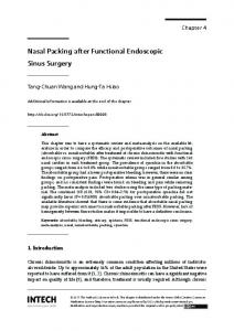
Figure 1. Radical surgery: superior and medial parts of the sinus mucosa (arrow) were removed.
256
infection were used in the present study. General anesthesia was induced by an intramuscular injection of 50 mg/kg of ketamine. An incision of the skin and of the periosteal was made in the cheek. The anterior maxillary wall was drilled under microscopic view and an antral window measuring 5 x 5 mm was made. An infection was induced by blocking the sinus opening with histoacryl (butyl-cyano-acrylate) on cotton wool and by introducing a 0.5 ml suspension of Streptococcus pneumoniae serotype 3 (108 Colony Forming Units). Four weeks after the induction (Day 28), the animals were re-anesthetized. The two surgical procedures compared were realized on each rabbit, FS on one side and RS on the other side. The natural ostium was reopened by removing both the cotton wool and the hystoacryl in order to mimic FS. The existence of efficient ventilation was verified by the injection of physiological serum in the sinus. In addition to reopening the natural ostium, both superior and medial parts of the sinus mucosa were removed when RS was performed (Figures 1 and 2). Such removed mucosa was submitted to both macroscopic and microscopic evaluation. The wound was closed with polyglycolic acid sutures. The animals were left to heal and were then sacrificed at varying intervals of 15 days, 1 month and 2 months.
Figure 2. Resection of the superior (A) and the medial (B) parts of the sinus mucosa according to the work of Forsgren (6).
Macroscopic study The volume of secretions found in the maxillary sinus was noted and graded as 0, +, ++ and +++. The mucosa was photographed by an endoscope (4 mm 0°; Karl-Stortz). The inflammatory reaction of the mucosa was conjointly evaluated in blind fashion by two independent observers according to the following parameters: normal (0), mild (+), moderate (++) and severe (+++). Inflammatory reaction corresponded to the presence of oedema, hyperaemia or granulations. Microscopic study After removal and dissection of the superior maxillary, the major part of the maxillary sinus, including superior and medial parts, was fixed in Bouin’solution for 48 hours. The specimen was then decalcified in a 24% EDTA (ethylenediaminetetraacetic acid) solution for 6 hours. The tissues were then
Guevara et al.
dehydrated in ethanol and embedded in paraffin. The 5 Ìm thick sections were colored with hematoxylin-and-eosine, Alcian blue and a trichrome stain. Different lesions of the sinus mucosa were quantified conjointly by two observers using a double-blind semi-analytic method. The following parameters were quoted as 0, +, ++, +++: infiltrates of chronic inflammatory cells (lymphoid and plasma cells), fibrosis, bone reaction (osteitis) and state of the epithelium (re-epithelialisation). The thickness of the basal membrane was noted as present (+) or absent (0). Statistic analysis Because the animals were used as their own control, we investigated the significance of macroscopic and microscopic evaluation between FS and RS by the Sign test for paired observations. All tests were two-sided and p values of ≤ 0.05 were considered as significant. RESULTS Experimental sinusitis’ model (Induced sinusitis - Day 28) All rabbits presented abundant sinus secretions. The macroscopic aspect showed an inflammatory mucosa associated with more or less severe polyposis lesions. The histological aspect of all specimens showed a severe and chronically evolving inflammatory reaction. The epithelium was most of the time abraded or was the site of superficial erosion. A focal hyperplasia of the epithelium was observed. The re-epithelialisation was noticed to begin in 50% of the specimens associated with the presence of basal cells or of a dedifferentiated epithelium. The goblet cells’ density was above the normal. When remaining, the basal membrane was thickened and hyalinized. A diffuse inflammatory cell infiltrate was noted in the lamina propria composed of a majority of both lymphoid and plasma cells, associated with some histiocytes and polymorphonuclear leukocytes. Moreover, a mild fibrosis with angiogenesis of capillaries was noted. Polypoid lesions were observed histologically in 50% of the specimens. Functional surgery versus radical surgery (Tables 1 and 2) The volume of secretions in the maxillary sinus and the macroscopic inflammatory reaction of the sinus mucosa were less important in FS than in RS at 15 days (secretion: [15 days – p=0,016]; [1 month - p=0,016]; Inflammatory reaction: [15 days - p=0,03]; [1 month - p=0,06]; [2 months - p=0,5]). FS had diminished chronic inflammatory criteria (lymphoid and plasma cells) faster that RS ([15 days (p=0,016)]; [1 month (p=0,03)]; [2 months (p=0,03)]). The mucosa fibrosis was more important after RS ([15 days (p=0,016)]; [1 month (p=0,03)]; [2 months (p=0,016)]). The FS never led to the thickening of the basal membrane conversely to RS ([15 days (p=0,016)]; [1 month (p=0,06)]; [2 months (p=0,5)]). The bone reaction was equivalent in both surgical procedures (p>0,9). The healing of epithelium was faster in FS than in RS ([15 days (p=0,016)]; [1 month – p=0,06]).
Experimental sinusitis’ efficiency treatment
Table 1. Macroscopic evaluation – Volume of secretions in the maxillary sinus and inflammatory reaction of the sinus mucosa at various stages of evaluation [functional surgery (FS) versus radical surgery (RS)] (* p ≤ 0,05). Secretions Inflammatory reaction 0 + ++ +++ 0 + ++ +++ 15 days FS 1* 2* 4* 0* 0* 7* 0* 0* RS 0* 1* 2* 4* 0* 1* 4* 2* 1 month FS 3* 3* 1* 0* 0 7 0 0 RS 0* 0* 6* 1* 0 2 5 0 2 months FS 3 4 0 0 5 2 0 0 RS 3 4 0 0 4 2 1 0
DISCUSSION In the present study, we demonstrated that FS accelerates healing with less fibrosis than RS in such pathological mucosa altered by a chronic inflammation. Our animal model has been the New-Zealand white rabbit. Actually, almost all experimental studies on infection responses or healing of sinus mucosa have been conducted on such animals (4-8). The rabbit’s maxillary sinus is very similar to of a human with a well-defined ostium and the aspect of its mucosa constituted of a pseudo stratified ciliated cylindrical epithelium (6). In order to conduct our study, we have induced a chronic sinusitis. An experimental sinusitis model had already been described in 1985 by Kumlien (3). Moreover, Johansson had shown the reproducibility of such model in 1988 (4). Other studies confirmed (i) that the ostial occlusion is the most important parameter in inducing a sinusitis and (ii) that the type and strength of the germ used are very important as regards the consequently induced mucosa histopathological changes (7). Various bacteria can be used to induce sinusitis: Streptococcus pneumoniae, Bacteroides fragilis, and Staphylococcus aureus (5,7). We opted for Streptococcus pneumoniae, which is less aggressive, according to the work of Westrin (9). Our goal was to reproduce human-like mucosa lesions in chronic sinusitis. In this regard, we produced both an inflammation and an infection by way of a mucosa trauma and an injection of Streptococcus pneumoniae, followed by obstruction of the ostium maintaining the pathological process.
257
We decided to intervene 4 weeks after the induction of the sinusitis so as to obtain sinus’ mucosa lesions very similar to those observed in chronic sinusitis. Westrin (9) has shown, after a 4 week-period, the diminution of the severe inflammatory phenomena along with the apparition of a more chronic infection characterized by (i) an oedema, (ii) an inflammatory cells’ infiltration, (iii) a thickening of the basal membrane, (iv) a mucoid cell hyperplasia and (v) sometimes, the apparition of polypes. Those changes correspond to what can be observed on mucosa biopsies in patients suffering from chronic sinusitis (10). Our results confirmed Westrin’s results (9), showing a very severe and chronically evolving inflammatory reaction. One major issue of the present study was the choice of the surgical procedures used to reproduce (i) a FS model and (ii) a RS model. In human, FS of the maxillary sinus corresponds to the middle meatal antrostomy, whereas RS is represented by CaldwellLuc’s intervention. The middle meatal antrostomy consists in restoring both a correct ventilation and drainage of the maxillary sinus by enlarging the physiological ostium of the maxillary sinus. On the contrary, the Caldwell-Luc technique consists mainly (i) in the complete ablation of the pathological mucosa of the entire maxillary sinus and (ii) in the restoration of the sinus’ drainage by an inferior meatal antrostomy. Such restoration of the sinus’ ventilation has an important impact on the healing of the sinus mucosa. In order to prevent our study from being biased, we had to limit the analysis to the “ablation of pathological mucosa or not” criteria and, in that respect, we did not intervene on the ostium nor did we carry out inferior meatal antrostomy because an efficient sinus reventilation had always been verified during the procedure. Mucosa resection was similar to that accomplished in Forsgren’s study (6). Hence, we have realized a simple model (presence of the mucosa versus ablation of the mucosa) without any other variable factor such as middle meatal antrostomy or inferior meatal antrostomy. The results regarding permeability or repetition of the sinusitis are contradictory to different previous studies (11,12). The last point constitutes one of the distinctions between our model and Forsgren’s study on the effects of FS (middle meatal antrostomy) and RS (ablation of the mucosa and re-ventilation)
Table 2. Microscopic evaluation – Chronic inflammatory parameters (lymphoid and plasma cells), fibrosis’ intensity, thickness of the basal membrane, bone reaction’s intensity and state of the epithelium at various stages of evaluation [functional surgery (FS) versus radical surgery (RS)]. (* p ≤ 0,05). Thickness Chronic of the inflammatory basal reaction Fibrosis membrane Bone reaction Epithelium 0 + ++ +++ 0 + ++ +++ 0 + 0 + ++ +++ 0 + ++ +++ 15 days FS 2* 5* 0* 0* 0* 3* 4* 0* 7* 0* 0 7 0 0 0* 5* 2* 0* RS 0* 1* 6* 0* 0* 0* 3* 4* 0* 7* 0 6 1 0 7* 0* 0* 0* 1 month FS 2* 5* 0* 0* 2* 5* 0* 0* 7 0 3 3 1 0 1 4 2 0 RS 0* 3* 4* 0* 0* 3* 4* 0* 2 5 3 3 1 0 4 3 0 0 2 months FS 0* 7* 0* 0* 3* 4* 0* 0* 7 0 6 1 0 0 0 0 0 7 RS 0* 1* 6* 0* 0* 3* 4* \0* 5 2 5 2 0 0 0 0 0 7
258
on a pathological mucosa (5). Forsgren had actually realized an enlargement of the maxillary sinus’s natural ostium so as to mimic FS. On the contrary, we did not find necessity to intervene on a non-pathological ostium, which may have modified the sinus physiology and the mucosa healing. Our study aimed at comparing different treatments in evaluating the mucosa’s healing. Consequently, using similar groups was a major issue and in that respect each rabbit was made to be its own control. Side-differences when inducing sinusitis were not as important as in inter-individual variations. The analysis of the results corresponds to several evaluations: - We realized a macroscopic analysis of the secretions and of the sinus’ mucosa. The operator alone evaluated the secretions. Nevertheless, in order to strengthen the objectivity of the study, the mucosa was blindly evaluated using photoendoscopic pictures. - The histological analysis was realized on the entire maxillary sinus, which had previously been removed. Such a technique is similar to the one used by Forsgren (5,6) in which the entire sinuso-nasal complex had been removed. As we had strictly limited the specimen to the maxillary sinus, we obtained shorter decalcification time (6 hours versus 3 months). We have consequently managed to conduct a histological analysis on a less altered mucosa through the action of EDTA, preventing at the same time the study from being biased by isolated mucosa biopsies. We had actually analyzed the entire medial part of the maxillary sinus whereas several authors had only realized simple mucosa biopsies in comparative studies of the treatment of experimental sinusitis (11). The histological analysis was conducted in a semi-quantitative way on most of the parameters used by Forsgren in his study on histological changes of both healthy and pathological mucosa after surgery (5,6). Such a process seemed more appropriate than a quantitative analysis (13) which, although it permits a better analysis of the mucosa, is much more complicated to realize.
Guevara et al.
ACKNOWLEDGMENT The authors thank Aurélie Senechal for advice and help in translating into English. We acknowledge the assistance of Frederic Berthier, MD (Department of Medical Information) for advice on medical statistics. The authors have no relevant financial interest in this article. REFERENCES 1 2
3
4
5
6
7
8
9
10 11
12
13
In our animal model, functional surgery turned out to be much more interesting than radical surgery, the secretions’ volume being definitely less important in the former. The complete removal of the mucosa had probably led to a major dysfunction of the mucociliar clearance as shown previously by Kennedy (8) and Min (14). We have also shown that even though both functional and radical surgeries had diminished inflammatory parameters, functional surgery proved to diminish them faster and more efficiently with a better healing (less important fibrosis). These results reinforce the trend, defended by most authors (1), that consists in preserving the mucosa in the treatment of chronic sinusitis and that meatal antrostomy may not be necessary, at least not at a relatively early stage of the disease.
14
Levine HL. Functional endoscopic sinus surgery: evaluation, surgery, and folow-up of 250 patients. Laryngoscope 1990; 100: 79-84. Forsgren K, Fukami M, Penttilä M, Kumlien J, Stierna P. Endoscopic and Caldwell-Luc approaches in chronic maxillary sinusitis: a comparative histopathologic study on preoperative and postoperative mucosal morphology. Ann Otol Rhinol Laryngol 1995; 104: 350-357. Kumlien J, Schiratzki H. Blood flow in the rabbit sinus mucosa during experimentally induced chronic sinusitis. Acta Otolaryngol (Stockh) 1985; 99: 630-636. Johansson P, Kumlien J, Carlsöö B, Drettner B, Nord CE. Experimental acute sinusitis in rabbits. Acta otolaryngol (Stockh) 1988; 105: 357-366. Forsgren K, Westrin KM; Fukami M, Stierna P. Effects of surgery on mucosal pathologic changes following experimental sinusitis in rabbit. Ann Otol Rhinol Laryngol 1998; 107: 155-163. Forsgren K, Stierna P, Kumlien J, Carlsöö B. Regeneration of maxillary sinus mucosa following surgical removal. Experimental study in rabbits. Ann Otol Rhinol Laryngol. 1993; 102: 459-466. Fukami M, Norlander T, Stierna P, Westrin KM, Carlsöö B, Nord CE. Mucosal pathology of the nose and sinuses: a study in experimental maxillary sinusitis in rabbits induced by Streptococcus pneumoniae, Bacteroides fragilis, and Staphylococcus aureus. Am J Rhinology 1993; 7: 125-132. Kennedy DW, Shaalan H. Reevaluation of maxillary sinus surgery: experimental study in rabbits. Ann Otol Rhinol Laryngol 1989; 105: 357-366. Westrin KM, Stierna P, Kumlien J, Carlsöö B, Nord CE. Induction, course, and recovery of maxillary sinusitis: a bacteriological and histological study in rabbits. Am J Rhinol. 1990; 4: 61-64. Stierna P, Carlsöö B. Histopathological observations in chronic maxillary sinusitis. Acta Otolaryngol (Stockh) 1990; 110: 450-458. Benninger MS, Kaczor J, Stone C. Natural ostiotomy vs. Inferior antrostomy in the management of sinusitis: An animal model. Otolaryngol Head Neck Surgery 1993; 109: 1034-1042. Arnes E, Anke IM, Mair IWS. A comparison between middle and inferior meatal antrostomy in the treatment of chronic maxillary sinus infection. Rhinology 1985; 23: 65-69. Mogensen C, Tos M. Quantitative histology of the maxillary sinus. Rhinology 1977; 15: 129-140. Min YG, Kim IT, Park SH. Mucociliary activity and ultrastructural abnormalities of regenerated sinus mucosa in rabbits. Laryngoscope 1994; 104: 1482-1486.
Dr. Nicolas Guevara Department of Otorhinolaryngology CHU de Nice, Hôpital Pasteur 30, avenue de la Voie Romaine B.P. 69. 06002 Nice Cedex 1 France Tel: +33-49-203 8452 Fax: +33-49-203 8464 E-mail:
[email protected]