Gene 610 (2017) 49–58
Contents lists available at ScienceDirect
Gene journal homepage: www.elsevier.com/locate/gene
Short Communication
A comprehensive computational study on pathogenic mis-sense mutations spanning the RING2 and REP domains of Parkin protein Ria Biswas, Angshuman Bagchi ⁎ Department of Biochemistry and Biophysics, University of Kalyani, Kalyani 741235, Nadia, India
a r t i c l e
i n f o
Article history: Received 8 December 2016 Received in revised form 17 January 2017 Accepted 6 February 2017 Available online 09 February 2017 Keywords: Parkin RING2 domain Mis-sense mutation E3 ligase Interaction energy Parkinson's disease Ubiquitination Protein docking
a b s t r a c t Various mutations in PARK2 gene, which encodes the protein parkin, are significantly associated with the onset of autosomal recessive juvenile Parkinson (ARJP) in neuronal cells. Parkin is a multi domain protein, the N-terminal part contains the Ubl and the C-terminal part consists of four zinc coordinating domains, viz., RING0, RING1, in between ring (IBR) and RING2. Disease mutations are spread over all the domains of Parkin, although mutations in some regions may affect the functionality of Parkin more adversely. The mutations in the RING2 domain are seen to abolish the neuroprotective E3 ligase activity of Parkin. In this current work, we carried out detailed in silico analysis to study the extent of pathogenicity of mutations spanning the Parkin RING2 domain and the adjoining REP region by SIFT, Mutation Accessor, PolyPhen2, SNPs and GO, GV/GD and I-mutant. To study the structural and functional implications of these mutations on RING2-REP domain of Parkin, we studied the solvent accessibility (SASA/RSA), hydrophobicity, intra-molecular hydrogen bonding profile and domain analysis by various computational tools. Finally, we analysed the interaction energy profiles of the mutants and compared them to the wild type protein using Discovery studio 2.5. By comparing the various analyses it could be safely concluded that except P437L and A379V mutations, all other mutations were potentially deleterious affecting various structural aspects of RING2 domain architecture. This study is based purely on computational approach which has the potential to identify disease mutations and the information could further be used in treatment of diseases and prognosis. © 2017 Elsevier B.V. All rights reserved.
1. Introduction Parkinson's Disease (PD) is the second most common neurodegenerative disorder affecting the motor neurons. It is caused due to progressive loss of dopaminergic neurons in the substantia nigra (SN) of midbrain (Klockgether, 2004). PD is a multi-factorial disease resulting from various genetic and environmental factors and could be categorized into sporadic, familial and symptomatic PD (Gasser, 2011). Six genes have been identified which are involved in the onset of PD. The genetic form of the disease can be autosomal dominant (arising out of the dysfunctions of PARK1/4, PARK8 genes) or autosomal recessive (arising out of the dysfunctions PARK2, PARK6, PARK7 and PARK8 genes) (Seirafi et al., 2015; Klein and Schlossmacher, 2006). Direct or accurate molecular causes of the disease are yet to be identified. Many of the symptoms of sporadic and familial PD overlap including the case of autosomal recessive juvenile Parkinson (ARJP), in which the disease onset occurs in patients before the age of 40 years. The presence of cytoplasmic protein aggregates termed Lewy body encoded by PARK1/4,
⁎ Corresponding author. E-mail address:
[email protected] (A. Bagchi).
http://dx.doi.org/10.1016/j.gene.2017.02.008 0378-1119/© 2017 Elsevier B.V. All rights reserved.
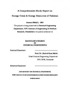
is the hallmark of PD (Beitz 2014). Surprisingly, these protein bodies are rarely seen in the patients affected with ARJP (Huynh 2003) Over 100 mutations in proteins related to the progression of PDs have been identified thus far. The point mutations are observed spanning all the domains of the protein but majority of them are located in the RING-IBR-RING domain (Oczkowska et al. 2014). Mutations in PARK2 gene encoding Parkin protein leads to the onset of the most common form of ARJP (Safadi et al., 2011). According to the information provided by the database PDmutDB (http://www.molgen.vib-ua.be/ PDMutDB), the RING2 and REP domains of the Parkin protein account for 16.4% and 17.2% of mutations respectively among PD patients (Fig. 1a). Among these mutations in the RING2 and REP (repressor element of RING) domains of the Parkin protein, 10.2% and 11% of mutations are known to be disease causing. The status of the remaining mutations is still unclear. Parkin is involved in a range of important cellular functions like mitophagy, cell cycle and vesicle trafficking (McLelland et al. 2014; Jin and Youle 2012; Durcan and Fon 2015; Konovalova et al., 2015). Parkin is an E3 ligase and functions by tagging the misfolded and unneeded proteins with ubiquitin molecule and guiding them to the proteasomal degradation pathway (Sandebring and Cedazo-Mínguez 2012). The ubiquitin-proteasomal system (UPS) plays important roles in regulating critical cellular processes of cell
50
R. Biswas, A. Bagchi Gene 610 (2017) 49–58
Fig. 1. Parkin protein and mutation: (a) distribution of mutations in various domains of Parkin protein. (b) location of the mutations spanning the RING2 and REP domains. The secondary structures within the domain are shown by red (helix), cyan (sheet), white (coil) and green (turn). Zinc ions are shown in grey. (c) N-terminal consists of ubiquitin like domain (Ubl) and C-terminal domain comprises of RBR (RING-in between RING-RING) domain. REP (repressor element of Parkin) connects the IBR with RING2. RING2 consists of a catalytic cysteine residue at position C431. Each RING domain co-ordinates two zinc ions. (d) Phylogenetic analysis; the mis-sense mutation residues are highly conserved throughout phylogeny. 379A seem to be less conserved which corresponds to low pathogenicity. (For interpretation of the references to colour in this figure legend, the reader is referred to the web version of this article.)
division and growth (Deshaies and Joazeiro 2009). Proper functionality of neurons is dependent on the efficient transport of mitochondria to the hot-spots of energy consumption. Accumulation of mitochondrial toxins in neurons results in neuronal degeneration and is known to trigger PD in humans and animals. Loss of E3 activity of Parkin leads to improper ubiquitin tagging and degradation of the target protein which results in protein accumulation and cellular toxicity (Winklhofer and Haass 2010). Various types of mutations like frame-shift mutations, exon-rearrangements, point mutations and mis-sense mutations are responsible for the onset of familial Parkinson's Disease (Kay et al., 2010; Kay et al., 2007). In 1998, the first Parkin mutation associated with autosomal recessive juvenile Parkinsonism (ARJP) was reported in natives of Japan (Shimizu et al. 1998). Some studies suggest that the frequency of mutations related to Parkin is comparatively lower in late onset PD than early-onset PD (Oliveira et al. 2003; Lücking et al. 2000; Bertoli-Avella et al. 2005). As Parkin linked PD is inherited recessively; a deleterious mutation is expected to affect both the alleles. Persons carrying heterozygous mutations shall be unaffected; however some clinical studies suggest a large proportion of patients carry single heterozygous mutations. Patients with heterozygous mutations are reported to suffer from reduced gene expression and enzymatic activity,
which results in predisposition of the disease (Oczkowska et al. 2014). Clark et al. (2006), showed that Parkin associated heterozygous mutations could influence the age of the disease onset by increasing early onset of PD. Mutations in Parkin are also linked with various forms of cancers like melanoma, pancreatic and lung cancers (Xu et al. 2014; Veeriah et al. 2010; Hu et al. 2016). Parkin is a member of RBR E3 ligase containing 465 amino acid residues. The architecture of Parkin consists of an N-terminal Ubl (Ubiquitin like domain) and four RING (Really Interesting New Gene) like domains (RING0, RING1, IBR and RING2) comprising eight zinc (Zn) atoms coordinated by cysteine and histidine residues (Fig. 1c). The Ubl domain amends the activity of Parkin by keeping it in an auto-inhibited state. Riley et al., 2013 synthesized crystals spanning the R0RBR domains. In the stabilized auto-inhibited state, the Ubl domain binds the RING1 domain and the RING0 domain binds the C-terminal catalytic domain. The RING1 domain is the only RING domain similar to the canonical criss-brace structure of RING-type E3 ligases (Riley et al. 2013). Earlier studies based on structural comparison of Parkin RING2 domain with canonical RING reveal very less similarity in protein folding. RING2 domain along with IBR and RING1 assists substrate interaction and ubiquitination (Beasley et al., 2007b). Parkin RING2 adopts a sequential
R. Biswas, A. Bagchi Gene 610 (2017) 49–58
Zn2+ co-ordination site as opposed to the criss-brace structure of RING E3s. RING2 of Parkin also takes on an elongated structure which is dissimilar to the compact canonical RING structure. The structure of Parkin RING2 resembles more closely to Parkin IBR domain (Beasley et al., 2007a). These results indicated that the Parkin RING2 may-not function as the canonical RING E3s and hence may not be able to drive the E2 dependent E3 ligase activity. An interesting research by Rankin et al., 2014 showed that RING2 possess a catalytic core and the isolated Parkin RING2 domain is self sufficient in carrying out the process of ubiquitination through E2 binding (Rankin et al. 2014). This makes the role of RING2 domain of Parkin very speculative. Mis-sense mutations spanning the RING2 domain have gathered considerable attention from biochemists and geneticists but the structural effect and the functional consequence resulting from the mutations are still lacking. In our current work, we carried out a detailed investigation of the effect of missense point mutations on RING2 and the adjoining REP (repressor element of RING) domain of Parkin protein by various bioinformatics tools. To attain its globular conformation, the RING2 domain of Parkin binds two Zn ions by motif rich in cysteine and histidine. Hence, the interactions of cysteine and histidine residues are essential for proper motif formation and functioning of the RING2 domain. As a next step of our study, we calculated the total and residue wise interaction energies of the cysteine and histidine residues for each mutant taking wild type as reference. We tried to predict the pathogenicty of the mutations by using different computational techniques. This report is the first bioinformatic work to relate the relative pathogenic characteristics of the mutations with the onset of PD. 2. Materials and methods 2.1. Isolation of mutations linked to REP and RING2 domains The mutations in Parkin protein were extracted from two different databases: 1. PDmutDB (http://www.molgen.vib-ua.be/PDMutDB/): This locus specific database was built in November 2009. This database collects all known up-to-date mutations and polymorphisms identified in patients with PD. The database comprises mutations in the gene associated with α-synuclein (SNCA), Leucine rich repeat kinase 2 (LRRK2), Parkin (PARK2), PTEN-induced putative kinase 1 (PINK1) and DJ-1 (PARK7). Information on a total of 535 different DNA variations and 2084 families are stored in this database. 2. Parkinson's Disease Mutation Database (http://www.thepi.org/ parkinson-s-disease-mutation-database/): The database sums up all disease associated mutations linked to PD. This database provides comprehensive details of known and recurrent mutations based on clinical test results of patients and from the available literature references. A total of seven mis-sense mutations associated with the RING2 and REP domains were collected which were known to be a causative of PD phenotype in patients. Some of the mutations are also associated with various cancers. Table 1 lists all the mutations with their corresponding domains.
51
2.2. Analysis of mutational effects on Parkin We analysed the following features of the wild type and mutated Parkin proteins to study the effects of mutations on the structure and function of the protein: a) Analysis of severity of mutations: for prioritizing the deleterious nature of mutations, they were checked with various bioinformatics tools like: Mutation Accessor, which is based on evolutionary conservation of the mutated amino acids in the homologous proteins (Reva et al., 2011), SIFT (Sorting Intolerant from Tolerant amino acid substitutions) which is based on sequence homology to determine whether a substitution will contribute to the disease state or not (Kumar et al., 2009), PolyPhen2 (Polymorphism Phenotyping) which is based on evolutionary conservation of amino acid which are frequent to mutate and structural property change on amino acid substitution (Adzhubei et al., 2013), SNPs and GO which is based on support vector machines to predict pathogenic mutations (Thusberg et al., 2011), Align GV (Grantham Variation)/GD(Grantham Deviation) and GV which determine the extent of biochemical changes upon amino acid substitution. GD reflects the biochemical distance between the mutated and the wildtype amino acid (Tavtigian et al. 2008). We used all the tools to generate a consensus result, shown in Table 1. Mutations showing positive implication with all used tools were considered the most pathogenic. Mutations which scored at least by two tools were considered moderate and remaining were considered low on pathogenicity. The stabilities of wild type and each mutated protein were further analysed using I-Mutant 2.0 (Capriotti et al., 2005). To find the effect of mutations on domain organisation, the wild type and mutated amino acid sequences were analysed in NCBI conserved domain search (Marchler-Bauer et al. 2015) and Pfam (Finn et al. 2014). The mutation G429E was found to acquire RRM (RNA recognition motif) domain instead of RING2 domain. To further investigate the feasibility of G429E mutant to bind RNA we performed molecular docking using Patchdock Beta 1.3 version (http://bioinfo3d.cs.tau.ac.il/PatchDock/) with default clustering RMSD of 4.0. For docking, heterogeneous ribonucleoprotein particle (hnRNP) with PDB ID 2MQO was taken as a reference structure. hnRNP is a RRM domain containing protein. The sequences of Parkin REP-RING2 domain and hnRNP were aligned using sequence alignment tool in Discovery Studio 2.5 (DS 2.5) to identify common amino acid residues which can bind RNA molecule. The 10 best docked poses were subjected to energy minimisation using steepest descent and conjugate gradient algorithms in DS 2.5 in GBSW (Knight and Brooks 2011) until an Root Mean Square gradient of 0.001 kcal/mol was reached using the CHARMm force-field (Brooks et al. 1983). Interaction energy calculation was done using calculate interaction energy protocol in DS 2.5. The pose with the least interaction energy was considered the best representative structure of the complex. b) Analysis of hydropathy profile: Hydropathy profiles of the wild type and each mutant were analysed using PROTPARAM server. The programme is based on Kyte-Dolittle algorithm which assigns hydrophobicity to each amino acid sequence (Kyte and Doolittle 1982).
Table 1 Overview of extent of pathology on PARK2 mutations associated with REP and RING2 domain. Mutation
Domain
Nucleotide change
Region of DNA
Reference
Mut. accessor
SIFT
PolyPhen2
SNPs&GO
GV/GD
Score
Inference
R402C P437L G429E C418R C441R R420H A379V
REP R2 R2 R2 R2 R2 REP
CNT CNT GNA TNC TNC GNA CNT
EX11 EX12 EX12 EX11 EX12 EX11 EX10
(Poorkaj et al. 2004) (Hedrich et al. 2002) (Brüggemann et al. 2009) (Bertoli-Avella et al. 2005) (Shyu et al. 2005) (Sun et al. 2006) (Veeriah et al. 2010)
Medium Low Low High High Neutral Low
Not tolerated Tolerated Tolerated Not tolerated Not tolerated Not tolerated Tolerated
Damaging Damaging Damaging Damaging Damaging Damaging Benign
Disease Neutral Disease Disease Disease Neutral Neutral
Class C65 Class C65 Class C65 Class C65 Class C65 Class C25 Class C65
Five out of five Two out of five Three out of five Five out of five Five out of five Two out of five One out of five
Very high Moderate High Very high Very high Moderate Low
52
R. Biswas, A. Bagchi Gene 610 (2017) 49–58
c) Calculation of solvent accessibility: The changes in relative solvent accessibility (RSA) of the mutated residues of Parkin were analysed using XSSP online server. The formula used for calculating percentage relative accessibility of wild type and each mutant was [(ACC number / Highest ACC number) × 100]. Here ACC number was the score obtained from the server and the highest ACC number was the highest score which could be obtained for the amino acid (Rost and Sander 1994). The overall Solvent Accessible Surface Area (SASA) of calculation of the protein was carried out using solvent accessibility tool in DS 2.5. ITasser server was employed to construct the structures of mutant proteins and to predict the overall changes in solvent accessibilities of individual residues upon single point mutation. d) Contact plot for intra-molecular hydrogen bonds: To study the changes in intra-molecular hydrogen bond profile in mutants as compared to the wild type protein, contact plots were generated using DS 2.5 platform. 2.3. Homology modeling of Parkin and construction of mutants Crystal structure of full length human Parkin is available with PDB ID 4I1H; however there are some missing residues in the region spanning the REP and RING2 domains. To rectify the missing residue problem we performed homology modeling using Parkin crystal structures 4K7D from Rat and 4I1H from human as templates in modeller programme in the DS 2.5 platform to build the model for the REP and RING2 domains of Parkin. 4K7D and 4I1H had query coverage of 100% and 99% respectively and sequence identity of 86% and 79% respectively. The target sequence for model construction was obtained from Uniprot with accession number O60260. The mutants were generated using build mutant tool of DS 2.5 using wild type as the reference. A model was constructed incorporating all the mutations to study the effect of multiple mutations on Parkin RING2 domain. Energy minimizations were applied as per the previously mentioned protocol. Structural verification of wild type and each mutant were done using Verify3D (Eisenberg et al., 1997) and RAMPAGE (Lovell et al. 2003). For interaction energy studies we focused on the RING2 and REP domain of human Parkin protein. 2.4. Analysis of changes in interaction energy (ΔG) pattern due to Parkin mutations To analyse the interaction energy pattern of cysteine and histidine residues with the Zn ion we calculated the total and individual residue interaction energy patterns for wild type and each mutated proteins in DS 2.5. Each structure was energy minimized and verified as mentioned before. 3. Results and discussion
regarding the mutations and its effect on interaction energy profile of zinc binding cysteine and histidine residues in the wild type and the mutants. 3.1. Effect on structure and function due to mutations in Parkin RING2/REP domain Clinical and pathogenic studies have already identified mutations mentioned in Table 1 but their molecular basis regarding the pathogenicity remains unclear. Proteins are known to perform diverse functions which are implemented by the residues at the catalytic sites. As the residues at catalytic sites are involved in highly specific reactions, they are often evolutionary highly conserved. The results from multiple sequence alignment (Fig. 1d) suggest that the residues of human Parkin, viz., R402, C418, G429 and C441 were highly conserved and R420 was highly similar in evolution. Proline was observed to be conserved in human, chimpanzee and mouse at positions 437, 437 and 436 respectively however it is replaced by threonine 455 in drosophila. Drosophila has long been used as a model organism to study the cellular effects of Parkin mutations. Drosophila is known to encode a homolog of human Parkin protein and our sequence alignment studies suggest that it has a significant sequence identity of 40.3% and sequence similarity of 60.1% with the human Parkin homolog. The characteristic domains Ubl, RING1, IBR and RING2 are known to be highly conserved in drosophila. Drosophila Parkin null mutants are known to exhibit clumped mitochondria with swollen and disrupted cristae in its dopaminergic neurons. The drosophila PD models exhibit multi-system defects such flight and climbing muscle degeneration, male sterility, defective morphology and decreased life-span which are similar to the defects observed in PD patients (Dawson et al., 2010; Pesah et al. 2004; Greene et al. 2005; Muñoz-Soriano and Paricio 2011; Guo 2012). On the basis of these evidences, it would be interesting to study the effect of mutation at this position. Alanine 379 was not found to be conserved. Also, mutations at position C418, G429, C441 and R420 in human Parkin protein may have detrimental effects on structural stability and function of the Parkin RING2 domain. Results from pathogenicity prediction tools suggested that, cysteine residues mutated to any other residue (C418R, C441R), have high inference of pathogenicity. Glycine mutated to acidic residue glutamic acid (G429E) and arginine mutated to cysteine (R402C) showed high inference of pathogenicity as well. I-mutant results (Table 2) suggested that mutations C418R, R420H, P437L and C441R were destabilizing mutations whereas A379V, R402C and G429E would correspond to stabilizing mutations. G429E appeared to be evolutionary very conserved but Mut Accessor and SIFT pathogenicity tool did not recognize it as the pathogenic mutation. This may be Table 3 RSA value of each mutant compared to the wildtype Parkin.
In the following work, we tried to collect and classify disease causing mutations in Parkin RING2 domain from two different online repositories for patients of PD. We selected all mis-sense mutations which were identified responsible for disease onset (Table 2) to find a plausible biochemical explanation for occurrence of the disease causing mutations on the basis of local interaction energy profile of the zinc binding residues. In the following section we will discuss about our outcomes Table 2 Effect on Parkin stability with respect to pathogenic mutations of REP/RING2 domains. Position
WT
New
Stability
379 402 418 420 429 437 441
A R C R G P C
V C R H E L R
Increase Increase Decrease Decrease Increase Decrease Decrease
Type
Position
Residue
ACC value 1
Max value 2
RSA (RelAcc)3 value
WT A379V WT R402C WT R420H WT C418R WT C441R WT G429E WT P437L
379 379 402 402 420 420 418 418 441 441 429 429 437 437
A V R C R H C R C R G E P L
26 32 148 65 156 129 1 21 6 15 51 174 98 160
106 142 248 135 248 184 135 248 135 248 84 194 136 164
24.5 22.5 59.67 48.14 62.9 70.1 0.74 8.46 4.44 6.04 60.71 89.69 72.05 97.56
1. ACC = solvent accessibility value (Å2) of a given residue which approximates the number of surrounding water molecues. 2. Max value = the maximum value which could be gained by a particular residue. Measured in Å2. 3. RelAcc(RSA) = Acc/Max value, gives the relative accessibility of a residue.
R. Biswas, A. Bagchi Gene 610 (2017) 49–58
because these algorithms are based on untrained datasets and also don't incorporate machine learning technique. Also they are known to cite distantly related orthologs and paralogs. (Ng and Henikoff 2003; Reva et al., 2011). On the other hand, tools like Poly-Phen2 incorporate machine learning methods and identify mutational consequences on the basis of selected mutations which are earlier known to be damaging or benign (Adzhubei et al., 2013). Solvent accessibility of the wild type and mutants was calculated by total calculating total SASA and residue wise SASA (RSA) (Fig. 3). SASA calculations of the wild type and mutants proved that mutations C418R, R420H, P437L and C441R would have an increase in total solvent accessibility which could reveal that these mutations might provoke unfolding and hence were destabilizing in nature. It is a well established fact that cysteine cross-links could stabilize the protein structure by decreasing the overall entropy and solvent accessibility (Ali et al. 2014), therefore an increase in SASA may result in protein unfolding and damage. SASA value for wild-type protein was 8037.27 Å2; R402C showed a
53
lower solvent accessibility of 7991.11 Å2 than the wild type and hence could be suggested to contribute to the compactness and stability of the protein. Increased compactness can further affect the normal functioning of the protein by hindering its ability to interact with its substrate proteins. Although I-mutant results showed that G429E mutation could stabilize the protein, the total solvent accessibility results showed otherwise with SASA 8383.97 Å2, which was higher than wild-type. This might be due to replacement of the non-polar Glycine residue by the negatively charged Glutamic acid. The introduction of a charged residue results in new electrostatic interactions with the solvent molecules (Pace et al., 2000). These charge-charge interactions may further result in protein denaturation and unfolding by reducing the net contribution of interactions within the core of the protein. Protein unfolding results in an increase of exposed amino acid residues and hence increase in SASA. Hence, mutation of non-polar Glycine residue with acidic Glutamic acid residue is resulting in an unfolded protein which is highly stable. C418R, R420H, P4,37 L and C441R resulted in
Fig. 2. Kyte-Dolittle hydrophobocity profile. The X-axis defines the window position and Y-axis defines the hydropathy score. Negative peaks correspond to possible surface regions of globular proteins. Window size is kept 9. Window size indicates the average hydropathy for the entire window.
54
R. Biswas, A. Bagchi Gene 610 (2017) 49–58
SASA values of 8395.25 Å2, 8268.65 Å2, 8086.52 Å2 and 8037.70 Å2 respectively. A379V mutant showed a SASA of 8047.42 Å. From the results we could safely conclude that mutations G429E, C418R and R420H might lead to the promotion of protein unfolding and domain injury. Furthermore, to study the changes in buried and exposed residue associated with the mutations, we calculated relative solvent accessibility (RSA) of each residue in the wild type and the corresponding mutated residue (Table 3). RSA defines the extent of surface exposure of protein residue to the solvent. In the folded state of protein, the polar and charged amino acids had higher solvent accessibility than the nonpolar side chains. This suggests an important role of hydrophobic core formation in proper folding of protein and hence its function. RSA could help in explaining the protein structure and functional relationship. The residues which reside on the inside of the protein are important for protein stability. The buried residues are seen to give rise to temperature sensitive mutations and hence the site of deleterious mutations (Chen 2005; Rost and Sander 1994). An increase in the RSA
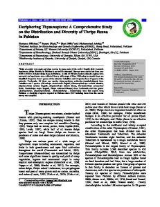
value corresponds to the formation of a local bulge in the protein surface which can make an unwanted residue to be accessible for protein-protein interaction whereas a decrease in the RSA value refers to the formation of a cavity which makes the residue buried and inaccessible to the solvent (Marsh and Teichmann 2011). The mutation involving exchange of basic arginine to cysteine residue (R402C) showed a decrease in RSA value which would correspond to cavity formation, whereas all other mutations showed increase in RSA which might infer unwanted contacts. This result could clearly indicate protein instability and dysfunction upon mutation. A379V mutant did not show significant deviation of RSA value from the WT protein. NCBI conserved domain database (CDD) search for identification of domain architecture revealed the loss of normal domain architecture and introduction of RRM (RNA binding) domain in the Parkin protein with G429E mutation. RRM domain is known to be involved in the post-transcriptional gene expression processes (Maris et al., 2005). Sequence alignment of hnRNP with Parkin REP-RING2 domain identified two residues of Parkin, Arg396 and
Fig. 3. Changes in solvent accessibility in RING2 and REP domains of Parkin upon mutation. Solvent accessibility is seen to change upon mutation in the RING2 and REP domain of Parkin protein. The model of the mutants was build using I-Tasser (http://zhanglab.ccmb.med.umich.edu/I-TASSER/). Red colour represents the residues with the least solvent accessibility or buried residues and the blue colour represents the most accessible residues or surface residues. Changes in the solvent accessibility of the mutant proteins are observed. (For interpretation of the references to colour in this figure legend, the reader is referred to the web version of this article.)
R. Biswas, A. Bagchi Gene 610 (2017) 49–58
55
Arg455 in RING2 domain to be conserved. Arg396 and Arg455 were specified as binding site residues for docking of the WT and G429E mutant Parkin proteins with RNA. Molecular docking and interaction energy studies of WT-RNA and G429E-RNA showed a higher affinity of the mutant protein (− 53.295 kcal/mol) to bind the single stranded RNA than the WT (− 35.641 kcal/mol). Interestingly, RING E3 ubiquitin ligases like MDM2 and Hdm2 are known to bind RNA/DNA by their RING domains (Cano et al., 2010). Hence overall it could be concluded that the single point mutation G429E converts the Parkin RING2 domain into RRM domain and increases the ability of the Parkin RING2 domain to interact with RNA oligonucleotide (Fig. 4). This could further affect the substrate binding affinity of the RING2 domain. The R402C, C441R and R420H mutations suggested the absolute loss of IBR domain of Parkin RING2. These results could reflect the important roles of the residues 420R, 441C, R402 and 429G in the proper domain architecture of Parkin protein. P437L and A379V did not result in any changes of domain architecture. Hydrophobicity of a protein is a critical factor for its stability (Biro 2006). Local hydrophobicity profile was found to be altered due to mutations (Fig. 2); this could clearly indicate alterations in the solubility of the protein which might further contribute to the onset of PD. The three dimensional conformation of proteins is stabilized by complex set-up of hydrogen bonds. Intra-molecular hydrogen bond constitutes main chain-main chain H-bond, side chain–side chain H-bond, main chain-side chain H-bond and multiple H-bond. The main chain-main chain H-bonds constitute the majority of (nearly 68%) of H-bonds in proteins and contribute mainly to the proper folding of evolutionary conserved secondary structures. The side chain-side chain and side chain-main chain H-bonds are approximately equally distributed (Stickle et al. 1992). The side chain-main chain H-bonds are known to be involved in initiation of helix and formation of turns and are majorly located at turns and at the termini of helices. Side chain-main chain H-bonds could confer protein conformational stability, as these H-bonds are buried in the interior of the protein where they bind to the polar groups of main chain and satisfy H-bonding potential of main chain atoms (Bordo and Argos 1994; Baker and Hubbard 1984). In case of mutations (Supplementary Fig. 1), the new structural rearrangements are introduced by substitution of one amino acid with another and subsequently the hydrogen bond order must be maintained in order to conserve the physico-chemical properties of the proteins. C418R and R420H showed difference in main chain-main chain and main chain-side chain interactions from wild type. The mutations R402C and G429E showed loss of main chain-side chain interactions. On the other hand, the mutations A379V, C441R and P437L showed no prominent change in the hydrogen bonding profile with respect to the wild type protein. 3.2. Local energy changes in Parkin RING2 due to pathogenic mutations Parkin protein belongs to the RBR E3 ligase family which is considered a hybrid of RING/HECT E3 ligases. The RING2 domain does not resemble the canonical RING E3 domain structure and as already mentioned, the cysteine and histidine residues are arranged in a sequential order rather than a criss-brace order. The RING2 domain is known to co-ordinate two Zn2+ ions in the native state. The substitution of cysteine residues is a causative of protein inactivity, making the interaction of cysteine residues with Zn2 + ion absolutely important for proper protein functionality(Metzger et al. 2014). Here, we studied the changes in local energy profile by calculating the total and residue wise interaction energy pattern of the wild type and each pathogenic mutant protein. In metalloproteins, metals bind to proteins to assist proper folding and to increase their functional properties. The binding of macromolecules to specific substrates is driven by the process of molecular recognition. Molecular recognition is the process by which macromolecules form specific complexes by interacting with self or other molecules by non-covalent interactions (Van der Waals and electrostatic interactions). The interaction energy of a protein-ligand complex is
Fig. 4. Effect on domain architecture in G429E mutation: G429E mutation replaces the RING2 domain with a RNA recognition motif (RRM) domain which is known to bind RNA. (a) and (b) shows interaction of the WT and G429E mutant with RNA. The interacting residues are highlighted in yellow. The mutant shows much higher affinity towards RNA than the WT. The purine and pyrimidine bases are colored red and pink respectively. For protein, helix, sheet, coil and turn are colored red, cyan, white and green respectively. (c) Interaction energies of wild type (WT_RNA) and G429E mutant (G429E_RNA) with RNA. (For interpretation of the references to colour in this figure legend, the reader is referred to the web version of this article.)
represented by a negative sign and the specificity and stability of the complex is determined by the magnitude of the interaction energy (Du et al. 2016; Agnieszka, 2011). The lower the interaction energy the more favorable and specific is the interaction between two molecules. The mutations C441R, C418R and G429E resulted in positive interaction energy which indicates changes in the binding of cysteine and histidine residues to the Zn2 + ions. Residues C441 and C418 are present in binding pocket of Zn2+ ion; hence substitution of these residues with the basic Arg residue may result in loosening of the binding. C418R mutation resulted in dramatic changes in interaction energies
56
R. Biswas, A. Bagchi Gene 610 (2017) 49–58
showing no interaction of the mutated residues with Zn2+ ion. The interaction energies with C421, C436, C446 and H461 increased which indicates less probability of binding of the residues with Zn2+. In C441R mutation, Arg residue displayed an unusually highly positive Van der Waal interaction (1.3 × 10⁸ kcal/mol) which results absolute unlikely interaction with Zn2+. This might result in loosening of the Zn2+ ion from the active site and collapse of RING2 domain architecture. The profile for individual interaction energy of binding site cysteine and histidine residues showed alterations in interaction compared to the wild type (Fig. 5a). To study how each mutation affected interaction of active site cysteine and histidine residues with Zn ion, we observed the interaction energy profile for Cys and His residue each for the wild type and the mutants (Fig. 5b). Mutant R402C showed decreased interaction
with residues C418, C421, C441, C446, C457 and H461, most prominent decrease was observed in the case of C421 and C441. In case of residue C449, mutant R402C showed slightly higher interaction than wild type. Mutant P437L resulted in higher interaction of residues C421, C436, C441, C449, C457 and H461. The mutation resulted in a decreased interaction of C446 residue. Mutant C441R resulted in non-favorable interaction of residue C421 and displayed no interaction with residue C449. Mutant C418R residues lowered interaction of residues C421, C436, C446 and His461. Except for C446 there was a drastic decrease of interaction of the other resides with Zn ion for C418R. C418R showed non-favorable interaction of residues C441 and C449, also there was a slight increase of interaction of residue C457. Mutant R420H showed prominent decrease of interaction energy of residues C418, C446 and H461.
Fig. 5. Total and residue wise interaction energy profile of RING2 domain of Parkin in wild type and mutants. (a) Total interaction energy of WT, A379V, R402C, P437L, C418R, R420H and G429E. Mutations C418R, R420H and G429E results in decreased interaction whereas interaction increases in case of P437L. For the ease of representation interaction energy of the mutant C441R was not plotted as the interaction energy for C441R was 1.3 × 10⁸ kcal/mol which is very high compared to others. (b) residue wise interaction energy profile of active site cysteine residues, (from left to right) C418, C421, C436, C441, C446, C449, C457 and histidine residue, H461 co-ordinating with Zn ion. Wild type(black), A379V(red), R402C (blue), P437L (dark green), C441R (cyan), C418R (purple), R420H(flurocsent green), G429E (olive green). (For interpretation of the references to colour in this figure legend, the reader is referred to the web version of this article.)
R. Biswas, A. Bagchi Gene 610 (2017) 49–58
The mutation resulted in unfavorable interaction of residues C436, C441 and C449. It also resulted in a higher interaction of residue C421. Mutation G429E showed drastic decrease of interaction of residues C446 and His461 and non-favorable interaction of residues C418, C436, C441 and C449. The mutation resulted in a higher interaction of residue C457. Mutation A379V did not result in much deviation from the interaction energy of WT. The multi-mutant model was subjected to interaction energy calculations to see whether the mutations when present together nullify the effects of each other and what would be the effect of such mutation on zinc binding ability of the protein. The multiple mutation model showed highly positive (35 kcal/mol) interaction energy with zinc ion, which means total loss of zinc interaction which will lead to protein dysfunction and collapse. Overall in silico mutation analysis showed that A379V had no significant effect on the structural and functional properties of Parkin RING2 domain. Also, P437L mutation might not be deleterious in nature as it increased total interaction energy and also resulted in no effect on hydrogen bonding or domain architecture of the RING2 domain. Solvent accessibility was also not prominently affected compared to the wild type. The mutations of the highly conserved active site cysteine residues C418 and C441 proved to be very deleterious resulting in the decrease in stability, changes in domain architecture and decreased interaction energy with Zn ion which may lead to domain collapse. C418R mutation resulted in changes in main chain-side chain hydrogen bonding patterns. R402C mutation in the REP domain seems to be deleterious in nature resulting in increased compactness, loss of main chain-side chain hydrogen bonding and loss of domain architecture of the protein. Although the total interaction energy showed no prominent change, residue wise interaction energy calculation showed prominent decrease of interaction of critical cysteine residues C421, C441 with Zn ion. Mutations R420H and G429E resulted in decreased stability, loss of domain architecture, decrease total interaction energy and alteration in hydrogen bonding pattern. All the results mirror the fact that all the missense mutations except A379V and P437L can result in severe structural distortion of Parkin RING2 domain and hence can be fatal. 4. Conclusion A mutation altering an amino acid with another may result in change of globular conformation of the protein. The new structural changes must keep the physico-chemical properties of the old residue intact. If physico-chemical properties are less affected upon mutation, the changes will be minimal but if the properties are very different there could be drastic structural changes which may further lead to protein unfolding and dysfunction. In our present study, we analysed mis-sense mutations spanning the Parkin RING2 and adjoining REP domain to study the structure-function relationship of human Parkin. The mutations except P437L and A379V resulted in protein structure deformation and functional loss of Parkin. These studies will in future help to understand the structural aspect of Parkin protein dysfunctioning upon RING2/ REP mutations and also will aid in drug development which could help in Parkin activation and recovery of E3 ligase activity in patients suffering from PD. Supplementary data to this article can be found online at http://dx. doi.org/10.1016/j.gene.2017.02.008. Conflict of interest None. Acknowledgements The authors acknowledge University of Kalyani, Kalyani (W.B.) India, DBT funded Bioinformatics Infrastructure Facility (BIF) and DST-FIST to provide infrastructural support. The authors would also like to thank the Department of Biotechnology (DBT, India) for the financial support
57
(SAN No. 102/IFD/SAN/1824/2015-2016). The authors would also like to extend their gratitude to the anonymous reviewers for their valuable suggestions. References Adzhubei, Ivan, Jordan, Daniel M., Sunyaev, Shamil R., 2013. Predicting functional effect of human missense mutations using PolyPhen-2. Current Protocols in Human Genetics. John Wiley & Sons, Inc., Hoboken, NJ, USA:pp. 7.20.1–7.20.41 http://dx.doi.org/10. 1002/0471142905.hg0720s76. Agnieszka, K., 2011. Thermodynamics of ligand-protein interactions: implications for molecular design. Thermodynamics - Interaction Studies - Solids, Liquids and Gases. InTech http://dx.doi.org/10.5772/19447. Ali, Syed Ausaf, Hassan, Md Imtaiyaz, Islam, Asimul, Ahmad, Faizan, 2014. A review of methods available to estimate solvent-accessible surface areas of soluble proteins in the folded and unfolded states. Curr. Protein Pept. Sci. 15 (5), 456–476. Baker, E.N., Hubbard, R.E., 1984. Hydrogen bonding in globular proteins. Prog. Biophys. Mol. Biol. 44 (2), 97–179. Beasley, S.A., Hristova, V.A., Shaw, G.S., 2007a. Structure of the Parkin in-between-ring domain provides insights for E3-ligase dysfunction in autosomal recessive Parkinson's disease. Proc. Natl. Acad. Sci. 104 (9):3095–3100. http://dx.doi.org/10.1073/pnas. 0610548104. Beasley, Steven A., Hristova, Ventzislava A., Shaw, Gary S., 2007b. Structure of the Parkin in-between-ring domain provides insights for E3-ligase dysfunction in autosomal recessive Parkinson's disease. Proc. Natl. Acad. Sci. 104 (9), 3095–3100. Beitz, Janice M., 2014. Parkinson's disease: a review. Frontiers in Bioscience (Scholar Edition) 6, 65–74. Bertoli-Avella, Aida M., Giroud-Benitez, José L., Akyol, Ali, Barbosa, Egberto, Schaap, Onno, van der Linde, Herma C., Martignoni, Emilia, et al., 2005. Novel Parkin mutations detected in patients with early-onset Parkinson's disease. Mov. Disord. 20 (4):424–431. http://dx.doi.org/10.1002/mds.20343. Biro, J.C., 2006. Amino acid size, charge, hydropathy indices and matrices for protein structure analysis. Theor. Biol. Med. Model. 3 (1):15. http://dx.doi.org/10.1186/ 1742-4682-3-15. Bordo, D., Argos, P., 1994. The role of side-chain hydrogen bonds in the formation and stabilization of secondary structure in soluble proteins. J. Mol. Biol. 243 (3):504–519. http://dx.doi.org/10.1006/jmbi.1994.1676. Brooks, Bernard R., Bruccoleri, Robert E., Olafson, Barry D., States, David J., Swaminathan, S., Karplus, Martin, 1983. CHARMM: a program for macromolecular energy, minimization, and dynamics calculations. J. Comput. Chem. 4 (2):187–217. http://dx.doi.org/ 10.1002/jcc.540040211. Brüggemann, Norbert, Mitterer, Manfred, Lanthaler, Andrea J., Djarmati, Ana, Hagenah, Johann, Wiegers, Karin, Winkler, Susen, et al., 2009. Frequency of heterozygous Parkin mutations in healthy subjects: need for careful prospective follow-up examination of mutation carriers. Parkinsonism Relat. Disord. 15 (6):425–429. http://dx. doi.org/10.1016/j.parkreldis.2008.11.014. Cano, Florencia, Miranda-Saavedra, Diego, Lehner, Paul J., 2010. RNA-binding E3 ubiquitin ligases: novel players in nucleic acid regulation. Biochem. Soc. Trans. 38 (6): 1621–1626. http://dx.doi.org/10.1042/BST0381621. Capriotti, Emidio, Fariselli, Piero, Casadio, Rita, 2005. I-Mutant2.0: predicting stability changes upon mutation from the protein sequence or structure. Nucleic Acids Res. 33 (Web Server issue): W306-10. 10.1093/nar/gki375. Chen, H., 2005. Prediction of solvent accessibility and sites of deleterious mutations from protein sequence. Nucleic Acids Res. 33 (10):3193–3199. http://dx.doi.org/10.1093/ nar/gki633. Clark, L.N., Afridi, S., Karlins, E., Wang, Y., Mejia-Santana, H., Harris, J., ... Waters, C., 2006. Case-control study of the parkin gene in early-onset Parkinson disease. Arch. Neurol. 63 (4), 548–552. Dawson, Ted, Ko, Han, Dawson, Valina, 2010. Genetic animal models of Parkinson's disease. Neuron 66 (5):646–661. http://dx.doi.org/10.1016/j.neuron.2010.04.034. Genetic. Deshaies, Raymond J., Joazeiro, Claudio A.P., 2009. RING domain E3 ubiquitin ligases. Annu. Rev. Biochem. 78 (1):399–434. http://dx.doi.org/10.1146/annurev.biochem. 78.101807.093809. Du, Xing, Li, Yi, Xia, Yuan-Ling, Ai, Shi-Meng, Liang, Jing, Sang, Peng, Ji, Xing-Lai, Liu, ShuQun, 2016. Insights into protein–ligand interactions: mechanisms, models, and methods. Int. J. Mol. Sci. 17 (2):144. http://dx.doi.org/10.3390/ijms17020144. Durcan, Thomas M., Fon, Edward A., 2015. The three ‘P's of mitophagy: PARKIN, PINK1, and post-translational modifications. Genes Dev. 29 (10):989–999. http://dx.doi. org/10.1101/gad.262758.115. Eisenberg, David, Lüthy, Roland, Bowie, James U., 1997. [20] VERIFY3D: assessment of protein models with three-dimensional profiles. :pp. 396–404 http://dx.doi.org/10. 1016/S0076-6879(97)77022-8. Finn, Robert D., Bateman, Alex, Clements, Jody, Coggill, Penelope, Eberhardt, Ruth Y., Eddy, Sean R., Heger, Andreas, et al., 2014. Pfam: the protein families database. Nucleic Acids Res. 42 (D1):D222–D230. http://dx.doi.org/10.1093/nar/gkt1223. Gasser, T., Hardy, J., Mizuno, Y., 2011. Milestones in PD genetics. Mov. Disord. 26 (6), 1042–1048. Greene, Jessica C., Whitworth, Alexander J., Andrews, Laurie A., Parker, Tracey J., Pallanck, Leo J., 2005. Genetic and genomic studies of drosophila Parkin mutants implicate oxidative stress and innate immune responses in pathogenesis. Hum. Mol. Genet. 14 (6):799–811. http://dx.doi.org/10.1093/hmg/ddi074. Guo, Ming, 2012. Drosophila as a model to study mitochondrial dysfunction in Parkinson's disease. Cold Spring Harbor Perspectives in Medicine 2 (11):1–17. http://dx.doi.org/10.1101/cshperspect.a009944.
58
R. Biswas, A. Bagchi Gene 610 (2017) 49–58
Hedrich, K., Marder, K., Harris, J., Kann, M., Lynch, T., Meija-Santana, H., Pramstaller, P.P., et al., 2002. Evaluation of 50 Probands with early-onset Parkinson's disease for Parkin mutations. Neurology 58 (8), 1239–1246. Hu, Hui-Han, Kannengiesser, Caroline, Lesage, Suzanne, André, Jocelyne, Mourah, Samia, Michel, Laurence, Descamps, Vincent, et al., 2016. PARKIN inactivation links Parkinson's disease to melanoma. J. Natl. Cancer Inst. 108 (3) djv340. 10.1093/jnci/djv340. Huynh, D.P., 2003. The autosomal recessive juvenile Parkinson disease gene product, Parkin, interacts with and ubiquitinates synaptotagmin XI. Hum. Mol. Genet. 12 (20):2587–2597. http://dx.doi.org/10.1093/hmg/ddg269. Jin, S.M., Youle, R.J., 2012. PINK1- and Parkin-mediated mitophagy at a glance. J. Cell Sci. 125 (4):795–799. http://dx.doi.org/10.1242/jcs.093849. Kay, Denise M., Moran, Dawn, Moses, Lina, Poorkaj, Parvoneh, Zabetian, Cyrus P., Nutt, John, Factor, Stewart A., et al., 2007. Heterozygous Parkin point mutations are as common in control subjects as in Parkinson's patients. Ann. Neurol. 61 (1):47–54. http:// dx.doi.org/10.1002/ana.21039. Kay, D.M., Stevens, C.F., Hamza, T.H., Montimurro, J.S., Zabetian, C.P., Factor, S.A., Samii, A., et al., 2010. A comprehensive analysis of deletions, multiplications, and copy number variations in PARK2. Neurology 75 (13):1189–1194. http://dx.doi.org/10.1212/WNL. 0b013e3181f4d832. Klein, Christine, Schlossmacher, Michael G., 2006. The genetics of Parkinson disease: implications for neurological care. Nat. Clin. Pract. Neurol. 2 (3):136–146. http://dx.doi. org/10.1038/ncpneuro0126. Klockgether, Thomas, 2004. Parkinson's disease: clinical aspects. Cell Tissue Res. 318 (1): 115–120. http://dx.doi.org/10.1007/s00441-004-0975-6. Knight, Jennifer L., Brooks, Charles L., 2011. Surveying implicit solvent models for estimating small molecule absolute hydration free energies. J. Comput. Chem. 32 (13): 2909–2923. http://dx.doi.org/10.1002/jcc.21876. Konovalova, E.V., Lopacheva, O.M., Grivennikov, I.A., Lebedeva, O.S., Dashinimaev, E.B., Khaspekov, L.G., Yu Fedotova, E., Illarioshkin, S.N., 2015. “Mutations in the Parkinson's disease-associated PARK2 Gene are accompanied by imbalance in programmed cell death systems.”. Acta Nat. 7 (4), 146–149. Kumar, Prateek, Henikoff, Steven, Ng, Pauline C., 2009. Predicting the effects of coding non-synonymous variants on protein function using the SIFT algorithm. Nat. Protoc. 4 (7):1073–1081. http://dx.doi.org/10.1038/nprot.2009.86. Kyte, J., Doolittle, R.F., 1982. A simple method for displaying the hydropathic character of a protein. J. Mol. Biol. 157 (1), 105–132. Lovell, Simon C., Davis, Ian W., Bryan Arendall, W., de Bakker, Paul I.W., Michael Word, J., Prisant, Michael G., Richardson, Jane S., Richardson, David C., 2003. Structure validation by Calpha geometry: phi,psi and Cbeta deviation. Proteins 50 (3):437–450. http://dx.doi.org/10.1002/prot.10286. Lücking, Christoph B., Dürr, Alexandra, Bonifati, Vincenzo, Vaughan, Jenny, De Michele, Giuseppe, Gasser, Thomas, Harhangi, Biswadjiet S., et al., 2000. Association between early-onset Parkinson's disease and mutations in the Parkin gene. N. Engl. J. Med. 342 (21):1560–1567. http://dx.doi.org/10.1056/NEJM200005253422103. Marchler-Bauer, Aron, Derbyshire, Myra K., Gonzales, Noreen R., Lu, Shennan, Chitsaz, Farideh, Geer, Lewis Y., Geer, Renata C., et al., 2015. CDD: NCBI's conserved domain database. Nucleic Acids Res. 43 (Database issue): D222-6. 10.1093/nar/gku1221. Maris, Christophe, Dominguez, Cyril, Allain, Frédéric H.-T., 2005. The RNA recognition motif, a plastic RNA-binding platform to regulate post-transcriptional gene expression. FEBS J. 272 (9):2118–2131. http://dx.doi.org/10.1111/j.1742-4658.2005.04653.x. Marsh, Joseph A., Teichmann, Sarah A., 2011. Relative solvent accessible surface area predicts protein conformational changes upon binding. Structure (London, England: 1993) 19 (6):859–867. http://dx.doi.org/10.1016/j.str.2011.03.010. McLelland, Gian-Luca, Soubannier, Vincent, Chen, Carol X., McBride, Heidi M., Fon, Edward A., 2014. Parkin and PINK1 function in a vesicular trafficking pathway regulating mitochondrial quality control. EMBO J. 33 (4):282–295. http://dx.doi.org/10. 1002/embj.201385902. Metzger, Meredith B., Pruneda, Jonathan N., Klevit, Rachel E., Weissman, Allan M., 2014. RING-Type E3 Ligases: Master Manipulators of E2 Ubiquitin-Conjugating Enzymes and Ubiquitination. Biochim. Biophys. Acta, Mol. Cell Res. 1:47–60 1843. Elsevier B.V.:. 10.1016/j.bbamcr.2013.05.026. Muñoz-Soriano, Verónica, Paricio, Nuria, 2011. Drosophila models of Parkinson's disease: discovering relevant pathways and novel therapeutic strategies. Parkinson's Disease 2011:520640. http://dx.doi.org/10.4061/2011/520640. Ng, Pauline C., Henikoff, Steven, 2003. SIFT: predicting amino acid changes that affect protein function. Nucleic Acids Res. 31 (13), 3812–3814. Oczkowska, Anna, Kozubski, Wojciech, Lianeri, Margarita, Dorszewska, Jolanta, 2014. Mutations in PRKN and SNCA genes important for the progress of Parkinson's disease. Curr. Genet. 14 (8):502–517. http://dx.doi.org/10.2174/1389202914666131210205839.
Oliveira, Sofia A., Scott, William K., Martin, Eden R., Nance, Martha A., Watts, Ray L., Hubble, Jean P., Koller, William C., et al., 2003. Parkin mutations and susceptibility alleles in late-onset Parkinson's disease. Ann. Neurol. 53 (5):624–629. http://dx.doi. org/10.1002/ana.10524. Pace, C.N., Alston, R.W., Shaw, K.L., 2000. Charge-charge interactions influence the denatured state ensemble and contribute to protein stability. Protein Sci. 9 (7): 1395–1398. http://dx.doi.org/10.1110/ps.9.7.1395. Pesah, Yakov, Pham, Tuan, Burgess, Heather, Middlebrooks, Brooke, Verstreken, Patrik, Zhou, Yi, Harding, Mark, Bellen, Hugo, Mardon, Graeme, 2004. Drosophila Parkin mutants have decreased mass and cell size and increased sensitivity to oxygen radical stress. Development (Cambridge, England) 131 2183–94. 10.1242/dev.01095. Poorkaj, P., Nutt, J.G., James, D., Gancher, S., Bird, T.D., Steinbart, E., Schellenberg, G.D., Payami, Haydeh, 2004. Parkin mutation analysis in clinic patients with early-onset Parkinson [corrected] disease. Am. J. Med. Genet. A 129A (1):44–50. http://dx.doi. org/10.1002/ajmg.a.30157. Rankin, Carolyn A., Nadezhda, A., Galeva, Kyeongmin Bae, Ahmad, Mirza Nayyar, Witte, Travis M., Richter, Mark L., 2014. Isolated RING2 domain of Parkin is sufficient for E2-dependent E3 ligase activity. Biochemistry 53 (1):225–234. http://dx.doi.org/10. 1021/bi401378p. Reva, B., Antipin, Y., Sander, C., 2011. Predicting the functional impact of protein mutations: application to cancer genomics. Nucleic Acids Res. 39 (17):e118. http://dx. doi.org/10.1093/nar/gkr407. Riley, B.E., Lougheed, J.C., Callaway, K., Velasquez, M., Brecht, E., Nguyen, L., Shaler, T., et al., 2013. Structure and function of Parkin E3 ubiquitin ligase reveals aspects of RING and HECT ligases. Nature Communications 4. Nature Publishing Group May. 1982. 10.1038/ncomms2982. Rost, B., Sander, C., 1994. Conservation and prediction of solvent accessibility in protein families. Proteins 20 (3):216–226. http://dx.doi.org/10.1002/prot. 340200303. Safadi, Susan S., Barber, Kathryn R., Shaw, Gary S., 2011. Impact of autosomal recessive juvenile Parkinson's disease mutations on the structure and interactions of the Parkin ubiquitin-like domain. Biochemistry 50 (13):2603–2610. http://dx.doi.org/10.1021/ bi200065g. Sandebring, Anna, Cedazo-Mínguez, Angel, 2012. Parkin - an E3 ubiquitin ligase with multiple substrates. J Alzheimers Dis Parkinsonism S10 (2):1–6. http://dx.doi.org/ 10.4172/2161-0460.S10-002. Seirafi, Marjan, Kozlov, Guennadi, Gehring, Kalle, 2015. Parkin structure and function. FEBS J. 282:1–13. http://dx.doi.org/10.1111/febs.13249. Shimizu, Nobuyoshi, Kitada, Tohru, Asakawa, Shuichi, Hattori, Nobutaka, Matsumine, Hiroto, Yamamura, Yasuhiro, Minoshima, Shinsei, Yokochi, Masayuki, Mizuno, Yoshikuni, 1998. No title. Nature 392 (6676):605–608. http://dx.doi.org/10.1038/ 33416. Shyu, Woei-Cherng, Lin, Shinn-Zong, Chiang, Ming-Fu, Pang, Cheng-Yoong, Chen, ShinYuan, Hsin, Yue-Loong, Thajeb, Peterus, Lee, Yih-Jing, Li, Hung, 2005. Early-onset Parkinson's disease in a Chinese population: 99mTc-TRODAT-1 SPECT, Parkin gene analysis and clinical study. Parkinsonism Relat. Disord. 11 (3):173–180. http://dx. doi.org/10.1016/j.parkreldis.2004.12.004. Stickle, D.F., Presta, L.G., Dill, K.A., Rose, G.D., 1992. Hydrogen Bonding in Globular Proteins. J. Mol. Biol. 226 (4), 1143–1159. Sun, Mei, Latourelle, Jeanne C., Frederick Wooten, G., Lew, Mark F., Klein, Christine, Shill, Holly A., Golbe, Lawrence I., et al., 2006. Influence of heterozygosity for Parkin mutation on onset age in familial Parkinson disease: the GenePD study. Arch. Neurol. 63 (6):826–832. http://dx.doi.org/10.1001/archneur.63.6.826. Tavtigian, Sean V., Greenblatt, Marc S., Lesueur, Fabienne, Byrnes, Graham B., 2008. In silico analysis of missense substitutions using sequence-alignment based methods. Hum. Mutat. 29 (11):1327–1336. http://dx.doi.org/10.1002/humu.20892. Thusberg, Janita, Olatubosun, Ayodeji, Vihinen, Mauno, 2011. Performance of mutation pathogenicity prediction methods on missense variants. Hum. Mutat. 32 (4): 358–368. http://dx.doi.org/10.1002/humu.21445. Veeriah, Selvaraju, Taylor, Barry S., Meng, Shasha, Fang, Fang, Yilmaz, Emrullah, Vivanco, Igor, Janakiraman, Manickam, et al., 2010. Somatic mutations of the Parkinson's disease-associated Gene PARK2 in glioblastoma and other human malignancies. Nat. Genet. 42 (1):77–82. http://dx.doi.org/10.1038/ng.491. Winklhofer, Konstanze F., Haass, Christian, 2010. Mitochondrial dysfunction in Parkinson's disease. Biochim. Biophys. Acta (BBA) - Mol. Basis Dis. 1802 (1):29–44. http://dx.doi.org/10.1016/j.bbadis.2009.08.013. Xu, Liang, Lin, De Chen, Dong, Yin, Phillip Koeffler, H., 2014. An emerging role of PARK2 in cancer. J. Mol. Med. 92 (1):31–42. http://dx.doi.org/10.1007/s00109013-1107-0.