A Computational Profiling of Changes in Gene Expression and Transcription Factors Induced by vFLIP K13 in Primary Effusion Lymphoma Vasu Punj1,2, Hittu Matta1, Preet M. Chaudhary1* 1 From Jane Anne Nohl Division of Hematology and Center for the Study of Blood Diseases, University of Southern California Keck School of Medicine, Los Angeles, California, United States of America, 2 Bioinformatics Core, Norris Comprehensive Cancer Center at USC Epigenome Center, University of Southern California Keck School of Medicine, Los Angeles, California, United States of America
Abstract Infection with Kaposi’s sarcoma associated herpesvirus (KSHV) has been linked to the development of primary effusion lymphoma (PEL), a rare lymphoproliferative disorder that is characterized by loss of expression of most B cell markers and effusions in the body cavities. This unique clinical presentation of PEL has been attributed to their distinctive plasmablastic gene expression profile that shows overexpression of genes involved in inflammation, adhesion and invasion. KSHVencoded latent protein vFLIP K13 has been previously shown to promote the survival and proliferation of PEL cells. In this study, we employed gene array analysis to characterize the effect of K13 on global gene expression in PEL-derived BCBL1 cells, which express negligible K13 endogenously. We demonstrate that K13 upregulates the expression of a number of NFkB responsive genes involved in cytokine signaling, cell death, adhesion, inflammation and immune response, including two NF-kB subunits involved in the alternate NF-kB pathway, RELB and NFKB2. In contrast, CD19, a B cell marker, was one of the genes downregulated by K13. A comparison with K13-induced genes in human vascular endothelial cells revealed that although there was a considerable overlap among the genes induced by K13 in the two cell types, chemokines genes were preferentially induced in HUVEC with few exceptions, such as RANTES/CCL5, which was induced in both cell types. Functional studies confirmed that K13 activated the RANTES/CCL5 promoter through the NF-kB pathway. Taken collectively, our results suggest that K13 may contribute to the unique gene expression profile, immunophenotype and clinical presentation that are characteristics of KSHV-associated PEL. Citation: Punj V, Matta H, Chaudhary PM (2012) A Computational Profiling of Changes in Gene Expression and Transcription Factors Induced by vFLIP K13 in Primary Effusion Lymphoma. PLoS ONE 7(5): e37498. doi:10.1371/journal.pone.0037498 Editor: Robert E. Means, Yale Medical School, United States of America Received December 15, 2011; Accepted April 23, 2012; Published May 18, 2012 Copyright: ß 2012 Punj et al. This is an open-access article distributed under the terms of the Creative Commons Attribution License, which permits unrestricted use, distribution, and reproduction in any medium, provided the original author and source are credited. Funding: This work was supported by grants from the United States National Institutes of Health (DE019811 and CA139119). The funders had no role in study design, data collection and analysis, decision to publish, or preparation of the manuscript. Competing Interests: The authors have declared that no competing interests exist. * E-mail:
[email protected]
The K13 protein was originally thought to protect KSHV-infected cells from apoptosis by preventing the activation of caspase 8/ FLICE and, as such, was classified as a viral FLICE inhibitory protein (vFLIP) [6]. However, subsequent work from our laboratory and others showed that K13 is a potent activator of the NF-kB pathway and manipulates this pathway to promote cellular survival, proliferation, transformation and cytokine secretion [7,8,9,10,11,12,13,14,15]. To understand how viruses subvert host molecular pathways and cause cellular transformation has been a fascinating and challenging task. The advent of microarray technology has made it feasible to perform whole genome expression profiling of different disease states [16]. In the conventional method microarray data analysis, only the top few individual genes that are highly differentially expressed between two phenotypes are analyzed [16]. Although such individual genes may prove to be relevant for KSHV infection, it is increasingly doubted whether large fold changes in individual genes have more biological relevance as compared to smaller but coordinated fold changes in a set of genes encoding proteins that belong to a single pathway [17]. As in biological processes, genes often cooperate in the so-called
Introduction Kaposi sarcoma-associated herpesvirus (KSHV) is directly associated with Kaposi sarcoma and several lymphoproliferative disorders (LPDs) including primary effusion lymphoma (PEL) and multicentric Castleman’s disease (MCD) [1]. PEL is a highly malignant plasmablastic tumor that most frequently affects body cavities such as the pericardial or pleural spaces [1]. This clonal B cell tumor is characterized by the lack of expression of most B and T cell markers and thus has a ‘‘null phenotype’’ [1]. PEL cells over-express genes involved in inflammation, cell adhesion and evasion, which is believed to contribute to their unique presentation in body cavities [2]. A hallmark of all herpesviruses is their ability to establish a lifelong latent infection. Five major KSHV proteins are present in the cells latently infected with the virus, including LANA (Latency associated nuclear antigen), vCyclin, vFLIP (viral FLICE inhibitory protein, also called K13), vIL6, and vIRF3 [3,4]. LANA, vCyclin and vFLIP K13 are transcribed from the same genomic region into a single tricistronic mRNA, which gets alternatively spliced into three transcripts [5]. The K13 gene encodes for a protein with homology to the prodomain of caspase 8/FLICE [6]. PLoS ONE | www.plosone.org
1
May 2012 | Volume 7 | Issue 5 | e37498
K13-Induced Genes in PEL
biological pathways, and therefore analyzing microarray data at the level of pathways might yield better insights into biological mechanisms associated with the pathogenesis of a particular disease [18]. In addition, integrating genes into functional sets allow consideration of all genomic information available from a microarray platform rather than focusing on individual genes passing a certain significance threshold [17,19,20]. Previous work from our laboratory and others has shown that ectopic expression of K13 in human umbilical vein endothelial cells (HUVECs) induces them to acquire a spindle cell phenotype, which is accompanied by exuberant production of pro-inflammatory cytokines and chemokines known to be involved in the pathogenesis of KS lesions [15,21]. Using gene expression analysis, we further demonstrated that K13 may account for change in the expression of a significant proportion of genes following KSHV infection of vascular endothelial cells [22]. Although KSHV is also associated with PEL and MCD very little is known about the effect of K13 on gene expression in lymphoid cells. As such, in this study, we have studied the effect of ectopic K13 expression on gene expression and activation of signaling pathways in PEL-derived BCBL1 cells, which express negligible K13 endogenously. Furthermore, to determine whether K13 affects gene expression differently between lymphoma and vascular endothelial cells, we have compared our newly generated dataset of BCBL1 with our publicly available microarray expression dataset of K13-expressing HUVECs.
Luciferase reporter assay A luciferase reporter plasmid containing the RANTES promoter was kindly provided by Dr. Robert Schleliner (Northwestern University). Expression constructs for K13 and K1358AAA and phosphorylation-resistant mutants of IkBa have been described previously [7,26]. 293T cells were transfected in a 24well plate with various test plasmids along with the RANTES luciferase reporter constructs (75 ng/well) and a pRSV/LacZ (aˆ??galactosidase) reporter construct (75 ng/well), as described previously [27]. Cells were lysed 24-36 hours later, and cell extracts were used to measure firefly luciferase and aˆ-galactosidase activities, respectively. Luciferase activity was normalized by the aˆ-galactosidase activity to control for differences in transfection efficiency.
Gene set enrichment analysis (GSEA) Nonparametric gene set enrichment analysis (GSEA) was performed using GSEA 2.0 (Broad Institute, Cambridge, MA) [19]. This method ranks genes according to their relative difference in expression (Student’s t-statistic) between two phenotypes of K13-ERTAM cell (with and without 4OHT treatment). GSEA compares this ranked list of genes to a large collection of pathway data gene sets and assigns an enrichment score, if gene is present in the dataset its score is increased and if it is absent the score is decreased. The enrichment statistics is the maximum deviation of running enrichment score from zero. The gene sets that significantly perform the random-class permutations are considered significant. A significance threshold was set at a nominal p-value of 0.05 and a false discovery rate (FDR),0.20, which is the estimated probability that a gene set with a given enrichment statistic represents a false-positive finding. The gene set with an FDR,0.20 indicates that the result is likely to be valid 8 out of 10 times.
Materials and Methods Cell lines and reagents 293T and BCBL1 cells were obtained from the American Type Culture Collection. The IL-6 dependent murine T1165 plasmacytoma cell line has been described earlier [23]. Polyclonal populations of BCBL1 cells expressing K13 and K13-ERTAM have been described previously [21,24]. FLAG antibody was purchased from Sigma and mouse 8F6 monoclonal antibody against full length K13 was generated in our lab as detailed previously [25]. NF-kB inhibitors Bay-11-7082, IKK inhibitor VI and PS-1145 were purchased from Calbiochem (San Diego, CA) and Arsenic trioxide (As2O3) was from Sigma-Aldrich (St. Louis, MO).
Analysis of enrichment of Transcription Factor Binding sites (TFBS) Transcription factor (TF) binding sites show the highest density around the transcription start site (TSS); therefore we restricted our analysis to 1000 bp upstream and downstream of TSS site. We used Biomart tool of Ensembl to retrieve 59 flanking regions of each transcript. The conserved non-coding regions of the promoters were searched for matches to all TFBS profiles both in TRANSFAC [28] and JASPAR databases [29]. Briefly, while searching both databases in TELiS (Transcription Element Listening System), the parameters were set in order to set an FDR,20% (TRANSFEC) or ,10% (JASPER) with corresponding p,0.01. We also validated the JASPAR and TRANSFAC predicted transcription factor binding sites in Matinspecter of Genomatrix (Genomatrix, Munchen) as well as oPossum data bases [30].
Gene chip human array We used the human genome HGU-133 plus 2.0 arrays (Affymetrix, Santa Clara, CA); an oligonucleotide-probe based gene array chip containing ,50,000 transcripts, which provides a comprehensive coverage of the whole human genome.
RNA isolation and hybridization to Gene arrays BCBL1 cells stably expressing empty vector MSCV or MSCV K13-ERTAM-encoding constructs were treated with 4OHT (20 nM) or solvent for 48 h. Total RNA was isolated using Qiagen RNeasy kit (Qiagen, Valencia, CA). Ten micrograms of total RNA was used to synthesize cDNA. T7 promoter introduced during the first strand synthesis was then used to direct cRNA synthesis, which was labeled with biotinylated deoxynucleotide triphosphate, following the manufacturer’s protocol (Affymetrix, San Diego, CA). After fragmentation, the biotinylated cRNA was hybridized to the gene chip array at 45uC for 16 h. The chip was washed, stained with phycoerytherin-streptavidin, and scanned with the Gene Chip Scanner 3000. After background correction, data analysis was done using PLIER16 (probe logarithmic intensity error) algorithm using Gene Spring GX11.0 (Agilent Technologies, Santa Clara, CA). The microarray data has been deposited with NCBI GEO database (GSE37355). PLoS ONE | www.plosone.org
Real-time PCR cDNA was synthesized from RNA samples by PCR RNA core kit (Applied Biosystems, Bedford, MA). Real time quantitative reverse transcript-polymerase chain reaction (qRT-PCR) was performed with SYBER Green, using gene-specific PCR primers listed in Table S1. Samples were run in triplicate, and PCR was performed by an ABI Step One Plus thermocycler (Applied Biosystems, Bedford, MA). To take into consideration any change in the reference housekeeping gene, we used 4 representative reference genes (GNBL, b-2-microglobulin, GAPDH and 18S RNA) to calculate normalization factor (NF) using geNorm [31] module in qbasePLUSsoftware [32]. In addition, the efficiency (E) 2
May 2012 | Volume 7 | Issue 5 | e37498
K13-Induced Genes in PEL
Table 1 enlists top 50 upregulated and 15 downregulated genes. Among the genes upregulated by K13 were several known NF-kBresponsive genes, such as BIRC3, TNFAIP3, EBI3, NFKBIA, FAS, RELB and NFKB2. In contrast, CD19, a B cell marker, was among the genes whose expression was down-regulated by K13. A complete list of genes differentially expressed in 4OHT-treated BCBL1-K13-ERTAM cells is provided as Table S2.
of PCR in each run was also determined. Both NF and E were used to report relative expression of the gene of interest using 22DD ct method as detailed earlier [33]. The statistical significance of expression was calculated by two sided paired t-test and Pearson Correlation coefficient between gene expression array and real time PCR was calculated using R statistical software.
Cell viability and cell-cycle assays Pathway enrichment in K13 expressing cells
To study the biological activity of K13-induced IL6 secretion, the supernatants from untreated and 4OHT-treated BCBL1 cells expressing vector and K13-ERTAM cells were analyzed for the growth of IL-6 dependent T1165 cells. T1165 cells from exponentially growing cultures were washed three times with hIL6-free medium and plated in a flat-bottom 96-well plate at a density of 56103 cells/well in the presence or absence of BCBL1 supernatants or hIL6. Cell viability was measured after 48 hours using the MTS reagent (3-4,5-dimethylthiazol-2yl)-5-(3-carboxymethoxyphenyl)-2-(4-sulfophenyl)-2H-tetrazolium, inner salt) following manufacturer’s instructions (Promega, Madison, WI). Absorbance of viable cells was measured at 490 nm with 600 nm as a reference wavelength. Percent cell survival was calculated based on the reading of cells grown in the presence of hIL6 as 100%. DNA content analysis was performed as described previously [7]. Briefly, cell pellets were fixed in 70% ethanol, and incubated at 4uC overnight. For staining, the cell pellets were resuspended in 0.5 ml of 0.05 mg/ml Propidium Iodide (PI) plus 0.2 mg/ml RNAseA and incubated at 37uC for 30 minutes. Cell cycle distribution was analyzed on a BD Biosciences LSR II flow cytometry instrument.
We used a unique approach of defining the various biological pathways affected by K13, where expression ratios of all the genes in the dataset were used to divide genes in various sets in terms of their biological relatedness. Gene set enrichment analysis (GSEA) using pathway definitions from Biocarta gene sets database revealed 9 significant gene sets specifically affected by K13 expression at a nominal p-value,0.05 and FDR,0.20. There are several well-known pathways affecting the cellular growth and cancer progression in these gene sets, such as Cytokine, Death, NF-kB and Inflammatory pathways (Table 2). In a complementary approach, GSEA using pathway definitions from Kyoto Encyclopedia of Genes and Genomes (KEGG) database revealed 10 gene sets significantly up-regulated in K13-expressing BCBL1 cells at a nominal p-value,0.05 and FDR,0.20. This analysis also identified several pathways linked to immune and inflammatory response and cell signaling, such as antigen processing and presentation, apoptosis, adipo-cytokine signaling, Toll like receptor signaling, cell adhesion molecules, epithelial cell signaling and cytokine-cytokine receptor interaction pathways (Table 2). Thus, despite the difference in definition of gene sets using the KEGG and Biocarta databases, the pathways identified to distinguish cellular responses attributed to K13 expression were comparable. These well accepted and widely used databases not only differ in their pathway definitions, but also offer different analyzable gene sets. For example, the newly discovered HIV-NEF, NKT and neutrophil-controlling FMLP pathways were not present, as such, in the KEGG database, which could probably explain why they were identified only in the GSEA using the Biocarta database.
Enzyme linked Immunosorbent assay (ELISA) Human CCL5 (RANTES) was measured in the cell culture supernatant using a CCL5-ELISA kit from R&D systems (Minneapolis, MN) and following the recommendations of the manufacturer.
Results
Comparative Analysis of K13 induced genes in BCBL1 and HUVEC cells
Differential gene expression levels in BCBL1 -K13 cells To study the impact of K13 on gene expression in PEL cells, we took advantage of the PEL-derived BCBL1 cells as they express negligible amount of K13 endogenously [24]. We had previously generated BCBL1 cells stably expressing a K13-ERTAM fusion construct in which K13 is fused in-frame to the ligand -binding domain of a mutated estrogen receptor [24]. The mutated estrogen receptor does not bind to its physiological ligand estrogen, but binds with very high affinity to the synthetic ligand 4OHT (4-hydroxytamoxifen) and regulates the activity of K13 in a 4OHT-dependent manner. The BCBL1 cells expressing either mock vector or K13-ERTAM were treated with 4OHT for 48 hours and RNA was harvested. High quality RNA was used to run gene expression array using Affymetrix HG-U133 plus 2 chip representing more than 50,000 annotated transcripts. Analysis of normalized fluorescence intensities indicated that in the cells expressing empty vector, expression ratios of most of the genes with and without 4OHT treatment remained close to 1 or changed slightly (#2), suggesting that 4OHT by itself does not have any significant effect on gene expression. In contrast, induction of K13-ERTAM expression by 4OHT treatment changed the expression ratio of a significant proportion of genes more than 2 fold (data not shown). Next, the gene array data was uploaded to Gene Spring GX11.0 software, and after median shift normalization the primary analysis was performed using PLIER16 algorithm, as recommended in the workflow of the software. PLoS ONE | www.plosone.org
We have previously generated K13-ERTAM expressing HUVEC and used them to study the effect of K13 on gene expression. We compared our newly generated gene expression data set of K13-ERTAM expressing BCBL1 with our publically available gene expression data set of K13-ERTAM expressing HUVEC (GSE16051) to determine if K13 affects genes differentially in different cell lineages, which could explain, in part, the distinct clinical presentations of clinical disorders associated with KSHV infection. Among the genes showing .2 fold induction following treatment with 4OHT, there were 42 genes that were shared between the BCBL1 and HUVEC data sets (Table S3). For example, Ubiquitin D was the most upregulated gene in 4OHTtreated BCBL1-K13-ERTAM cells and the second most upregulated gene in 4OHT treated HUVEC-K13-ERTAM cells. Additionally, genes belonging to the NF-kB, Cytokine and Inflammatory, HIV-NEF and NKT pathways were enriched in both datasets. Representative enrichment plots showing t-test for each correlated gene in the ranked dataset for Cytokine, NF-kB and inflammatory pathways are shown in Figure 1. Although there was an overlap in the pathways activated by K13 in BCBL1 and HUVEC cells, there was a significant difference in the types and magnitude (fold induction) of genes induced by K13 in the two cell types. Thus, 4OHT treatment resulted in .20 fold induction of 14 genes in HUVEC-K133
May 2012 | Volume 7 | Issue 5 | e37498
K13-Induced Genes in PEL
Table 1. Summary of differentially regulated gene clusters in 4OHT-treated K13-ERTAM-transduced BCBL1 cells.
S.No.
Entrez Gene
Gene Symbol
Gene Title
RefSeq Transcript ID
Fold change Regulation
1.
10537
UBD
ubiquitin D
NM_001470
26.07
Up
2.
5328
PLAU
plasminogen activator, urokinase
NM_001145031
19.66
Up
3.
5645
PRSS2
protease, serine, 2 (trypsin 2)
NM_002770
14.84
Up
4.
330
BIRC3
baculoviral IAP repeat-containing 3
NM_001165
13.51
Up
5.
5644
PRSS1
protease, serine, 1 (trypsin 1)
NM_002769
13.24
Up
6.
4050
LTB
lymphotoxin beta (TNF superfamily, member 3)
NM_002341
12.98
Up
7.
7128
TNFAIP3
tumor necrosis factor, alpha-induced protein 3
NM_006290
12.85
Up
8.
154754
PRSS1
protease, serine, 1 (trypsin 1)
NM_002769
12.56
Up
9.
7262
PHLDA2
pleckstrin homology-like domain, family A, member 2
NM_003311
10.51
Up
10.
10148
EBI3
Epstein-Barr virus induced 3
NM_005755
9.90
Up
11.
4914
NTRK1
neurotrophic tyrosine kinase, receptor, type 1
NM_001007792
9.51
Up
12.
6352
CCL5
chemokine (C-C motif) ligand 5
NM_002985
9.42
Up
13.
4792
NFKBIA
nuclear factor of kappa light polypeptide gene enhancer in B-cells inhibitor, alpha
NM_020529
8.33
Up
14.
2015
EMR1
egf-like module containing, mucin-like, hormone receptor-like 1
NM_001974
8.10
Up
15.
355
FAS
Fas (TNF receptor superfamily, member 6)
NM_000043
7.94
Up
16.
972
CD74
CD74 molecule, major histocompatibility complex, class II invariant chain
NM_001025158
7.02
Up
17.
3117
HLA-DQA1
major histocompatibility complex, class II, DQ alpha 1
NM_002122
6.73
Up
18.
259307
IL4I1
interleukin 4 induced 1
NM_152899
6.58
Up
19.
51700
CYB5R2
cytochrome b5 reductase 2
NM_016229
6.44
Up
20.
1543
CYP1A1
cytochrome P450, family 1, subfamily A, polypeptide 1
NM_000499
6.01
Up
21.
25907
TMEM158
transmembrane protein 158
NM_015444
5.81
Up
22.
7273
TTN
Titin
NM_003319
5.21
Up
23.
84033
OBSCN
obscurin, cytoskeletal calmodulin and titininteracting RhoGEF
NM_001098623
5.17
Up
24.
7412
VCAM1
vascular cell adhesion molecule 1
NM_001078
5.07
Up
25.
9294
S1PR2
sphingosine-1-phosphate receptor 2
NM_004230
5.03
Up
26.
5971
RELB
v-rel reticuloendotheliosis viral oncogene homolog B
NM_006509
4.97
Up
27.
5996
RGS1
regulator of G-protein signaling 1
NM_002922
4.97
Up
28.
9308
CD83
CD83 molecule
NM_001040280
4.94
Up
29.
3383
ICAM1
intercellular adhesion molecule 1
NM_000201
4.93
Up
30.
25801
GCA
grancalcin, EF-hand calcium binding protein
NM_012198
4.92
Up
31.
9235
IL32
interleukin 32
NM_001012631
4.81
Up
32.
8651
SOCS1
suppressor of cytokine signaling 1
NM_003745
4.75
up
33.
11226
GALNT6
UDP-N-acetyl-alpha-D-galactosamine: polypeptide N-acetylgalactosaminyltransferase 6
NM_007210
4.70
Up
34.
1999
ELF3
E74-like factor 3 (ets domain transcription factor, epithelial-specific)
NM_001114309
4.68
Up
35.
3109
HLA-DMB
major histocompatibility complex, class II, DM beta
NM_002118
4.50
Up
36.
115019
SLC26A9
solute carrier family 26, member 9
NM_001142600
4.49
Up
37.
3561
IL2RG
interleukin 2 receptor, gamma (severe combined immunodeficiency)
NM_000206
4.48
Up
38.
4261
CIITA
class II, major histocompatibility complex, transactivator
NM_000246
4.34
Up
PLoS ONE | www.plosone.org
4
May 2012 | Volume 7 | Issue 5 | e37498
K13-Induced Genes in PEL
Table 1. Cont.
S.No.
Entrez Gene
Gene Symbol
Gene Title
RefSeq Transcript ID
Fold change Regulation
39.
4791
NFKB2
nuclear factor of kappa light polypeptide gene enhancer in B-cells 2 (p49/p100)
NM_001077493
4.29
Up
40.
718
C3
complement component 3
NM_000064
4.29
Up
41.
285025
CCDC141
coiled-coil domain containing 141
NM_173648
4.29
Up
42.
100132999
LOC100132999
hypothetical protein LOC100132999
XM_001722799
4.16
Up
43.
6892
TAPBP
TAP binding protein (tapasin)
NM_003190
4.14
Up
44.
6236
RRAD
Ras-related associated with diabetes
NM_001128850
4.12
Up
45.
27074
LAMP3
lysosomal-associated membrane protein 3
NM_014398
3.99
Up
46.
2232
FDXR
ferredoxin reductase
NM_004110
3.98
Up
47.
3119
HLA-DQB1
major histocompatibility complex, class II, DQ beta 1
NM_002123
3.77
Up
48.
114294
LACTB
lactamase, beta
NM_032857
3.70
Up
49.
64319
FBRS
Fibrosin
NM_001105079
3.70
Up
50.
780
DDR1
discoidin domain receptor tyrosine kinase 1
NM_001954
3.68
Up
51.
11124
FAF1
Fas (TNFRSF6) associated factor 1
NM_007051
3.31
Down
52.
55916
NXT2
nuclear transport factor 2-like export factor 2
NM_018698
2.74
Down
53.
51616
TAF9B
TAF9B RNA polymerase II, TATA box binding protein (TBP)-associated factor
NM_015975
2.71
Down
54.
2162
F13A1
coagulation factor XIII, A1 polypeptide
NM_000129
2.63
Down
55.
3753
KCNE1
potassium voltage-gated channel, Isk-related family, member 1
NM_000219
2.53
Down
56.
55737
VPS35
vacuolar protein sorting 35 homolog (S. cerevisiae)
NM_018206
2.45
Down
57.
51474
LIMA1
LIM domain and actin binding 1
NM_001113546
2.43
Down
58.
135932
TMEM139
transmembrane protein 139
NM_153345
2.38
Down
59.
2844
GPR21
G protein-coupled receptor 21
NM_005294
2.34
Down
60.
930
CD19
CD19 molecule
NM_001770
2.32
Down
61.
6711
SPTBN1
spectrin, beta, non-erythrocytic 1
NM_003128
2.30
Down
62.
2744
GLS
Glutaminase
NM_014905
2.29
Down
63.
3890
KRT84
keratin 84
NM_033045
2.23
Down
64.
54704
PPM2C
protein phosphatase 2C, magnesiumdependent, catalytic subunit
NM_018444
2.23
Down
65.
1131
CHRM3
cholinergic receptor, muscarinic 3
NM_000740
2.22
Down
doi:10.1371/journal.pone.0037498.t001
ERTAM cells (Table S4). In contrast, only one gene, Ubiquitin D, was induced .20 fold in 4OHT-treated BCBL1-K13-ERTAM cells (26.07 fold induction) (Table 1). Interestingly, although ubiquitin D was the most upregulated gene in BCBL1-K13-ERTAM cells and the second most upregulated gene in HUVEC-K13-ERTAM cells, there was a significant difference in the magnitude of induction (26.07 fold vs 61.02 fold) for this gene in the two cell types. Furthermore, a majority of genes whose expression was strongly induced by K13 in HUVECs were chemokines such as CCL2 (73.04 fold), CCL20 (63.8 fold), CCL5 (49.7 fold), CXCL10 (53.32 fold), IL8 (18.4 fold), CX3CL1 (30.48 fold) and IL6 (17.57 fold) (Table S4). In contrast, CCL5 (9.4 fold) and IL32 (4.8 fold) were the only chemokines whose expression was upregulated greater than 4-fold by K13 in BCBL1 cells (Table 1). Interestingly, suppressor of cytokine signaling 1 (SOCS1), a gene known to be involved in dampening of inflammatory response was upregulated 4.75 fold by K13 in BCBL1 cells but not in HUVECs (Table S4). Taken collectively, the above results demonstrate that although K13 activates the NF-??eˆB pathway in both BCBL1 cells and HUVEC, it affects gene expression differentially in the two cell PLoS ONE | www.plosone.org
lineages and strongly upregulates genes belonging to proinflammatory chemokines mainly in HUVECs.
Transcriptional control of K13-induced gene expression It has been well established that genes with common transcription factor (TF) binding sites have higher likelihood of sharing similar expression profiles [34,35]. Indeed, it is a widely accepted hypothesis that genes that are co-expressed share common regulatory motifs [36,37,38]. Therefore, we next examined if genes induced by 4OHT treatment in K13-ERTAM expressing BCBL1 and HUVECs were coordinately regulated by common TFs. For this purpose, we searched the JASPER database for TFs with binding sites over-represented in promoters of genes upregulated by K13 in BCBL1 and HUVECs. In the newly generated gene expression data set of 4OHT-treated BCBL1 K13ERTAM -expressing cells, there were 6 transcription factors whose binding sites were significantly over-represented in the promoters of K13-induced genes according to the JASPER database (p,0.01, FDR,0.10) (Table 3). Among these, three belonged to
5
May 2012 | Volume 7 | Issue 5 | e37498
K13-Induced Genes in PEL
Table 2. Gene sets enriched in BCBL-K13 and HUVEC-K13 cells (a) Biocarta gene sets, (b) KEGG (Kyoto Encyclopedia of Genes and Genomes) gene sets.
S.No.
Gene Set
No. of genes in set
ES
NES
NOMp-value
FDR q-value
RANKAT MAX
BCBL-K13 (a)-Biocarta 1
Cytokine pathway
20
20.70
21.82
0.007
0.087
1546
2
Death pathway
33
20.62
21.79
0.000
0.066
1824
3
NF-kB pathway
23
20.68
21.76
0.005
0.062
1824
4
HIV Nef pathway
55
20.55
21.74
0.002
0066
2079
5
Inflammatory pathway
29
20.61
21.71
0.005
0.072
1546
6
NKT pathway
26
20.57
21.53
0.042
0.271
1709
7
FMLP pathway
35
20.52
21.53
0.028
0.237
2728
8
Stress pathway
24
20.56
21.51
0.025
0.240
3078
9
NTHI pathway
21
20.59
21.50
0.049
0.227
2824
Antigen processing and presentation
75
20.62
22.06
0.000
0.002
1538
BCBL-K13 (b)-KEGG 1 2
Diabetes mellitus
41
20.65
21.92
0.000
0.010
2777
3
Apoptosis
83
20.57
21.89
0.000
0.011
2661
4
Adipocytokine signaling pathway
72
20.56
21.86
0.000
0.015
3555
5
Toll like receptor signaling pathway
100
20.51
21.76
0.000
0.041
2889
6
Cell adhesion molecules
125
20.49
21.74
0.000
0.044
3225
7
Small cell lung cancer
87
20.50
21.69
0.003
0.062
2889
8
Thyroid cancer
29
20.59
21.62
0.014
0.107
3555
9
Epithelial cell signaling
65
20.47
21.54
0.009
0.200
560
10
Cytokine cytokine receptor interaction
244
20.40
21.53
0.001
0.196
1950
HUVEC-K13 (a)-Biocarta 1
Inflammatory pathway
29
20.86
22.21
0.000
0.000
486
2
Cytokine pathway
20
20.83
21.99
0.000
0.003
138
3
IL-1Receptor pathway
31
20.74
21.99
0.002
0.002
959
4
NF-kB pathway
23
20.80
21.99
0.000
0.002
959
5
DC pathway
21
20.78
21.92
0.002
0.008
224
6
Toll pathway
33
20.68
21.81
0.006
0.029
1298
7
NTHI pathway
21
20.76
21.80
0.002
0.029
959
8
Death pathway
33
20.66
21.80
0.002
0.026
1712
9
HIV NEF pathway
55
20.61
21.78
0.002
0.029
2101
10
NKT pathway
26
20.69
21.75
0.013
0.036
800
11
IL-6 pathway
21
20.70
21.72
0.011
0.042
1766
12
IL-12 pathway
20
20.68
21.63
0.037
0.088
1446
13
Raccycd pathway
22
20.62
21.59
0.045
0.112
928
14
Stress pathway
24
20.60
21.51
0.074
0.180
949
1
Cytokine cytokine receptor interaction
244
20.73
22.69
0.000
0.000
863
2
Antigen processing and presentation
75
20.80
22.48
0.000
0.000
1246
3
Toll like receptor signaling pathway
100
20.74
22.38
0.000
0.000
1053
4
Epithelial cell signaling
65
20.65
22.01
0.000
0.002
1170
5
Diabetes mellitus
41
20.73
22.00
0.000
0.001
1192
HUVEC-K13 (b)-KEGG
6
JAK STAT pathway
151
20.58
21.99
0.000
0.001
1766
7
Cell adhesion molecules
125
20.59
21.99
0.000
0.001
1025
PLoS ONE | www.plosone.org
6
May 2012 | Volume 7 | Issue 5 | e37498
K13-Induced Genes in PEL
Table 2. Cont.
S.No.
Gene Set
No. of genes in set
ES
NES
NOMp-value
FDR q-value
RANKAT MAX
8
Hematopoietic cell lineage
84
20.61
21.93
0.000
0.003
713
9
Adipocytokine signaling pathway
72
20.60
21.84
0.000
0.010
1766
10
Small cell lung cancer
87
20.58
21.83
0.000
0.010
928
11
Aminosugars metabolism
29
20.66
21.69
0.019
0.051
1425
12
Apoptosis
83
20.54
21.69
0.004
0.047
1161
13
Natural killer cell mediated cytotoxicity
127
20.49
21.65
0.004
0.062
2245
14
T cell receptor signaling pathway
93
20.49
21.59
0.004
0.101
1755
doi:10.1371/journal.pone.0037498.t002
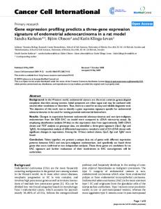
Figure 1. Gene set enrichment analysis. For Gene set enrichment analysis of signatures genes from BCBL1-K13 (top panel) and HUVEC-K13 (lower panel), the t-test was graphed for each correlated gene in the ranked dataset. Three GSEA enrichment plots for representative biological pathways (Cytokine, NF-kB and Inflammatory) enriched in 4OHT-treated BCBL1-K13-ERTAM and HUVEC-K13-ERTAM are shown. The top portion of each GSEA plot shows the running enrichment score for validated genes specific for particular pathway as it moves down the ranked list. The bottom portion of each plot shows the value of ranking matrices as it moves down the list of ranked genes. The red horizontal bar which terminate with blue color indicate shift from positively correlated genes (red) to negatively correlated genes (blue). Further detailed interpretation about these plots can be found at Broad Institute web site (http://www.broadinstitute.org/gsea/index.jsp). doi:10.1371/journal.pone.0037498.g001
PLoS ONE | www.plosone.org
7
May 2012 | Volume 7 | Issue 5 | e37498
K13-Induced Genes in PEL
Table 3. Transcription factors with binding sites over-represented in promoters of genes upregulated by K13 in BCBL1 and HUVECs.
JASPAR database in BCBL1-K13 cells S.No.
Sequence motif
Description
p- value
% of input
1
Jaspar NF-kappaB
REL
2.16E-6
29.6
2
Jaspar p65
REL
0.0001
20.1
3
Jaspear Irf-1
TRP-cluster
0.0025
17.6
4
Jaspar CFI-USP
Nuclear receptor
0.0027
16.4
5
Jaspar p50
REL
0.001
15.1
6
Jaspar COUP-TF
Nuclear receptor
0.0030
4.4
7
Jaspar Irf-2
TRP-cluster
0.0088
2.6
TRANSFEC database in BCBL1-K13 cells 1
V$NFKAPPAB-01: NF-kappaB
NF-kappaB
1.07E-5
27.7
2
V$NFKAPPAB65-01: NF-kappaB
NF-kappaB (p65)
3.16E-5
24.5
3
V$NFKAPPAB50-01: NF-kappaB
NF-kappaB (p50)
0.0002
17
4
V$NFKB-Q6: NF-kappaB
NF-kappaB
0.0001
12
5
V$IK3-01: Ik-3
Ikaros 3
0.0012
8.8
6
V$COUP-01: COUP-TF
COUP/HNF-4 heterodimer
0.0031
3.0
7
V$PAX2-01: Pax-2
Pax-2
0.0024
2.6
8
V$NRSF-01: NRSF
neuron-restrictive silencer factor
9.56E-6
1.2
JASPAR database in HUVEC-K13 1
Jaspar c-REL
REL
1.00E-10
37.8
2
Jaspar NF-kappaB
REL
3.99E-6
32.7
3
Jaspar Irf-1
TRP-Cluster
1.00E-10
26.5
4
Jaspar p65
REL
1.00E-10
22.5
5
Jaspar Dorsal-2
REL
4.84E-6
20.4
6
Jaspar p50
REL
0.0006
19.4
7
Jaspar c-FOS
bZIP
0.0004
14.3
8
Jaspar Irf-2
TRP-Cluster
1.00E-10
11.2
9
Jaspar Brachyury
T-BOX
0.0006
1.2
10
Jaspar COUP-TF
Nuclear receptor
0.0088
1.1
TRANSFEC database in HUVEC-K13 1
V$NFKAPPAB-01: NF-kappaB
NF-kappaB
2.19E-6
38.7
2
V$CREL-01: c-Rel
c-Rel
0.0003
37.8
3
V$AP1-C: AP-1
AP-1 binding site
0.0008
37.7
4
V$STAT-01: STATx
signal transducers and activators of transcription
0.0003
32.8
5
V$NFKAPPAB65-01: NF-kappaB
NF-kappaB (p65)
1.00E-10
28.6
6
V$NFKB-C: NF-kappaB
NF-kappaB binding site
1.00E-10
19.4
7
V$NFKAPPAB50-01: NF-kappaB
NF-kappaB (p50)
0.0005
19.4
8
V$NFKB-Q6: NF-kappaB
NF-kappaB
1.00E-10
18.4
9
V$IRF1-01: IRF-1
interferon regulatory factor 1 1.00E-10
16.3
10
V$IRF2-01: IRF-2
interferon regulatory factor 2 1.53E-6
9.2
11
V$ISRE-01: ISRE
interferon-stimulated response 0.0022 element
5.1
12
V$VMAF-01: v-Maf
v-Maf
0.0013
3.1
13
V$TAXCREB-02: Tax/CREB
Tax/CREB complex
1.00E-10
1.1
14
V$MEF2-01: MEF-2
myogenic enhancer factor 2
4.83E-5
1.1
(Genes upregulated more than 1.5-fold were analyzed using the JASPAR and TRANSFEC databases. Results with a p-value of less than 0.05 are shown. % input refers to the number of gene promoters bearing the specific motif compared to total number screened. doi:10.1371/journal.pone.0037498.t003
PLoS ONE | www.plosone.org
8
May 2012 | Volume 7 | Issue 5 | e37498
K13-Induced Genes in PEL
Figure 2. Validation of gene array data by qRT-PCR. (A) Twenty five genes from NF-kB, cytokine, and inflammatory pathways were randomly selected and their relative mRNA levels in mock and 4OHT-treated vector and K13-ERTAM-expressing BCBL1 cells were examined using qRT-PCR. Realtime PCR reactions were performed in triplicate and the data presented as fold change mean 6S.E in target gene expression (*p,0.05; Student’s ttest). (B) Pearson Correlation coefficient between gene expression array and real time PCR showed a significant agreement (Correlation coefficient 0.88; p,0.0001). doi:10.1371/journal.pone.0037498.g002
addition to NF-kB, several other TF binding sites were also overrepresented among the promoters of genes upregulated by K13.
the members of the NF-kB/REL family. To confirm the results, we also searched the TRANSFAC database and similarly found over-representation of binding sites for the members of the NFkB/REL family among the promoters of the genes induced by K13 in BCBL1 cells. We next repeated the analysis with our previously published HUVEC dataset. There were 10 sites that were over-represented in JASPAR database using stringent parameters (p,0.01, FDR,0.10), while 14 binding sites were over-represented in TRANSFAC (p,0.01, FDR,0.15) database. Similar to the BCBL1 dataset, NF-kB/REL binding sites were again over-represented among the promoters of the genes induced by K13 in HUVEC in both databases. Thus, consistent with our published studies [9,27] and the results of the pathway analysis, NF-kB/REL is the major transcription factor responsible for gene induction by K13 in both BCBL1 and HUVECs. However, in
PLoS ONE | www.plosone.org
Validation of in silico results We validated our gene array data by determining mRNA expression of twenty-five highly expressed genes (TRADD, TNFSRF25, TNFSRF1B, RANTES/CCL5, SELE, CXCL10, BID, NFKB1A, NFKB1, LTbR, VCAM, LMNB2, CTSS, ALCAM, IL15, IL9, IL6, BIRC3, CIITA, HLADMB, IFNG, HLADQB1, HLADQ1, GAS2 and CD74) belonging to the cytokine, NF-kB, cell death, cell adhesion and antigen processing pathways (Table S5). We found a good correlation with the expression of the above as determined by real-time RT-PCR and gene array data (Figure 2). As a control for the reliability of qRTPCR experiments, the mRNA expression of various housekeeping genes such as GAPDH, 18S, GNBL, and b-2-microglobulin was 9
May 2012 | Volume 7 | Issue 5 | e37498
K13-Induced Genes in PEL
Figure 3. NF-kB inhibitors block K13-regulated genes. BCBL1 K13-ERTAM cells were treated with two NF–kB inhibitors (2 mM Bay 11-7082 or 2 mM As2O3) for 2 hours followed by 4OHT treatment for additional 24 hours and total RNA was isolated as described in the Materials and Method section. Nine genes were randomly picked and their relative mRNA levels were examined using real-time RT-PCR as explained in Figure 2A. doi:10.1371/journal.pone.0037498.g003
RANTES/CCL5 as a representative example since its expression was also induced by K13 in HUVECs. RANTES is an important protein involved in immunoregulatory, inflammatory and cell proliferation pathways [40], making its mechanism of upregulation by K13 of biological and clinical significance. We began by validating the results of gene expression and qRT-PCR analyses by checking the effect of K13 on RANTES expression at the protein level. As shown in Figure 4A, we confirmed that expression of RANTES was up-regulated in the supernatant of BCBL1-K13ERTAM cells upon treatment with 4OHT, as determined by ELISA. To investigate the mechanism by which K13 upregulates RANTES expression, human embryonic kidney 293T cells were transfected with a luciferase-based reporter construct containing the RANTES gene promoter. K13 strongly activated the RANTES promoter as compared to empty vector-transfected cells (Figure 4B). Since NF-kB pathway was identified as the major
investigated and their expression level in both mock cells as well K13-ERTAM -expressing cells remained unaltered (data not shown). K13-induced NF-kB pathway is known to be effectively blocked by Bay-11-7082 and arsenic trioxide [13,39]. To examine the role of the NF-kB pathway in the expression of K13-regulated genes in BCBL1 cells, we studied the effect of Bay-11-7082 and arsenic trioxide on 4OHT induced gene induction in BCBL1-K13-ER cells by qRT-PCR analysis. As shown in Figure 3, pretreatment of BCBL1 K13-ERTAM cells with Bay-11-7082 and arsenic trioxide effectively blocked the induction of TNFSRF1B, SELE, CXCL10, NFKB1A, LMNB2, IL6, IFNG, CIITA, CTSS and CD74 genes by 4OHT.
Mechanism of K13-induced RANTES/CCL5 upregulation To study the mechanism by which K13 upregulates the expression of genes in BCBL1 cells in greater detail, we selected
PLoS ONE | www.plosone.org
10
May 2012 | Volume 7 | Issue 5 | e37498
K13-Induced Genes in PEL
Figure 4. K13-induced NF-kB activity is critical for the activation of RANTES. (A) K13-induced up regulation of RANTES at protein level. Cellular supernatant from BCBL1 vector and K13-ERTAM cells treated with and without 4OHT were collected and used to measure the secretion of RANTES by ELISA. The values shown are averages (mean 6 SE) of one representative experiment out of three in which the level of RANTES secretion was measured in triplicate. (B–C) 293T cells were transfected with an empty vector, wild-type K13 (B) or K13-58AAA (C) (250 ng/well) along with a RANTES promoter-driven luciferase constructs (75 ng/well) and a pRSV/LacZ (b-galactosidase) reporter construct (75 ng/well), and the reporter assay performed as described under the Materials and Methods section. (D) Dominant-negative mutants of IkBa (IkBaDN and IkBaSS32/36AA) block K13induced RANTES promoter activity. 293T cells were transfected either with the indicated plasmids and reporter assay performed as described above. The amount of IkBa mutant plasmids (500 ng/well) was five times the amount of vector or K13 (100 ng/well) plasmid and the total amount of transfected DNA was kept constant by adding empty vector. (E) Pharmacologic inhibitors of NF-kB block K13-induced RANTES promoter activation. 293T cells were transfected with an empty vector or a vector encoding K13 along with RANTES-Luc and pRSV/LacZ reporter constructs. Approximately 3 hours after transfection, cells were treated with dimethyl sulfoxide (vehicle) or the indicated compounds for 18 hours before cell lysis and measurement of reporter activities. Reporter assay was performed as described for Figure 4B–C. doi:10.1371/journal.pone.0037498.g004
pathway induced by K13 by both the pathway analysis and JASPAR/TRANFAC database analysis, we next examined the involvement of this pathway in K13-induced RANTES transcriptional activation. For this purpose, we took advantage of a mutant of K13, K13-58AAA, which lacks the ability to activate the NF-kB pathway [7]. As shown in Figure 4C, K13-58AAA mutant failed to activate the RANTES promoter. Furthermore, K13-induced RANTES activity was effectively blocked by two phosphorylation-resistant mutants of IkBa (IkBa SS32/36AA and IkBaDN) (Figure 4D–E) that are known to block the NF-kB pathway [27], and by treatment with chemical inhibitors of the NF-kB pathway, including Bay-11-7082 [13], IKK inhibitor VI [41], PS1145 [42] and arsenic trioxide [39]. Collectively, these results confirm the involvement of the NF-kB pathway in K13-induced up-regulation of RANTES.
PLoS ONE | www.plosone.org
Biological consequences of K13-induced gene expression We have previously shown that ectopic expression of K13 in Rat1 fibroblast cells stimulates cellular proliferation [8] and its shRNA-mediated silencing results in a block in cell proliferation [9]. To study the biological consequences of K13 in PEL, we studied the effect of 4OHT on cell cycle progression in BCBL1K13-ERTAM cells. As shown in Figure 5A, 4OHT treatment of serum-starved BCBL1-K13-ERTAM cells resulted in an increase in cells in the S-phase as compared to the untreated cells (24.6% vs 14.8%), suggesting that K13 stimulates cell-cycle progression from G1 to S phase. Treatment with 4OHT had no significant effect on cell cycle progression in the BCBL1-vector cells (data not shown). The stimulatory effect of 4OHT on G1 to S phase transition was blocked by treatment with Bay-11-7082, thereby confirming the role of K13-induced NF-kB activation in this process (Figure 5A).
11
May 2012 | Volume 7 | Issue 5 | e37498
K13-Induced Genes in PEL
Figure 5. Biological assays to study K13-induced IL6 production and cell cycle analyses. (A) A DNA content analysis showing significant increase in S-phase of cell cycle by 4OHT treatment in serum-starved BCBL1 K13-ERTAM cells and inhibition of this increase in S-phase cells by pretreatment with Bay-11-7082 (2 mM). DNA content analysis was performed as described previously [7] and explained in Materials and Method section. (B) Cell survival of T1165 cells with supernatants from 4OHT-treated BCBL1-K13-ERTAM cells. BCBL1 vector- and K13-ERTAM cells were pretreated with 2 mM Bay-11-7082 for 2 hours followed by treatment with 4OHT for additional 72 hours and supernatants from these cells were collected and filtered. T1165 cells were treated in triplicate in a 96 well plate (100 ml/well) with 20 ml of cell-free supernatant collected from cells described above. Seventytwo hours post-treatment, cell viability of T1165 cells was measured by MTS assay as described in Materials and Method section. The values (Mean6SEM) shown are from a representative of three independent experiments performed in triplicate. doi:10.1371/journal.pone.0037498.g005
In addition to stimulating the proliferation of KSHV-infected cells, K13 induced cytokine upregulation could contribute to the pathogenesis of KSHV-associated malignancies by stimulating the survival and proliferation of neighboring uninfected cells by acting in a paracrine fashion. To test this hypothesis, we examined the ability of conditioned supernatant from untreated and 4OHTtreated BCBL1-K13-ERTAM cells to support the survival of murine T1165 plasmacytoma cell line that requires IL6 for survival and proliferation [43]. As shown in Figure 5B, survival of T1165 cells was reduced to approximately 6% when grown in IL6free medium for 72 hours. The addition of conditioned supernatant from untreated or 4OHT-treated BCBL1-vector cells afforded minor protection against IL6-withdrawal-induced cell death (approximately 25% cell survival), reflecting low-level constitutive IL6 secretion from the BCBL1 cells. The conditioned supernatant from untreated BCBL1-K13-ERTAM cells similarly resulted in only partial rescue from IL6-withdrawal-induced cell death (Figure 5B). In contrast, conditioned supernatant from 4OHT-treated BCBL1-K13-ERTAM cells markedly improved the survival (77% survival) of T1165 cells, reflecting upregulation of IL6 secretion by induction of K13 activity (Figure 5B). The survival advantage conferred by the conditioned medium from 4OHT-treated BCBL1-K13-ERTAM cells, however, disappeared if the cells were treated with both 4OHT and Bay-11-7082, confirming a role of K13-induced NF-kB in this process (Figure 5B).
PLoS ONE | www.plosone.org
Discussion PEL is a rare subset of non-Hodgkin’s lymphoma found in patients with HIV/AIDS that predominantly grows in the body cavities as neoplastic effusions, usually without a contiguous tumor mass [44]. Morphologically, PEL shows features that bridge immunoblastic and anaplastic large-cell lymphomas, and frequently displays some degree of plasma cell differentiation [45]. We and others have demonstrated that K13 plays a key role in constitutive NF-kB activity observed in PEL cells [9,46] and is an oncogenic protein which contribute to lymphoproliferative disorders [11,47,48]. In the present investigation, we provide a comprehensive picture of global transcriptional changes induced by K13 in PEL-derived BCBL1 cells. BCBL1 cells are infected with KSHV but express very little K13 endogenously and have negligible constitutive NF-kB activity [24,49]. As such, we chose BCBL1 cells as a physiological relevant cell line to study the effect of ectopic K13 expression on gene expression in lymphoid cells. We observed that K13 upregulated the expression of a number of NF-kB-responsive genes in BCBL1 cells. This observation was confirmed by GSEA, qRT-PCR as well as analysis of JASPER and TRANSFAC databases. Nuclear factor-kB (NF-kB) is a critical transcription factor involved in the regulated expression of several genes involved in the inflammatory and immune response [50]. Although many dimeric forms of NF-kB have been described, the classical NF-kB complex is a heterodimer of the p65/RelA and p50 subunits and is found in most cells in association with a family 12
May 2012 | Volume 7 | Issue 5 | e37498
K13-Induced Genes in PEL
progression of PEL and KS via generation of Tregs with suppressive functions. Another cytokine whose expression was upregulated by K13 in BCBL1 cells was IL-32. A recent study demonstrated that the IL32 promoter contains NF-kB binding sites [64], which suggests that, similar to the situation with RANTES/CCL5, K13 might upregulate IL-32 through NF-kB activation. Interestingly, there is a good correlation between IL-32 levels and HIV-1 replication in lymphatic tissues and IL-32 was recently shown to play an immunosuppressive role in lymphatic tissues during HIV-1 infection [65]. IL-32 was shown to induce the expression of immunosuppressive molecules IDO (indole-amine-2,3-dioxygenase) and Ig-like transcript 4 in immune cells, which not only block immune activation but also impair host defenses [65]. It is conceivable that the dampening of anti-viral immune response by K13-induced IL-32 induction may enhance HIV-1 replication and persistence, thereby resulting in a synergistic interaction between HIV-1 and KSHV. A comparative analysis of K13-induced genes in BCBL1 and HUVEC revealed that although K13 primarily induced NF-kB responsive genes in both cell types, there were significant qualitative and quantitative differences. In particular, genes belonging to cytokines and chemokines were highly and differentially induced in HUVEC but not in the BCBL1 cells, with few exceptions, such as RANTES/CCL5 and CXCL10. The differential and robust induction of proinflammatory chemokines and cytokines by K13 in HUVEC may account for the presence of intense inflammatory infiltrate in KSHV associated KS. The underlying reasons for the differential gene induction by K13-induced NF-kB in the two cell types are not clear, but may reflect the presence of lineage-specific positive and negative regulators or epigenetic alterations. It is interesting to note in this regard that SOCS1, a negative regulator of the cytokine signaling, was upregulated only in the K13-expressing BCBL1 cells. In addition to the NF-kB pathway, genes belonging to a number of other pathways, such as cytokine, death, inflammatory, and antigen processing pathways, were found to be enriched among K13-expressing BCBL1 cells and HUVECs. Similarly, JASPER and TRANSFAC databases revealed the enrichment of a number of transcriptional factors in addition to NF-kB among the promoters of genes upregulated by K13 in BCBL1 and HUVECs. However, there is considerable overlap among the genes induced by the different signaling pathways and transcription factors. In particular, NF-kB pathway is well known for its ability to induce genes belonging to cytokine, death, antigen processing and inflammatory pathways, and to work in concert with other transcription factors, such as AP1 [66]. Therefore, it is conceivable that enrichment of genes belonging to signaling pathways other than the NF-kB pathway may simply reflect this overlap. We also observed that K13 upregulates the expression of a number of chemokines and cytokines, especially in HUVECs, which could work in an autocrine/paracrine fashion to activate a number of secondary signaling pathways and transcription factors, providing an alternative explanation for our results. Studies are in progress to delineate the role of K13 in activation of signaling pathways other than the NF-kB pathway. PEL display a gene expression profile that is distinct from all non-Hodgkin lymphomas of immunocompetent hosts and AIDSassociated NHL [67]. The gene expression profile of PEL has been defined as plasmablastic as it shares features of both immunoblasts and plasma cells [67]. Interestingly, increased expression of a number of genes found to be induced by K13 in the BCBL1 cells in the present study, including SOCS1, TNFAIP3, NFKB1A,
of inhibitory proteins called IkBs [51,52]. We have previously shown that K13 activates the classical NF-kB pathway by activating a multi-subunit IkB kinase (IKK) complex [53], which contains two catalytic subunits, IKK1/IKKa and IKK2/IKKb, and a regulatory subunit, NEMO/IKKc [52,54]. Activation of the IKK complex by K13 results in phosphorylation of IkB proteins which leads to their ubiquitination and proteasomal-mediated degradation, allowing the classical NF-kB subunits to enter the nucleus and turn on the expression of their target genes [53]. We have also shown that K13 can also activate an alternate NF-kB pathway [9] that involves IKK1/IKKa-mediated phosphorylation of p100/NFKB2 and its slow proteasome-mediated processing into the active p52/p49 subunit that culminates in kinetically slower nuclear translocation of the p52-RelB NF-kB complex [54,55]. Interestingly, NFKB2/p100 and RelB, the two NF-kB subunits involved in alternate NF-kB pathway, were among the genes upregulated by K13 in BCBL1 cells (Table 1). These results suggest that, in addition to IKK1-induced p100 phosphorylation, transcriptional upregulation of NFKB2 and RelB may also contribute to the activation of the alternate NF-kB pathway by K13. We also observed significant upregulation of TNFAIP3 (A20) and NFKB1A (IkBa) by K13 in BCBL1 cells. A20 is a deubiquitinating enzyme that is known to act as a tumor suppressor in several subtypes of non-Hodgkin and Hodgkin lymphomas [56]. We have recently demonstrated that A20 blocked K13-induced NF-kB activity and K13-induced upregulation of proinflammatory cytokines in a negative-feedback fashion [49]. A20 was also shown to block K13- induced cellular transformation [49]. In addition to A20, K13 upregulated the expression of NFKB1A, which codes for IkBa, the negative regulator of the classical NF-kB pathway that serves to keep the p65/p50 heterodimers sequestered in an inactive state in the cytoplasm [57]. Finally, the expression of SOCS1 (suppressor of cytokine signaling 1), a negative regulator of the JAK/STAT [58] signaling was significantly induced in the K13-expressing BCBL1 cells. The uncontrolled activation of the NF-kB and JAK/STAT pathways has the potential of resulting in uncontrolled inflammatory response. Therefore, induction of A20, IkBa and SOCS1 by K13 in lymphoid cells may serve to attenuate the inflammatory response and thus help maintain a balance between the virus and the host. In addition to the positive and negative regulators of the NF-kB pathway, expression of RANTES/CCL5 was significantly upregulated in K13-expressing BCBL1 and HUVECs. Further studies utilizing a RANTES promoter-driven luciferase reporter construct demonstrated that K13 upregulates RANTES through the NF-kB pathway. RANTES is a powerful chemoattractant for blood monocytes, memory T helper cells and eosinophils [59] and as such may contribute to the inflammatory infiltrate present in KSHV-associated malignancies. Additionally, RANTES/CCL5 has been shown to promote tumor proliferation, invasion, metastases and angiogenesis [60] [61], which may also contribute to the development of PEL and KS. K13 also upregulated the expression of Epstein-Barr virusinduced gene-3 (EBI-3), which associates with p28 to form interleukin-27 (IL-27) or with IL-12 p35 to form IL-35. Both IL27 and IL-35 have immunosuppressive properties [62]. In particular, IL-35 has been implicated in the suppressive function of regulatory T cells (Treg), which contributes to infection tolerance and tumor progression [62,63]. Thus, it is conceivable that K13-induced upregulation of EBI-3 may not only contribute to immune tolerance of KSHV-infected cells but also promote
PLoS ONE | www.plosone.org
13
May 2012 | Volume 7 | Issue 5 | e37498
K13-Induced Genes in PEL
LTB, IL2RG, RELB, RRAD, CCL5, PHLDA2, CIITA, RGS1 and FAS, have been associated with the plasmablastic phenotype in human lymphomas [68]. K13 also upregulated the expression of IL6, a plasma cell growth factor that has been also shown to stimulate the proliferation of PEL cells [69]. PEL cells are typified by their lack of expression of B cell markers [48]. Interestingly, CD19, a B cell marker, was one of the genes downregulated by K13 in BCBL1 cells. Finally, PEL cells are defined by the overexpression of genes involved in inflammation, cell adhesion and invasion, which is believed to contribute to their presentation in the body cavities [2]. Interestingly, in the present study, we observed significant upregulation of genes belonging to all the above categories upon K13 expression. Thus, K13 may contribute to the unique gene expression profile and presentation in body cavities that are characteristic of KSHV-associated PEL.
Summary of differentially regulated gene clusters in 4OHT-treated K13-ERTAM-transduced BCBL1 cells. (DOC)
Supporting Information
Acknowledgments
Table S1 List of primers used for qRT-PCR (mRNA expression). (DOC)
We thank Dr. Robert Schleliner (Northwestern University) for the RANTES reporter constructs, Dr. Steven Cole (University of California at Los Angeles) for helpful discussions regarding transcription factor analysis and Dr. Ciaren Graham for a critical review of the manuscript.
Table S2
Table S3 List of common differentially regulated genes in HUVECs and BCBL1 cells. (DOC) Table S4 List of genes upregulated .20 fold in HUVECs
dataset. (DOC) Table S5 Summary of representative genes corresponding to pathways identified by GSEA analysis and validated in qRT-PCR. (DOCX)
Author Contributions Conceived and designed the experiments: PMC VP. Performed the experiments: VP HM. Analyzed the data: VP HM PMC. Contributed reagents/materials/analysis tools: VP PMC. Wrote the paper: VP PMC.
References 1. 2.
3. 4.
5.
6.
7.
8.
9.
10.
11.
12. 13.
14.
15.
Carbone A, Gloghini A (2007) HHV-8-associated lymphoma: state-of-the-art review. Acta Haematol 117: 129–131. Jenner RG, Maillard K, Cattini N, Weiss RA, Boshoff C, et al. (2003) Kaposi’s sarcoma-associated herpesvirus-infected primary effusion lymphoma has a plasma cell gene expression profile. Proc Natl Acad Sci U S A 100: 10399–10404. Ganem D (2006) KSHV infection and the pathogenesis of Kaposi’s sarcoma. Annual Review of Pathology-Mechanisms of Disease 1: 273–296. Chaudhary PM, Nicholas J (2008) Molecular Biology of Human Herpesvirus 8 neoplasia. In: Nicholas J, Jeang KT, Wu TC, eds. Human Cancer Viruses Principles of Transformation and Pathogenesis. Basel: Karger. pp 186–210. Dittmer D, Lagunoff M, Renne R, Staskus K, Haase A, et al. (1998) A cluster of latently expressed genes in Kaposi’s sarcoma-associated herpesvirus. J Virol 72: 8309–8315. Thome M, Schneider P, Hofmann K, Fickenscher H, Meinl E, et al. (1997) Viral FLICE-inhibitory proteins (FLIPs) prevent apoptosis induced by death receptors. Nature 386: 517–521. Sun Q, Matta H, Chaudhary PM (2003) The human herpes virus 8-encoded viral FLICE inhibitory protein protects against growth factor withdrawalinduced apoptosis via NF-kappa B activation. Blood 101: 1956–1961. Sun Q, Zachariah S, Chaudhary PM (2003) The human herpes virus 8-encoded viral FLICE-inhibitory protein induces cellular transformation via NF-kappaB activation. Journal of Biological Chemistry 278: 52437–52445. Matta H, Chaudhary PM (2004) Activation of alternative NF-kappa B pathway by human herpes virus 8-encoded Fas-associated death domain-like IL-1 betaconverting enzyme inhibitory protein (vFLIP). Proc Natl Acad Sci U S A 101: 9399–9404. Sun Q, Matta H, Lu G, Chaudhary PM (2006) Induction of IL-8 expression by human herpesvirus 8 encoded vFLIP K13 via NF-kappaB activation. Oncogene 25: 2717–2726. Chugh P, Matta H, Schamus S, Zachariah S, Kumar A, et al. (2005) Constitutive NF-kappaB activation, normal Fas-induced apoptosis, and increased incidence of lymphoma in human herpes virus 8 K13 transgenic mice. Proc Natl Acad Sci U S A 102: 12885–12890. Guasparri I, Keller SA, Cesarman E (2004) KSHV vFLIP is essential for the survival of infected lymphoma cells. J Exp Med 199: 993–1003. Matta H, Surabhi RM, Zhao J, Punj V, Sun Q, et al. (2007) Induction of spindle cell morphology in human vascular endothelial cells by human herpesvirus 8encoded viral FLICE inhibitory protein K13. Oncogene 26: 1656–1660. Xu Y, Ganem D (2007) Induction of chemokine production by latent Kaposi’s sarcoma-associated herpesvirus infection of endothelial cells. J Gen Virol 88: 46–50. Grossmann C, Podgrabinska S, Skobe M, Ganem D (2006) Activation of NFkappaB by the latent vFLIP gene of Kaposi’s sarcoma-associated herpesvirus is
PLoS ONE | www.plosone.org
16.
17. 18. 19.
20.
21.
22.
23.
24.
25.
26.
27.
28.
14
required for the spindle shape of virus-infected endothelial cells and contributes to their proinflammatory phenotype. J Virol 80: 7179–7185. Lockhart DJ, Dong H, Byrne MC, Follettie MT, Gallo MV, et al. (1996) Expression monitoring by hybridization to high-density oligonucleotide arrays. Nat Biotechnol 14: 1675–1680. Bild AH, Potti A, Nevins JR (2006) Linking oncogenic pathways with therapeutic opportunities. Nat Rev Cancer 6: 735–741. Vogelstein B, Kinzler KW (2004) Cancer genes and the pathways they control. Nat Med 10: 789–799. Subramanian A, Tamayo P, Mootha VK, Mukherjee S, Ebert BL, et al. (2005) Gene set enrichment analysis: a knowledge-based approach for interpreting genome-wide expression profiles. Proc Natl Acad Sci U S A 102: 15545–15550. Crijns AP, Fehrmann RS, de Jong S, Gerbens F, Meersma GJ, et al. (2009) Survival-related profile, pathways, and transcription factors in ovarian cancer. PLoS Med 6: e24. Matta H, Mazzacurati L, Schamus S, Yang T, Sun Q, et al. (2007) Kaposi’s Sarcoma-associated Herpesvirus (KSHV) Oncoprotein K13 Bypasses TRAFs and Directly Interacts with the IkappaB Kinase Complex to Selectively Activate NF-kappaB without JNK Activation. J Biol Chem 282: 24858–24865. Punj V, Matta H, Schamus S, Chaudhary PM (2009) Integrated microarray and multiplex cytokine analyses of Kaposi’s Sarcoma Associated Herpesvirus viral FLICE Inhibitory Protein K13 affected genes and cytokines in human blood vascular endothelial cells. BMC Med Genomics 2: 50. Yang Y, Groshong JS, Matta H, Gopalakrishnan R, Yi H, et al. (2011) Constitutive NF-kappaB Activation Confers Interleukin 6 (IL6) Independence and Resistance to Dexamethasone and Janus Kinase Inhibitor INCB018424 in Murine Plasmacytoma Cells. J Biol Chem 286: 27988–27997. Zhao J, Punj V, Matta H, Mazzacurati L, Schamus S, et al. (2007) K13 Blocks KSHV Lytic Replication and Deregulates vIL6 and hIL6 Expression: a Model of Lytic Replication Induced Clonal Selection in Viral Oncogenesis. PLoS ONE 2: e1067. Matta H, Punj V, Schamus S, Mazzacurati L, Chen AM, et al. (2008) A nuclear role for Kaposi’s sarcoma-associated herpesvirus-encoded K13 protein in gene regulation. Oncogene 27: 5243–5253. Punj V, Matta H, Schamus S, Yang T, Chang Y, et al. (2009) Induction of CCL20 production by Kaposi sarcoma-associated herpesvirus: role of viral FLICE inhibitory protein K13-induced NF-kappaB activation. Blood 113: 5660–5668. Chaudhary PM, Jasmin A, Eby MT, Hood L (1999) Modulation of the NFkappa B pathway by virally encoded death effector domains-containing proteins. Oncogene 18: 5738–5746. Matys V, Fricke E, Geffers R, Gossling E, Haubrock M, et al. (2003) TRANSFAC: transcriptional regulation, from patterns to profiles. Nucleic Acids Res 31: 374–378.
May 2012 | Volume 7 | Issue 5 | e37498
K13-Induced Genes in PEL
29. Sandelin A, Alkema W, Engstrom P, Wasserman WW, Lenhard B (2004) JASPAR: an open-access database for eukaryotic transcription factor binding profiles. Nucleic Acids Res 32: D91–94. 30. Ho Sui SJ, Fulton DL, Arenillas DJ, Kwon AT, Wasserman WW (2007) oPOSSUM: integrated tools for analysis of regulatory motif over-representation. Nucleic Acids Res 35: W245–252. 31. Vandesompele J, De Preter K, Pattyn F, Poppe B, Van Roy N, et al. (2002) Accurate normalization of real-time quantitative RT-PCR data by geometric averaging of multiple internal control genes. Genome Biol 3: RESEARCH0034. 32. Hellemans J, Mortier G, De Paepe A, Speleman F, Vandesompele J (2007) qBase relative quantification framework and software for management and automated analysis of real-time quantitative PCR data. Genome Biol 8: R19. 33. Punj V, Matta H, Schamus S, Tamewitz A, Anyang B, et al. (2010) Kaposi’s sarcoma-associated herpesvirus-encoded viral FLICE inhibitory protein (vFLIP) K13 suppresses CXCR4 expression by upregulating miR-146a. Oncogene 29: 1835–1844. 34. Sinha S, Adler AS, Field Y, Chang HY, Segal E (2008) Systematic functional characterization of cis-regulatory motifs in human core promoters. Genome Res 18: 477–488. 35. Warner JB, Philippakis AA, Jaeger SA, He FS, Lin J, et al. (2008) Systematic identification of mammalian regulatory motifs’ target genes and functions. Nat Methods 5: 347–353. 36. Roider HG, Kanhere A, Manke T, Vingron M (2007) Predicting transcription factor affinities to DNA from a biophysical model. Bioinformatics 23: 134–141. 37. Hestand MS, van Galen M, Villerius MP, van Ommen GJ, den Dunnen JT, et al. (2008) CORE_TF: a user-friendly interface to identify evolutionary conserved transcription factor binding sites in sets of co-regulated genes. BMC Bioinformatics 9: 495. 38. Kim SY, Kim Y (2006) Genome-wide prediction of transcriptional regulatory elements of human promoters using gene expression and promoter analysis data. BMC Bioinformatics 7: 330. 39. Matta H, Sun Q, Moses G, Chaudhary PM (2003) Molecular genetic analysis of human herpes virus 8-encoded viral FLICE inhibitory protein-induced NFkappaB activation. Journal of Biological Chemistry 278: 52406–52411. 40. Appay V, Rowland-Jones SL (2001) RANTES: a versatile and controversial chemokine. Trends Immunol 22: 83–87. 41. Baxter A, Brough S, Cooper A, Floettmann E, Foster S, et al. (2004) Hit-to-lead studies: the discovery of potent, orally active, thiophenecarboxamide IKK-2 inhibitors. Bioorg Med Chem Lett 14: 2817–2822. 42. Hideshima T, Chauhan D, Richardson P, Mitsiades C, Mitsiades N, et al. (2002) NF-kappa B as a therapeutic target in multiple myeloma. J Biol Chem 277: 16639–16647. 43. Nordan RP, Potter M (1986) A macrophage-derived factor required by plasmacytomas for survival and proliferation in vitro. Science 233: 566–569. 44. Carbone A, Cesarman E, Spina M, Gloghini A, Schulz TF (2009) HIVassociated lymphomas and gamma-herpesviruses. . pp 1213–1224. 45. Wen KW, Damania B (2010) Kaposi sarcoma-associated herpesvirus (KSHV): molecular biology and oncogenesis. Cancer Lett 289: 140–150. 46. Keller SA, Schattner EJ, Cesarman E (2000) Inhibition of NF-kappaB induces apoptosis of KSHV-infected primary effusion lymphoma cells. Blood 96: 2537–2542. 47. Ahmad A, Groshong JS, Matta H, Schamus S, Punj V, et al. (2010) Kaposi’s sarcoma associated herpesvirus-encoded viral FLICE inhibitory protein (vFLIP) K13 cooperates with Myc to promote lymphoma in mice. Cancer Biol Ther 10. 48. Ballon G, Chen K, Perez R, Tam W, Cesarman E (2011) Kaposi sarcoma herpesvirus (KSHV) vFLIP oncoprotein induces B cell transdifferentiation and tumorigenesis in mice. J Clin Invest 121: 1141–1153. 49. Matta H, Gopalakrishnan R, Punj V, Yi H, Suo Y, et al. (2011) A20 is induced by Kaposi sarcoma-associated herpesvirus-encoded viral FLICE inhibitory
PLoS ONE | www.plosone.org
50. 51. 52. 53.
54. 55. 56.
57. 58.
59.
60.
61.
62.
63.
64.
65.
66. 67.
68.
69.
15
protein (vFLIP) K13 and blocks K13-induced nuclear factor-kappaB in a negative feedback manner. J Biol Chem 286: 21555–21564. Aggarwal BB (2004) Nuclear factor-kappaB: the enemy within. Cancer Cell 6: 203–208. Hayden MS, Ghosh S (2004) Signaling to NF-kappaB. Genes Dev 18: 2195–2224. Karin M, Ben-Neriah Y (2000) Phosphorylation meets ubiquitination: the control of NF-[kappa]B activity. Annu Rev Immunol 18: 621–663. Liu L, Eby MT, Rathore N, Sinha SK, Kumar A, et al. (2002) The Human Herpes Virus 8-encoded Viral FLICE Inhibitory Protein Physically Associates with and Persistently Activates the Ikappa B Kinase Complex. J Biol Chem 277: 13745–13751. Bonizzi G, Karin M (2004) The two NF-kappaB activation pathways and their role in innate and adaptive immunity. Trends Immunol 25: 280–288. Pomerantz JL, Baltimore D (2002) Two pathways to NF-kappaB. Molecular Cell 10: 693–695. Honma K, Tsuzuki S, Nakagawa M, Tagawa H, Nakamura S, et al. (2009) TNFAIP3/A20 functions as a novel tumor suppressor gene in several subtypes of non-Hodgkin lymphomas. Blood 114: 2467–2475. Chiao PJ, Miyamoto S, Verma IM (1994) Autoregulation of I kappa B alpha activity. Proc Natl Acad Sci U S A 91: 28–32. Yoshikawa H, Matsubara K, Qian GS, Jackson P, Groopman JD, et al. (2001) SOCS-1, a negative regulator of the JAK/STAT pathway, is silenced by methylation in human hepatocellular carcinoma and shows growth-suppression activity. Nat Genet 28: 29–35. Crawford A, Angelosanto JM, Nadwodny KL, Blackburn SD, Wherry EJ (2011) A role for the chemokine RANTES in regulating CD8 T cell responses during chronic viral infection. PLoS Pathog 7: e1002098. Karnoub AE, Dash AB, Vo AP, Sullivan A, Brooks MW, et al. (2007) Mesenchymal stem cells within tumour stroma promote breast cancer metastasis. Nature 449: 557–563. Suffee N, Richard B, Hlawaty H, Oudar O, Charnaux N, et al. (2011) Angiogenic properties of the chemokine RANTES/CCL5. Biochem Soc Trans 39: 1649–1653. Tong H, Miyazaki Y, Yamazaki M, Hara H, Waldmann H, et al. (2010) Exacerbation of delayed-type hypersensitivity responses in EBV-induced gene-3 (EBI-3)-deficient mice. Immunol Lett 128: 108–115. Collison LW, Chaturvedi V, Henderson AL, Giacomin PR, Guy C, et al. (2010) IL-35-mediated induction of a potent regulatory T cell population. Nat Immunol 11: 1093–1101. Pan X, Cao H, Lu J, Shu X, Xiong X, et al. (2011) Interleukin-32 expression induced by hepatitis B virus protein X is mediated through activation of NFkappaB. Mol Immunol 48: 1573–1577. Smith AJ, Toledo CM, Wietgrefe SW, Duan L, Schacker TW, et al. (2011) The immunosuppressive role of IL-32 in lymphatic tissue during HIV-1 infection. J Immunol 186: 6576–6584. Karin M, Lin A (2002) NF-kappaB at the crossroads of life and death. Nat Immunol 3: 221–227. Klein U, Gloghini A, Gaidano G, Chadburn A, Cesarman E, et al. (2003) Gene expression profile analysis of AIDS-related primary effusion lymphoma (PEL) suggests a plasmablastic derivation and identifies PEL-specific transcripts. Blood 101: 4115–4121. Qi CF, Zhou JX, Lee CH, Naghashfar Z, Xiang S, et al. (2007) Anaplastic, plasmablastic, and plasmacytic plasmacytomas of mice: relationships to human plasma cell neoplasms and late-stage differentiation of normal B cells. Cancer Res 67: 2439–2447. Hengge UR, Ruzicka T, Tyring SK, Stuschke M, Roggendorf M, et al. (2002) Update on Kaposi’s sarcoma and other HHV8 associated diseases. Part 2: pathogenesis, Castleman’s disease, and pleural effusion lymphoma. Lancet Infect Dis 2: 344–352.
May 2012 | Volume 7 | Issue 5 | e37498