arXiv:1706.04970v1 [q-bio.QM] 15 Jun 2017
A convolutional autoencoder approach for mining features in cellular electron cryo-tomograms and weakly supervised coarse segmentation Xiangrui Zeng1 , Miguel Ricardo Leung2 , Tzviya Zeev-Ben-Mordehai2 , and Min Xu∗1 1
Computational Biology Department, School of Computer Science, Carnegie Mellon University, Pittsburgh, 15213, USA 2 Division of Structural Biology, Wellcome Trust Centre for Human Genetics, University of Oxford, Oxford, OX3 7BN, UK
Abstract Cellular electron cryo-tomography enables the 3D visualization of cellular organization in a nearnative state at submolecular resolution. However, the content of a cellular tomogram is often complex, making it difficult to automatically isolate different in situ cellular components. In this paper, we propose a convolutional autoencoder-based unsupervised approach to provide a coarse characterization of 3D patches extracted from tomograms. We demonstrate that the autoencoder can be used for the efficient and coarse characterizing of features that correspond to macromolecular complexes and surfaces, like membranes. In addition, it can be used to detect non-cellular features related to sample preparation and data collection like carbon edges from the grid, and tomogram boundaries. The autoencoder is also able to detect patterns that may indicate spatial interactions between cell components. Furthermore, we demonstrate that our autoencoder can be used for weakly supervised semantic segmentation of cellular components requiring very small amount of manual annotation.
1
Introduction
Recent developments in cellular electron cryo-tomography (CECT) now enable the 3D visualization of cellular organization at submolecular resolution in a near-native state. At this resolution, subcellular components can be systematically analyzed at unprecedented levels of detail. The 3D visualization in situ has made possible the discovery of numerous important structural features in both prokaryotic and eukaryotic cells as well as in viruses [17, 7, 14, 18]. As CECT develops, high quality data produced by this approach continues to yield valuable insights into the structural organization of the cell. In principle, a tomogram of a cell contains structural information of all cellular components within the field of view. However, given that cellular structures are packed within a small volume at high density, an important problem that CECT continues to grapple with is how to systemically extract cellular structural information. This is due in part to the crowded nature of these intracellular structures. In addition, imaging limits, including low signal to noise ratio (SNR) and missing wedge effects further increase the difficulty of systematic recovery of such information. Currently, many CECT structural identification / characterization and segmentation tasks are performed by visual inspection and manual labeling / annotation, which can be very laborious. Consequently, the labor-intensive nature of these analyses has become a bottleneck for CECT studies. Particle structural separation approaches may be used for systematic automatic characterization of structural or image features. A particle is represented as a subtomogram, which is an image patch of cubic shape. Reference-free subtomogram classification [e.g. 5, 40, 11, 37] and de novo structural pattern mining [44] have ∗ Corresponding
author email:
[email protected]
1
been developed for structural separation of particles. Nevertheless, such approaches are designed for recovering structures of large macromolecular complexes. The steps for subtomogram alignment or integration over all rigid transformations in those approaches are very computationally intensive, therefore limited their scalability. To increase scalability, we and others have developed 3D rotational invariant feature [41, 42, 10] and pose normalization [44] approaches. However, they are limited to subtomograms whose shapes are cubic to allow rotation operations. Recently, we developed a supervised deep structural feature extraction approach [43] that can be used for characterization of structural or image features. Nevertheless, this method employs a supervised approach that relies on training using annotated data. In this paper, we complement existing approaches by developing an unsupervised approach for automatic characterization of tomogram features. Automatic characterization of image features (represented as 3D image patches) is very useful for separating heterogeneous image patches into homogeneous image patch sets. Unlike subtomograms, 3D image patches are small sub-volumes extracted from a tomogram whose shape does not necessarily have to be cubic. Such homogeneous sets of image patches is useful to simplify the structural mining process by separating structures with different shapes or orientations. Although resulting image patch sets are not labeled, image feature clues are provided to guide the identification of representative structures. Unknown structures of the same type and orientation are likely to be clustered in the same image patch set, which helps the identification of the structure and spatial organization in a systematic fashion. Specifically, we propose a 3D convolutional autoencoder model for efficient unsupervised encoding image features (Figure 1a). A convolutional autoencoder is a type of Convolutional Neural Network (CNN) designed for unsupervised deep learning. The convolution layers are used for automatic extraction of an image feature hierarchy. The training of the autoencoder encodes image features (represented as 3D image patches) into compressed representations. The encoded image features are clustered using k-means clustering. An image patch set is then characterized by the decoded cluster center. With the omission of the image rotation operation, and with GPU acceleration, the separation process is significantly more scalable than the subtomogram classification [e.g. 40] and pattern mining [44] approaches. As a result, it is particularly suitable for unsupervised structural mining among large amounts of image patches and identifying representative structures with representative orientations. Through testing our approach on experimental cellular cryo-tomograms, we are able to efficiently encode and cluster tens of thousands of image patches using a single GPU. We identified 1) surface features such as membranes, carbon edges, and tomogram boundaries of certain orientations and 2) large globular features corresponding to macromolecular complexes likely to be ribosomes. Both the surface features and the large globular features were validated by embedding these patterns back into the tomograms. We further identified a spatial interaction pattern between cellular components , which is not straightforward to identify through visual inspection.
2
(a)
(b)
(1)
Input
(2)
Input
Decoder3D Network
Encoder3D Network
EDSS3D Segmentation Network
K-means Output Horizontal surface
(2)
(1)
Neural network
Image patch
Blob segmentation
Blob segmentation
Surface segmentation
Surface segmentation
Output Diagonal surface
Blob
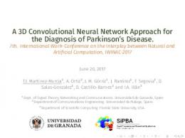
Figure 1: (a) Autoencoder for characterization of image patches (Section 2.2). (b) Encoder-decoder network for image patch semantic segmentation (Section 2.4). Manual segmentation of 2D images is laborious, manual segmentation of tomograms is even more so. To facilitate structural segmentation, automatic or semi-automatic approaches have been developed for segmenting specific structures. Such approaches use manually designed rules for 1) extraction of image features characterizing the specific ultrastructure and 2) segmentation based on combinations of extracted image features. All feature extraction and segmentation rules are specifically designed for particular types of image features or ultrastructures, such as membrane [e.g. 6, 27, 28, 13] and actin filaments [36, 45]. However, only very few generic and unified approaches exist for segmenting various structures [e.g. 9, 26]. Generic and unified approaches come with the advantage of being easily extended to segmenting new types of structures through automatic learning rules instead of manually designing them. The learning of rules is often done through a supervised fashion. In recent years, deep learning based approaches [e.g. 25] emerge as the dominant approaches for supervised generic segmentation in computer vision applications due to its superior performance in the presence of large amount of training data. Recently, deep learning has also been used for generic segmentation of ultrastructures [9] in cellular tomograms. Supervised segmentation approaches often rely on training data prepared through manual segmentation of images. Manual segmentation of tomograms is a voxel level annotation, and can be very laborious. Therefore, it is beneficial to develop approaches to reduce the amount of supervision (in terms of manual annotation) to speed up the automation of training data preparation. To complement existing approaches through reducing the amount of supervision, here we further demonstrate that the cluster groups generated from our autoencoder can be used to train a 3D CNN model for semantic segmentation in a weakly supervised fashion. In particular, after simple manual selection and grouping the clusters, the groups of clusters are used to train dense classifiers for voxel level classification for supervised (semantic) segmentation of tomograms. In the whole segmentation pipeline, the laborious manual voxel-wise segmentation of 3D images is no longer needed. The only step that require manual intervention is manual selection and grouping of image feature clusters among a number (such as 100) of candidate clusters, based on decoded cluster centers and location of the image features in the clusters. Therefore, the whole pipeline is weakly supervised with minimal manual processing. Our preliminary tests on experimental tomograms demonstrates the efficacy of our approach.
3
2 2.1
Methods Background
Deep learning is one of the dominant computer vision techniques used today across a broad array of applications [22]. Specifically, Convolutional Neural Network (CNN) [23] has achieved high performance and accuracy in computer vision tasks such as image classification [21] and semantic segmentation [25]. CNN is a feedforward artificial neural network inspired by the hierarchical organization of animal visual cortex. A CNN model is a combination of layers in sequence and each layer consists of a certain number of neurons with receptive fields on the previous layer. The learnable unit of each layer, called a neuron, connects with part of a previous layer, called a receptive field orPfilter. For example, a 1D convolution of input x, output y, and filter size 2m + 1 is defined as m yi = j=−m wi xi−j , where wj is the jth weight of the convolutional filter. The weight of a neuron is updated after every training epoch based on the loss of the prediction. There exists different types of CNN layers: convolutional layer, fully connected layer, up sampling layer, pooling layer, etc. A convolutional layer is defined as a layer with neurons of certain filter sizes. A fully connected layer is a layer in which every neuron is connected to every neuron in the previous layer. A pooling layer is a type of non-linear down sampling layer to reduce the spatial representation size. Such pooling often outputs the local maximum (max pooling) or average (average pooling) of its receptive field. For example, a 1D max pooling of input x, output y, and pooling size m is defined as yi = max(i−1)m