Bouzourene et al / MSI DETECTION IN COLORECTAL CANCER ..... radic cancers, such as colon, endometrial, and stomach.2,4,5,22,23. In HNPCC, germline ...
Anatomic Pathology / MSI DETECTION IN COLORECTAL CANCER
A Cost-Effective Algorithm for Hereditary Nonpolyposis Colorectal Cancer Detection Hanifa Bouzourene, MD,1 Lorenzo Taminelli, MD,1 Pascal Chaubert, MD,1 Christian Monnerat, MD,2 Walter Seelentag, MD,1 Dominique Sandmeier, MD,1 Snejana Andrejevic, MD,1 Maurice Matter, MD,3 Fred Bosman, MD, PhD,1 and Jean Benhattar, PhD1 Key Words: Colorectal cancer; Microsatellite instability; Hereditary nonpolyposis colorectal cancer; HNPCC; Immunohistochemistry; hMLH1; Methylation DOI: 10.1309/B0AFDT52ETMKEJBE
Abstract Colorectal cancer with microsatellite instability (MSI) may occur sporadically or be inherited in cases of hereditary nonpolyposis colorectal cancer (HNPCC) syndrome. However, there is no consensus as to which patients must be tested and how to test MSI. In this study, MSI was tested by immunohistochemical analysis and by polymerase chain reaction in 148 cases of colorectal cancer, and methylation of the hMLH1 promoter was examined. MSI status was correlated with tumor phenotype. We found that localization, tumor infiltrating lymphocytes, and mucinous differentiation were predictive of high-frequency MSI (MSI-H) colorectal cancer and might be used to select cases for MSI analysis. Immunohistochemical analysis detected most MSI-H colorectal cancer and might constitute the first step in MSI detection. Absence of hMLH1 promoter methylation in MSI-H colorectal cancer could be predictive of hereditary colorectal cancer, and, hence, methylation analysis might constitute the second step in the identification of patients with HNPCC.
Colon cancer results from the progressive accumulation of genetic and epigenetic alterations that lead to the transformation of normal colonic epithelium to adenocarcinoma of the colon. However, adenocarcinomas of the colon and rectum arise through at least 2 molecular genetic pathways. The majority of colorectal carcinomas (CRCs; 85%) arise through chromosomal instability, which involves the wnt signaling pathway, whereas 10% to 15% of tumors arise through microsatellite instability (MSI).1-5 The most common genetic alterations in tumors with chromosomal instability are allelic losses, chromosomal amplifications, and translocations that affect mainly tumor-suppressor genes (APC, p53, and SMAD4) and some oncogenes (k-ras).3 Cases with MSI display frameshift mutations and base-pair (bp) substitutions in microsatellites.2,4,5-7 These errors normally are controlled and repaired by the DNA mismatch repair (MMR) genes, including hMLH1, hMSH2, hMSH6, hPMS2, and hMSH3. Deficient MMR can arise sporadically or be inherited in an autosomal dominant manner in cases of the hereditary nonpolyposis colorectal cancer (HNPCC) syndrome.1,8,9 In most HNPCC cases, the cause is an inherited germline mutation of 1 of the 3 main MMR genes (hMLH1, hMSH2, and hMSH6), leading to deficient MMR and, ultimately, cancer.10,11 However, about 15% of CRCs show MSI without evidence of germline abnormalities. In these cases, the cause is biallelic methylation of CpG islands in the promoter sequence of hMLH1, an epigenetic change that leads to a deficiency of MMR genes.12-14 During the last few years, the detection of MSI has become increasingly more important. Not only does the detection of MSI permit the detection of familial colorectal cancer, but it also seems that the response of MSI cancers to Am J Clin Pathol 2006;125:823-831
© American Society for Clinical Pathology 823
DOI: 10.1309/B0AFDT52ETMKEJBE
823 823
Bouzourene et al / MSI DETECTION IN COLORECTAL CANCER
chemotherapy is different from the response of cancers with chromosomal instability. As a consequence, the demand for MSI testing is increasing sharply. However, because systematic screening for MSI in CRC is not realistic, the development of a cost-effective strategy to detect HNPCC is more essential. This study addresses the following questions: (1) Does morphologic assessment allow reliable prediction of MSI CRC? (2) Does immunohistochemical staining for hMLH1, hMSH2, and hMSH6 allow reliable prediction of MSI CRC? (3) Which proportion of MSI CRCs show methylation of the promoter of the hMLH1 gene? (4) Which combination of these approaches allows reliable and cost-effective identification of HNPCC?
Materials and Methods Cases Our cohort consisted of 148 consecutive CRC cases. Tumor specimens were obtained from surgical resection specimens performed at the Centre Hospitalier Universitaire Vaudois, Lausanne, Switzerland, between February 2002 and June 2003. The present analysis was in accordance with the original informed consent signed by all patients. Histopathologic Assessment All tumor samples were reviewed by 2 pathologists (H.B. and L.T.) who were blinded to the molecular results. The following variables were assessed: tumor location, tumor TNM stage, tumor grade,15 growth pattern, and lymphocytic host response. Tumors located before the splenic flexure were considered as right-sided and after as left-sided. The tumor growth pattern at the invasion front was examined at low power to determine whether the tumor grew with a pushing pattern or an infiltrative pattern. The invasion front of the tumor also was assessed for the presence of a Crohnlike inflammatory response. For this, at least 3 lymphoid aggregates per section were required. Tumor infiltrating lymphocytes (TILs) were identified on H&E-stained sections, and only lymphocytes infiltrating between tumor cells were counted. TILs were scored as absent or present when there were fewer or more than 4 lymphocytes per 100 epithelial cells.16 Immunohistochemical Analysis for hMLH1, hMSH2, and hMSH6 Protein For each case, 1 paraffin-embedded block with tumor tissue and normal colonic tissue was selected for the detection of hMLH1, hMSH2, and hMSH6 protein. Tissue sections, 4 µm, were mounted on aminopropylmethoxysilane-coated glass slides, deparaffinized in xylene, taken through to absolute alcohol, and 824 824
Am J Clin Pathol 2006;125:823-831 DOI: 10.1309/B0AFDT52ETMKEJBE
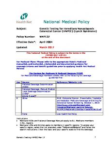
blocked for endogenous peroxidase with 0.3% hydrogen peroxide in methanol (45 minutes). They were subjected to microwave oven heating for 15 minutes in 10 mmol/L of citrate buffer, pH 6.0, and rinsed in Tris-buffered saline (TBS; 0.05 mol/L of tris(hydroxymethyl)aminomethane [Tris] and sodium chloride, 0.9%, pH 7.6). Then, sections were incubated for 30 minutes at room temperature with the primary monoclonal antibody (hMLH1, Pharmingen, Basel, Switzerland; hMSH2, Pharmingen; and hMSH6, Becton Dickinson, Basel; diluted 1:50, 1:600, and 1:100, respectively, in TBS containing 5% nonfat dry milk). After rinsing with TBS, the slides were incubated with the EnVision+/mouse system (DAKO, Carpinteria, CA), directed against mouse monoclonal antibodies for 30 minutes at room temperature. Peroxidase activity was revealed with 55,diaminobenzidine as the chromogen, and the sections were counterstained in Mayer acid-free hematoxylin. For a negative control sample, the first-step monoclonal antibody was replaced by a hybridoma supernatant of similar isotype but without reactivity for the tissue examined. A positive reaction showed unequivocal nuclear staining of epithelial cells, including neoplastic cells ❚Image 1❚. Tumor cells without nuclear staining in the presence of a positive internal control were considered deficient for the antigen (Image 1). MSI Analysis For MSI analysis, microdissection was performed after identification of tumor tissue by a pathologist (H.B.). DNA was extracted from tumor tissue and from normal mucosa from 2 blocks to avoid contamination.17 Extracted DNA was amplified by polymerase chain reaction (PCR), using the reference panel of microsatellite primers recommended for CRC by the National Cancer Institute, which includes the markers BAT25, BAT26, D5S346 (APC), D2S123 (hMSH2), and D17S250 (p53).18 The presence of additional bands in the PCR products from tumor DNA that were not observed in DNA from corresponding normal tissue was scored as unstable at that particular locus. Tumor samples were classified as displaying high-frequency MSI (MSI-H) when instability was observed for 2 or more of the loci screened, low-frequency MSI (MSI-L) when fewer than 2 of the loci screened, or microsatellite stability (MSS) when stability was present at all the loci tested. Methylation-Sensitive Single-Strand Conformation Analysis of the hMLH1 Promoter We selected 15 tumor samples with MSI-H status and loss of immunostaining of hMLH1 for promoter methylation analysis. After deparaffinization and staining in 0.1% toluidine blue, histologically selected areas in tissue sections were microdissected manually.17 Only the tumor cells were retained, and final histologic control before collection of the © American Society for Clinical Pathology
Anatomic Pathology / ORIGINAL ARTICLE
A
B
C
D
E
F
❚Image 1❚ Immunohistochemical staining for mismatch-repair proteins in colorectal adenocarcinomas. A, Positive staining for hMLH1 (×200). B, Negative staining for hMLH1 (×200). C, Positive staining for hMSH2 (×200). D, Negative staining for hMSH2 (×200). E, Positive staining for hMSH6 (×200). F, Negative staining for hMSH6 (×200).
Am J Clin Pathol 2006;125:823-831
© American Society for Clinical Pathology 825
DOI: 10.1309/B0AFDT52ETMKEJBE
825 825
Bouzourene et al / MSI DETECTION IN COLORECTAL CANCER
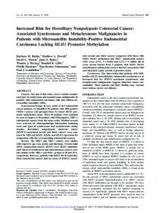
tumor cells confirmed that contamination with other cells was negligible. Extracted DNA was modified with sodium bisulfite as previously described.19 A 178-bp fragment of the hMLH1 gene promoter was amplified by nested PCR using the following primers: forward, 5'-GATTTTTTAAGGTTAAGAG-3', and reverse, 5'-ATAAAACCCTATACCTAATC-3', for the outer PCR; and forward, 5'-TTTTTAGGAGTGAAGGAG-3', and reverse, 5'-AAACCCTATACCTAATCTATC-3', for the inner PCR amplification. The outer PCR amplification was performed with 2 µL of modified DNA in a total volume of 20 µL for 40 cycles. For the inner PCR, 20 cycles were performed. Amplification was confirmed by analysis on a 2% agarose gel. Single-strand conformation analysis was performed as previously described.20 The percentage of methylated alleles was estimated semiquantitatively by comparing the intensity of the methylated and unmethylated bands ❚Image 2❚. Statistical Analysis Statistical analyses were carried out by using SPSS software, version 7.5 (SPSS, Chicago, IL). Quantitative data are expressed as mean ± SD. Group comparisons were made by using the χ2 test for categorical variables and the t test for continuous variables. Appropriate binary variables were generated to identify each subgroup of interest. The parameters used in this analysis were as follows: age of the patients, sex, location of the tumor (proximal or distal colon), histologic grade, invasive vs pushing tumor edge, absence or presence of TILs, absence or presence of Crohn-like reaction, absence of expression of one of the DNA MMR genes (hMLH1, hMSH2, and hMSH6), and MSI status (MSI-H vs MSI-L or MSS). A logistic regression model was used in multivariate analysis to identify independent predictors of MSI, and variables in the final model included all variables statistically significant at a P value of less than .05 in the univariate analysis.
Methylation scale 1
3
4
5
[
Unmethyl CpGs
2
[ 0
50
100
100 100
0
0
Methyl CpGs
100
Percentage of Methylated Alleles
❚Image 2❚ Methylation analysis of the hMLH1 promoter gene region by methylation-sensitive single-strand conformation analysis. Square brackets indicate the migration of the unmethylated DNA (Unmethyl CpGs) and the methylated DNA (Methyl CpGs).
826 826
Am J Clin Pathol 2006;125:823-831 DOI: 10.1309/B0AFDT52ETMKEJBE
Results Correlations between MSI status and clinicopathologic factors are summarized in ❚Table 1❚. In our series of 148 CRC cases, the mean age of the patients was 76.2 years; the pTNM stage was I in 6.7% of the cases, II in 16.8%, III in 45.2%, and IV in 31.0%. Of the 148 tumors, 55 (37.2%) were proximal to the splenic flexure, and 93 (62.8%) were distal. Most tumors were classified as conventional adenocarcinomas (117 [79.1%]); 25 (16.9%) were of the mucinous type; 1 (0.7%) was a signet-ring cell carcinoma; and 5 (3.4%) were undifferentiated carcinomas. Of 148 tumor samples tested for MSI, 21 (14.2%) demonstrated MSI-H, 3 (2.0%) were MSI-L, and 124 (83.8%) were MSS. Tumor Characteristics Related to Microsatellite Status High-Frequency MSI Of 21 MSI-H tumors, 5 had 4 unstable markers, 11 had 3 unstable markers, and 5 had 2 unstable markers ❚Table 2❚. All but 1 MSI-H tumor showed instability in the BAT25 and
❚Table 1❚ Comparison of Clinicopathologic Variables of 148 Colorectal Cancers Related to Microsatellite Status by Univariate Analysis* Microsatellite Status Variable Age (y)