SHORT COMMUNICATION Annals of Nuclear Medicine Vol. 20, No. 8, 569–573, 2006
A feasibility study of [11C]SA4503-PET for evaluating sigma1 receptor occupancy by neuroleptics: the binding of haloperidol to sigma1 and dopamine D2-like receptors Kiichi ISHIWATA,* Kenji ODA,** Muneyuki SAKATA,*** Yuichi KIMURA,* Kazunori KAWAMURA,* Keiichi ODA,* Toru SASAKI,* Mika NAGANAWA,*,**** Kunihiro CHIHARA,*** Yoshiro OKUBO***** and Kenji ISHII*
*Positron Medical Center, Tokyo Metropolitan Institute of Gerontology **Section of Liaison Psychiatry and Palliative Medicine, Graduate School of Tokyo Medical and Dental University ***Graduate School of Information Science, Nara Institute of Science and Technology ****JSPS Research Fellow, Tokyo *****Department of Psychiatry, Nippon Medical School
We investigated feasibility of positron emission tomography (PET) with [11C]SA4503 for evaluating the sigma1 receptor occupancy rate by neuroleptics. Haloperidol, which is well known to bind dopamine D2-like receptor (D2R) as well as to be a representative non-selective antagonist for sigma1 receptor (σ1R), was selected as a model drug. Three healthy male subjects underwent 60min [11C]raclopride-PET and 90-min [11C]SA4503-PET scans successively at a 120-min interval twice in a day for baseline measurement and on another day for haloperidol-loading measurement 16 hours after peroral administration of 3 mg of haloperidol. Binding potential (BP) of [11C]raclopride and [11C]SA4503 was quantitatively evaluated and the σ1R and D2R occupancy rates were determined. D2R occupancy rates by haloperidol were 64% and 62% in the caudate and putamen, respectively, 16 h after the administration, while σ1R occupancy rates were approximately 80% in all seven regions investigated including the caudate, putamen and cerebellum 18 h after the administration, suggesting that the σ1R receptor occupancy rate by haloperidol was slightly larger than the D2R receptor occupancy rate. We concluded that [11C]SA4503-PET can be used for evaluating the σ1R occupancy rates by neuroleptics or other drugs. Key words: [11C]SA4503, sigma1 receptor, receptor occupancy, haloperidol, positron emission tomography
INTRODUCTION In vivo evaluation of receptor occupancy by antipsychotic and antihistamineric drugs in the human brain has been investigated extensively by positron emission tomography (PET) and single-photon emission computed tomography (SPECT) with appropriate radioligands.1,2 These in vivo techniques are very useful for evaluating the therReceived May 18, 2006, revision accepted July 21, 2006. For reprint contact: Kiichi Ishiwata, Ph.D., Positron Medical Center, Tokyo Metropolitan Institute of Gerontology, 1–1 Nakacho, Itabashi-ku, Tokyo 173–0022, JAPAN. E-mail:
[email protected]
Vol. 20, No. 8, 2006
apeutic effects of the drugs, for determining appropriate dosages and for developing new drugs. So far, the occupancy of drugs for dopamine, serotonin and histamine receptors were mainly evaluated.1–3 It is also well known that a number of neuroleptics possess moderate to high affinity for sigma binding sites, suggesting the possibility that sigma receptors mediate some of the antipsychotic effects of neuroleptics.4,5 The physiological and pathophysiological roles of the sigma receptors remain under investigation and are considered as targets of pharmaceuticals for several diseases.6 However, the sigma receptor occupancy rates by the therapeutic drugs have not been evaluated in humans by PET or SPECT, because no in vivo selective radioligand was available. Recently,
Short Communication 569
Table 1 Binding potential (BP) of [11C]SA4503 and [11C]raclopride in the baseline and haloperidol-loading conditions and receptor occupancy rates by haloperidol [11C]SA4503 Binding potential*
Caudate Putamen Cerebellum Frontal lobe Temporal lobe Occipital lobe Thalamus
[11C]Raclopride Sigma1 receptor occupancy
Dopamine D2 receptor occupancy
Binding potential*
Baseline
Haloperidol
%
Baseline
Haloperidol
%
16.0 ± 1.4 18.5 ± 3.2 29.0 ± 1.9 20.8 ± 0.8 26.1 ± 4.1 20.3 ± 2.1 20.9 ± 2.1
3.0 ± 0.5 3.2 ± 0.5 5.7 ± 0.7 3.8 ± 0.8 4.7 ± 1.2 3.9 ± 1.3 4.8 ± 0.7
81.2 ± 4.9 81.9 ± 6.2 80.4 ± 3.1 81.5 ± 3.9 81.4 ± 7.0 81.1 ± 4.7 76.9 ± 2.7
3.3 ± 0.5 3.9 ± 0.6
1.2 ± 0.3 1.5 ± 0.3
64.4 ± 8.9 62.3 ± 7.8
Data show mean ± SD (n = 3). *Binding potential was evaluated based on a 2-tissue 3-compartment model for [11C]SA45039 and a reference tissue model for [11C]raclopride.15
we have developed [11C]SA4503 as a selective PET ligand for sigma1 receptor (σ1R),7–9 and clinically applied it to measuring σ1Rs of patients with Alzheimer’s and Parkinson’s disease.6,10 Previously, we investigated in mice using a tissue dissection technique whether [11C]SA4503 is available as an in vivo probe for evaluating the σ1R occupancy rates by neuroleptics using PET.11 We selected haloperidol and two other dopamine D2-like receptor (D2R) ligands which had high affinity for D2Rs and different affinity for sigma receptors. In the present study, we measured the σ1R occupancy rate by haloperidol in the human brain by [11C]SA4503-PET as a feasibility study that [11C]SA4503PET can be applied to evaluating σ1R occupancy rates by therapeutics. We also performed [11C]raclopride-PET in the same subjects for evaluating D2R occupancy rates by haloperidol. MATERIALS AND METHODS The study protocol was approved by the Institutional Ethical Committee. Three male volunteers (24 ± 4 years old) who were healthy according to the history and clinical investigations and showed no abnormality on brain MRI participated in the present study, with written informed consent obtained from each. All subjects underwent two PET scans with [11C]raclopride and [11C]SA4503 twice on two separate days: first for baseline measurement and 2–6 weeks later for haloperidol-loading conditions. On each day [11C]raclopride-PET was started late morning and two hours later [11C]SA4503-PET was followed, because [11C]raclopride shows a faster clearance rate from the brain than [11C]SA4503. On the second day the subjects were perorally given 3 mg of haloperidol 16 hours before [11C]raclopride-PET. The PET camera used was SET-2400W (Shimadzu, Kyoto, Japan), which has an axial field-of-view of 20 cm
570
Kiichi Ishiwata, Kenji Oda, Muneyuki Sakata, et al
and acquires 63 slices at a center-to-center interval of 3.125 mm.12 The injected doses of [11C]raclopride13 were 336 ± 17 MBq/5.6 ± 2.9 nmol (specific activity 74 ± 36 TBq/mmol) and those of [11C]SA45037 were 512 ± 160 MBq/13.6 ± 9.3 nmol (specific activity 51 ± 26 TBq/ mmol). After transmission scanning with a rotating [68Ge]/ [68Ga] line source to correct for attenuation, [11C]raclopride was intravenously injected into the subject, and then a 60min PET scanning in 2D mode (10 sec × 6 frames, 30 sec × 3 frames, 60 sec × 5 frames, 150 sec × 5 frames, and 300 sec × 8 frames) was performed without arterial blood sampling. In the second PET scan with [11C]SA4503, a 90-min dynamic scan in 2D mode (10 sec × 6 frames, 30 sec × 3 frames, 60 sec × 5 frames, 150 sec × 5 frames, and 300 sec × 14 frames) was carried out together with arterial blood sampling at 10, 20, 30, 40, 50, 60, 70, 80, 90, 100, 110, 120, 135 and 150 sec and 3, 5, 7.5, 10, 15, 20, 30, 40, 50, 60, 70, 80 and 90 min. The radioactivity levels in plasma were measured for radioactivity for gamma-counter and the time-activity curve (TAC) of plasma was calculated as Bq/ml and SUV. Metabolites of [11C]SA4503 in the plasma sampled at 3, 10, 20, 30, 40 and 60 min were analyzed by high-performance liquid chromatography as previously described.7 The tomographic images were reconstructed using a Fourier rebinning algorithm14 and a filtered backprojection method with Butterworth filter (cutoff frequency 1.25 cycle/cm and order 2). The data were collected in a 128 × 128 × 63 matrix, and the voxel size was 2 × 2 × 3.125 mm. Voxel counts were calibrated to activity concentration (Bq/ml). Regions of interest (ROIs) were placed over the caudate, putamen, cerebellum, frontal lobe, temporal lobe, occipital lobe and thalamus and on the PET images with reference to MRI. TACs of these ROIs were calculated as Bq/ml and SUV. For quantitative measurement of the radioligand-receptor binding, the binding potential (BP) of [11C]raclopride was calculated by the RPM with the
Annals of Nuclear Medicine
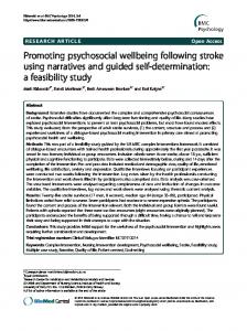
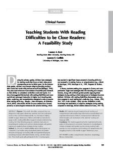
Fig. 1 PET images of [11C]SA4503 (A) and [11C]raclopride (B) in the same healthy subject in the baseline (upper images) and haloperidol-loading (lower images) conditions. The images of [11C]SA4503 and [11C]raclopride were acquired for 60–90 min and 40–60 min, respectively. White arrows show the striatum.
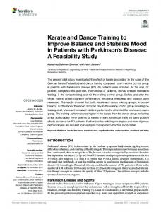
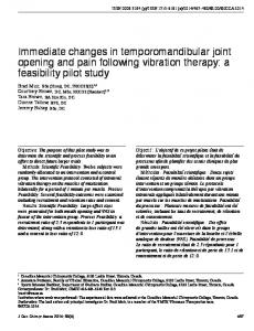
Fig. 2 Time-activity curves course of [11C]SA4503 (A) and [11C]raclopride (B) in the human brain and plasma in the baseline and haloperidol-loading conditions. Three regions of the brain were representatively selected: striatum (including caudate and putamen), cerebellum and frontal lobe.
cerebellum as a reference region.15 For the binding of [11C]SA4503, using the TACs of tissues and the metabolite-corrected TAC of plasma, the kinetic analysis was carried out based on a 2-tissue 3-compartment model
Vol. 20, No. 8, 2006
having 4 parameters: K1, influx rate constant from plasma to brain tissue; k2, efflux rate constant from tissue to plasma; k3, association rate constant between [11C]SA4503 and sigma1 receptor; and k4, dissociation rate constant of
Short Communication 571
[11C]SA4503-receptor complex.9 The BP was expressed as k3/k4. The detailed validation of kinetic analysis of [11C]SA4503 will be described elsewhere. The receptor occupancy (%) was defined as 100 × [(BP in baseline) – (BP in haloperidol-loading)]/(BP in baseline). RESULTS AND DISCUSSION Static PET images of [11C]SA4503 and [11C]raclopride in the baseline and haloperidol-loading conditions are represented in Figure 1. In the baseline, [11C]SA4503 was taken up in all regions, whereas [11C]raclopride was highly concentrated in the striatum. By the haloperidolloading, the uptake of [11C]SA4503 was reduced in all brain regions, while that of [11C]raclopride was blocked in the striatum. The TACs in the striatum, cerebellum, frontal lobe and plasma are represented in Figure 2. The levels of radioactivity in plasma rapidly decreased to a similar extent in the two conditions. In the baseline measurement of [11C]SA4503, the levels of radioactivity increased in all seven regions of the brain over 90 min in the case of Figure 2, or reached an apparent equilibrium state for 60 to 90 min in the other case (not shown), whereas the levels reached a plateau for 15–30 min and then decreased in the haloperidol-loading. The mean SUV values of [11C]SA4503 for 60–90 min in the haloperidolloading ranged approximately 55–65% of those in the baseline in the seven regions. BP was greatly reduced and the σ1R occupancy rates by haloperidol were approximately 80% in the seven regions investigated (Table 1). In the case of [11C]raclopride, the striatal uptake was greatly reduced by haloperidol-loading, while the levels of radioactivity in the other regions were not influenced by the haloperidol-treatment. The D2R occupancy rates based on BP values were 64% and 62% in the caudate and putamen, respectively (Table 1). A number of neuroleptics possess moderate to high affinity for sigma binding sites, suggesting the possibility that sigma receptors mediate some of the antipsychotic effects of neuroleptics.4 Frieboes et al. reported that haloperidol and the specific sigma ligand panamesine have similar antipsychotic properties regarding immunomodulation and sleep-electroencephalographic changes.16 These findings suggest indirectly the binding of haloperidol to sigma receptors. The present study directly demonstrated the binding of haloperidol to σ1Rs in the human brain by [11C]SA4503-PET. It is notable that the σ1R occupancy rates by haloperidol were larger than the D2R occupancy rates. A similar finding was previously observed in mice using a tissue dissection technique.11 However, the present and previous studies indicated no association between σ1R occupancy rates and antipsychotic efficacy of haloperidol. Thus, evaluation of the σ1R occupancy rates by haloperidol or other antipsychotic drugs in relation to behavioral potency in humans by [11C]SA4503-PET is of great interest.
572
Kiichi Ishiwata, Kenji Oda, Muneyuki Sakata, et al
In the present study, we performed quantitative evaluation of the binding of [11C]SA4503 to σ1Rs using a standard method based on a 2-tissue 3-compartment model having 4 parameters. We also applied the graphical analysis using a Logan plot to the quantitative evaluation.17 The Logan plot is a suitable method for parametric imaging of ligand-receptor binding because of its algorithmic simplicity and fast calculation speed, but it provides total distribution volume including both specific and nonspecific binding, but not BP. However, the results showed good agreement in the receptor occupancy rates between BP and DVt, and it implied that the receptor density of σ1Rs could be visualized using Logan plot. The details will be described elsewhere. In conclusion, [11C]SA4503-PET can be used for evaluating σ1R occupancy rates by neuroleptics or other drugs. The technique could be valuable for developing new drugs and for evaluating the therapeutic effects of drugs in term of σ1R occupancy. ACKNOWLEDGMENTS This work was supported by Grants-in-Aid for Scientific Research (B) No. 13557077 and Scientific Research (C) No. 18591373 and for JSPS Fellows No. 18-6916 from the Japan Society for the Promotion of Science. The authors thank Ms. Miyoko Ando for her care of the subjects undergoing PET measurement.
REFERENCES 1. Kasper S, Tauscher J, Kufferle B, Barnas C, Pezawas L, Quiner S. Dopamine- and serotonin-receptors in schizophrenia: results of imaging-studies and implications for pharmacotherapy in schizophrenia. Eur Arch Psychiatry Clin Neurosci 1999; 249 (Suppl 4): 83–89. 2. Nyberg S, Nilsson U, Okubo Y, Halldin C, Farde L. Implications of brain imaging for the management of schizophrenia. Int Clin Psychopharmacol 1998; 13 (Suppl 3): S15– S20. 3. Tashiro M, Mochizuki H, Sakurada Y, Ishii K, Oda K, Kimura Y, et al. Brain histamine H1 receptor occupancy of orally administered antihistamines measured by positron emission tomography with 11C-doxepin in a placebocontrolled crossover study design in healthy volunteers: a comparison of olopatadine and ketotifen. Br J Clin Pharmacol 2006; 61: 16–26. 4. Debonnel G, de Montigny C. Modulation of NMDA and dopaminergic neurotransmissions by sigma ligands: possible implications for the treatment of psychiatric disorders. Life Sci 1996; 58: 721–734. 5. Walker JM, Bowen WD, Walker FO, Matsumoto RR, de Costa B, Rice KC. Sigma receptors: biology and function. Pharmacol Rev 1990; 42: 355–402. 6. Hashimoto K, Ishiwata K. Sigma receptor ligands: Possible application as therapeutic drugs and as radiopharmaceuticals. Curr Pharm Design 2006; 12: 3915–3928. 7. Kawamura K, Ishiwata K, Tajima H, Ishii S, Matsuno K, Homma Y, et al. In vivo evaluation of [11C]SA4503 as a PET
Annals of Nuclear Medicine
8.
9.
10.
11.
12.
ligand for mapping CNS sigma1 receptors. Nucl Med Biol 2000; 27: 255–261. Kawamura K, Ishiwata K, Shimada Y, Kimura Y, Kobayashi T, Matsuno K, et al. Preclinical evaluation of [11C]SA4503: radiation dosimetry, in vivo selectivity and PET imaging of sigma1 receptors in the cat brain. Ann Nucl Med 2000; 14: 285–292. Kawamura K, Kimura Y, Tsukada H, Kobayashi T, Nishiyama S, Kakiuchi T, et al. An increase of sigma1 receptors in the aged monkey brain. Neurobiol Aging 2003; 24: 745–752. Mishina M, Ishiwata K, Ishii K, Kitamura S, Kimura Y, Kawamura K, et al. Function of sigma 1 receptors in Parkinson’s disease. Acta Neurologica 2005; 112: 103– 107. Ishiwata K, Kawamura K, Kobayashi T, Matsuno K. Sigma1 and dopamine D2 receptors occupancy in the mouse brain after a single administration of haloperidol and two dopamine D2-like receptor ligands. Nucl Med Biol 2003: 30: 429–434. Fujiwara T, Watanuki S, Yamamoto S, Miyake M, Seo S, Itoh M, et al. Performance evaluation of a large axial fieldof-view PET scanner: SET-2400W. Ann Nucl Med 1997; 11: 307–313.
Vol. 20, No. 8, 2006
13. Langer O, Någren K, Dolle F, Lundkvist C, Sandell J, Swahn CG, et al. Precursor synthesis and radiolabelling of the dopamine D2 receptor ligand [11C]raclopride from [11C]methyl triflate. J Label Compds Radiopharm 1999; 42: 1183–1193. 14. Defrise M, Kinahan PE, Townsend DW, Michel C, Sibomana M, Newport DF. Exact and approximate rebinning algorithms for 3-D PET data. IEEE Trans Med Imaging 1997; 16: 145–158. 15. Gunn RN, Lammertsma AA, Hume SP, Cunningham VJ. Parametric imaging of ligand-receptor binding in PET using a simplified reference region model. NeuroImage 1997; 6: 279–287. 16. Frieboes RM, Murck H, Antonijevic I, Kraus T, HinzeSelch D, Pollmacher T, et al. Characterization of the sigma ligand panamesine, a potential antipsychotic, by immune response in patients with schizophrenia and by sleep-EEG changes in normal controls. Psychopharmacology (Berl) 1999; 141: 107–110. 17. Logan J, Fowler JS, Volkow ND, Wolf AP, Dewey SL, Schlyer DJ, et al. Graphical analysis of reversible radioligand binding from time-activity measurements applied to [N11C-methyl]-(–)-cocaine PET studies in human subjects. J Cereb Blood Flow Meta 1990; 10: 740–747.
Short Communication 573