A FINITE ELEMENT MODEL OF THE C2-C3 SEGMENT – GROSS RESPONSES UNDER PHYSIOLOGICAL LOADS Vee-Sin Lee1, Ee-Chon Teo2*, Kim-Kheng Lee2, Kok-Yong Seng1, Hong-Wan Ng2 and Lih-Duen Neo1 1 Defence Medical Research Institute, Defence Science & Technology Agency, Singapore 2 School of Mechanical & Production Engineering, Nanyang Technological University, Singapore * Corresponding author E-mail:
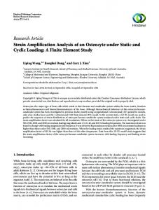
[email protected] stiffness was discernable between 200 – 400 Nmm in the extension direction.
INTRODUCTION The human cervical spine can be sub-divided into four units, each with a unique morphology that determines its kinematics and its contribution to the functions of the complete cervical spine. While literature delineating the biomechanical characteristics of the atlas, axis and the typical cervical spine abound, there remain a dearth of studies with regards to the biomechanics of the C2-C3 junction, from which the typical cervical spine commences. The present study of developing and exercising a finite element (FE) model of the C2-C3 segment was undertaken to partially fill the gap in the literature by quantifying its gross behavior under various physiological loads. MATERIALS AND METHODS A 3-dimensional FE model of the C2-C3 segment based on a 68-year old male cadaveric spine was developed. The model was developed based on previously reported digitizing techniques, modeling methodology and material models. The C2-C3 segment was subjected to bending moments in the anatomic sagittal, frontal and transverse planes. Five incremental loads of 300 Nmm each were applied uniformly across the superior articular surfaces of C2, while the inferior vertebral body of C3 was rigidly constrained in all directions. The rotational components of relative motion between the C2 and C3 segments under increment moment to maximal moments of 1500 Nmm were computed.
Figure 2: C2-C3 Responses under various loads. Under axially applied incremental torsion loads, the model exhibited gradual stiffening. Coupled motion – in the form of lateral bending in the frontal plane – was observed. In lateral bending exertions, the model predicted a maximum angular motion of 3o at 1400 Nmm, and no variation in stiffness was noted throughout the loading history. The model also predicted coupled motion in the transverse plane. The simulation of the 450-inclined facet joints via moving contact elements greatly influenced the kinematics of the C2C3 segment, while the spinal ligaments and the intervertebral disc controlled the range of motion in all the body planes considered in the present study.
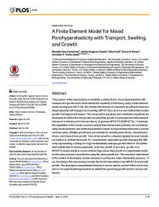
RESULTS AND DISCUSSION SUMMARY The FE model of the C2-C3 segment (Fig.1) comprised 4580 "8-noded" solid elements for the definition of the bony structures and the intervertebral disc, and 181 tension cable elements represented the spinal ligaments. Surface-to-surface contact elements were used to simulate the joints between the C2 inferior and C3 superior articulating surfaces. Figure 1: The FE model of C2-C3 The gross biomechanical response of the C2-C3 segment displayed varying non-linear and stiffening characteristics under different loads (Fig 2). Under applied moments in the sagittal plane, the C2-C3 model exhibited greater stiffness in the extension direction at high loads (>400 Nmm), but at low loads (0 – 400 Nmm), the spinal segment was stiffer under flexion. The variation in stiffness was insignificant for flexion, but the increase in
A 3D FE model of the C2-C3 spinal segment was developed and reported for the first time based on previously published methodology and materials for the lower cervical spine. As such, appreciable levels of confidence in the aforementioned results are expected. The non-linear response and range of motion of the C2-C3 segment were observed to be the concerted efforts of all spinal components and the orientation of articular facets. The model has the potential to supplement various in vitro and in vivo studies to further the understanding of human cervical spine biomechanics under different loaddisplacement vectors. REFERENCES Bogduk, N., Mercer, S. (2000) Clin. Biomechanics, 15, 633648. Teo, E.C., Ng, H.W. (2001) J. Biomechanics, 34, 13-21. Teo, E.C., Ng, H.W. (2001) Med. Eng. Phys, 23, 155-164. Ng, H.W., Teo, E.C. (2001) J. Spinal Disord, 14, 201-210.