CLINICAL REVIEWS
A Global, Evidence-Based Consensus on the Definition of Gastroesophageal Reflux Disease in the Pediatric Population Philip M. Sherman, MD1, Eric Hassall, MD2, Ulysses Fagundes-Neto, MD3, Benjamin D. Gold, MD4, Seiichi Kato, MD5, Sibylle Koletzko, MD6, Susan Orenstein, MD7, Colin Rudolph, MD8, Nimish Vakil, MD9,10 and Yvan Vandenplas, MD11
OBJECTIVES:
We sought to develop an international consensus on the definition of gastroesophageal reflux disease (GERD) in the pediatric population.
METHODS:
Using the Delphi process, a set of statements was developed and voted on by an international panel of eight pediatric gastroenterologists. Statements were based on systematic literature searches using Medline, EMBASE, and CINAHL. Voting was conducted using a six-point scale, with consensus defined a priori as agreement in 75% of the group. The strength of each statement was assessed using the GRADE system.
RESULTS:
There were four rounds of voting. In the final vote, consensus was reached on 98% of the 59 statements. In this vote, 95% of the statements were accepted by seven of eight voters. Consensus items of particular note are: (i) GERD is present when reflux of gastric contents causes troublesome symptoms and/or complications, but this definition is complicated by unreliable reporting of symptoms in children under the age of ~8 years; (ii) histology has limited use in establishing or excluding a diagnosis of GERD; its primary role is to exclude other conditions; (iii) Barrett’s esophagus should be defined as esophageal metaplasia that is intestinal metaplasia, positive or negative; and (iv) extraesophageal conditions may be associated with GERD, but for most of these conditions causality remains to be established.
CONCLUSIONS: The consensus statements that comprise the Definition of GERD in the Pediatric Population
were developed through a rigorous process. These statements are intended to be used for the development of future clinical practice guidelines and as a basis for clinical trials. Am J Gastroenterol advance online publication, 7 April 2009; doi:10.1038/ajg.2009.129
INTRODUCTION The Montreal Definition of gastroesophageal reflux disease (GERD) was developed as an international consensus document based on evidence reviewed from studies that were undertaken in adults (1). These guidelines are now being used for setting research priorities and as a basis for patient man-
agement guidelines in many parts of the world. However, in recognition of the special needs of the pediatric population, the work of this group (1) was limited to adults. A variety of health care practitioners encounter GERD in the pediatric age group, comprised of, infants, children, and adolescents. In a recent survey of the knowledge, attitudes and
1 Gastroenterology-Pediatric, Hospital for Sick Children, University of Toronto, Toronto, Canada; 2Division of Gastroenterology, British Columbia Children’s Hospital/ University of British Columbia, Vancouver, Canada; 3Disciplina de Gastroenterologia, Departamento de Pediatria, Escola Paulista de Medicina, Universidade Federal de São Paulo, São Paulo, Brasil; 4Division of Pediatric Gastroenterology, Hepatology and Nutrition, Emory University School of Medicine, Atlanta, Georgia, USA; 5Department of Pediatrics, Tohoku University School of Medicine, Sendai, Japan; 6Dr von Haunersches Kinderspital, Ludwig Maximilians University, Munich, Germany; 7University of Pittsburgh School of Medicine, Pittsburgh, Pennsylvania, USA; 8Division of Pediatric Gastroenterology and Nutrition, Medical College of Wisconsin, Milwaukee, Wisconsin, USA; 9University of Wisconsin School of Medicine and Public Health, Madison, Wisconsin, USA; 10Marquette University College of Health Science, Milwaukee, Wisconsin, USA; 11Department of Pediatrics, UZ Brussel Kinderen, Vrije Universiteit Brussel, Brussels, Belgium. Correspondence: Philip M. Sherman, MD, Gastroenterology-Pediatric, Hospital for Sick Children, University of Toronto, Room 8409, 555 University Avenue, Toronto, Ontario, Canada M5G 1X8. E-mail:
[email protected] Received 3 December 2008; accepted 22 January 2009
© 2009 by the American College of Gastroenterology
The American Journal of GASTROENTEROLOGY
1
REVIEW
nature publishing group
REVIEW
2
Sherman et al.
practice styles of pediatric health care providers, general pediatricians and subspecialists reported that a variety of terms and definitions of GERD are employed both in literature and in daily practice (2). This variety and inconsistent nomenclature contributes to wide variations in patient management. In addition, the lack of clear definitions of GERD and its manifestations in infants, children, and adolescents leads to confusion for the clinician, both when interpreting clinical trial literature and when trying to understand how to employ the available diagnostic tests. Therefore, there is a need to establish a uniform definition of GERD that could be used in these age groups. The collaborative updating of GERD guidelines by the North American and European Societies for Pediatric Gastroenterology, Hepatology and Nutrition, also highlights the need to develop internationally accepted criteria and support recommendations with a systematic review (3). The Montreal Definition of GERD, and the process used to develop it, provided a conceptual framework on which the definition of reflux disease for use in the pediatric setting is based. Thus, the aim of this process was to develop an international consensus regarding the definition and classification of GERD in pediatrics. This is different from the GERD guidelines, which provide management recommendations. These consensus statements are intended to be used for the development of future clinical practice guidelines and as a basis for clinical trials, which may include patient- and surrogate-reported GERDrelated outcomes, and to support the improved management of GERD in pediatric patients.
METHODS A modified Delphi technique, analogous to that used in developing the Montreal Definition of GERD in adults (1), was used to develop statements for the definition of GERD in the pediatric population. The main steps in this process were as follows: (i) selection of an international consensus group; (ii) development of draft statements; (iii) systematic literature review to identify and critically evaluate the evidence relevant to each of the statements; and (iv) anonymous voting, discussion, and repeated anonymous voting by the consensus group on a series of iterations of each of the draft statements. A non-voting liaison (NV), an adult gastroenterologist who was the Chair of the Montreal consensus committee, provided perspectives from the Montreal process and issues relevant to adults. In addition, he reviewed materials for statements and provided input for the paper revisions. The group members agreed that it was preferable to apply adult definitions (1), whenever relevant, in order to prevent confusion over conflicting terminologies. However, it was recognized that differences between pediatric patients and adults, and within the pediatric age group itself (infants, children, and adolescents), make age-specific definitions necessary. Accordingly, three age groups were employed comprising: newborns and infants (0–12 months), toddlers and children (1–10 years), and adolescents (11–18 years). The American Journal of GASTROENTEROLOGY
Selection of the consensus group
A non-voting Chair (PMS) led the consensus group. Members of the group were selected by the Chair based on the following criteria: recognized expertise in GERD in pediatrics as evidenced by relevant peer-reviewed publications, research activities, participation in national or regional activities, and expertise in the development of clinical practice guidelines. The consensus group choice was also influenced by the need for international representation and a diversity of views and expertise in the field. Systematic searches of the biomedical literature
Relevant English language studies on humans published from January 1980 to December 2007 were identified through systematic searches of Medline, EMBASE, and CINAHL. The Chair formulated numerous search strings (available on request), reviewed the search results, and selected articles for further review. Individual group members contributed further literature searches. Articles were added during the process, and distributed to all members of the consensus group. Grades of evidence
The strength of evidence was assessed using the GRADE system (4), which is as follows: High: Further research is unlikely to change our confidence in the estimate of effect. Moderate: Further research is most likely to have an important impact on our confidence in the estimate of effect and may change the estimate. Low: Further research is very likely to have an important impact on our confidence in the estimate of effect and may change the estimate. Very low: Any estimate of effect is uncertain. The designation “not applicable” was employed for situations, in which these grades of evidence were not relevant for a particular statement. A preliminary grade of evidence was assigned to each statement before the final iteration. Each was reviewed by the Committee after the final vote, and again in drafts of the paper. Voting on consensus statements
The group voted on four iterations of each of the consensus statements. Between each of the four votes, statements were revised based on feedback from the group, outside experts (a family doctor, academic general pediatrician, neonatologist, pediatric pulmonologist, pediatric otolarygologists, and pediatric general surgeons), and further critical review of the available literature. Additional statements were added on pediatric topics that had not been addressed earlier, particularly those conditions which have signs and/or symptoms that are similar to, or could be confused with GERD, such as food allergy. The final vote was held at a face-to-face meeting, which included extended discussion time. Voting was anonymous, conducted using electronic touch pads, and voting percentages were shown on-screen after each vote. A six-point scale was used: (i) agree strongly (A + ), (ii) agree moderately (A), (iii) just www.amjgastro.com
Definition of GERD in Children
Level of agreement
Agreement (% of statements)
A+
Agree strongly
40
A
Agree moderately
40
A−
Just agree
16
D−
Just disagree
2
D
Disagree moderately
1
D+
Disagree strongly
1
agree (A − ), (iv) just disagree (D − ), (v) disagree moderately (D), and (vi) disagree strongly (D + ). Agreement with the statement (the sum of voting for A + , A, or A − ) by three-quarters (i.e., ≥75%) of the voting members was defined a priori as consensus. The level of agreement in the final vote that was given for each statement, expressed as percentages, is shown in Table 1. Workshop organization and funding sources
Two face-to-face workshops were organized and supported financially by a third party, INSINC Consulting (Guelph, ON), using an unrestricted grant from AstraZeneca Research and Development (Mölndal, Sweden). AstraZeneca had no input into the content and conduct of the workshops nor into the development of the statements and this paper. Dr Catherine Henderson (Oxford PharmaGenesis Ltd, Oxford, UK) managed the literature database, prepared minutes from the face-to-face meetings, and assimilated written commentaries contributed by each consensus group member into a single document, which she copyedited. On the basis of this document, the Chair prepared a first draft of the paper, which was revised through several iterations by one member of the consensus group (EH), with input from the committee members. The Chair and committee members created the content of the paper, for which they are fully and solely responsible. Disclosures of potential conflict of interest of committee members were documented before the first meeting of the committee, and distributed to all the members. Similar to the Montreal process, the goal of the endeavor was to establish definitions of disease, not to advocate for or against any treatment or medical device (5). The major points emerging from this initiative were presented in a poster forum at a symposium at the Third World Congress of Pediatric Gastroenterology, Hepatology and Nutrition held at Iguassu Falls, Brazil in August 2008.
RESULTS AND DISCUSSION Overview of the voting on statements
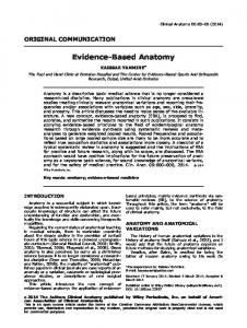
In this paper, Figure 1 shows an overview of the statement development process and the voting results. For the first © 2009 by the American College of Gastroenterology
43 Montreal Definition statements; 7 revised Montreal Definition statements; 12 new statements Number of Proportion of statements statements with consensus
Vote 1 (e-mail), July 2007
62
76%
Vote 2 (1st workshop), Sept 2007
117
67%
Vote 3 (e-mail), Nov 2007
86
66%
Vote 4 (2nd workshop), Dec 2007
59
98%
2 Montreal Definition statements; 29 revised Montreal Definition statements; 28 new statements
Figure 1. Consensus development process and voting results.
vote, 62 statements were presented, based on the 50 Montreal Definition statements (1) and 12 additional statements with particular pediatric relevance proposed by the Chair. Consensus was reached on 47 (76%) of the 62 statements on the initial vote. For the second vote, the number of statements increased substantially, to 117, mainly because of the group’s decision to vote on many of the statements separately for each of three pediatric age groups (newborns and infants, toddlers and children, and adolescents). Consensus was reached for 78 (67%) of 117 statements. For the third round of voting, 86 statements were considered, with the group reaching consensus on 57 (66%). In the final vote, there were 59 statements: consensus was reached for 98% (58) and an agreement by more than 87% of the voting group was attained for 95% (56). The strength of agreement in the final vote is shown in Table 1.
A GLOBAL DEFINITION OF GERD IN THE PEDIATRIC POPULATION: STATEMENTS 1. GERD in pediatric patients is present when reflux of gastric contents is the cause of troublesome symptoms and/or complications. Agree: 100% (A + , 87.5%; A, 12.5%; Grade of evidence: not applicable). The approach, adopted in the Montreal Definition of GERD in adults, was to use a patient-centered, symptom-based definition of the point at which the symptoms become sufficiently troublesome, so as to have a measurable impact on the quality of life of the patient (1). This approach also applies to children, but with several caveats. Child development and communicative The American Journal of GASTROENTEROLOGY
REVIEW
Table 1. Average strength of agreement with statements considered in the final vote
3
REVIEW
4
Sherman et al.
abilities mandate an age-group-specific approach to definitions. In children, the definition of when the symptoms become troublesome—and who defines that the symptoms are troublesome—is complicated by several factors. Although the verbal child can communicate pain, descriptions of the intensity, location, and severity may be unreliable until the age of at least 8 years, and sometimes even later (6,7). Younger children are generally more suggestible; so queries from parents or clinicians regarding a specific symptom may be biased toward affirmative responses, thereby decreasing the reliability of the symptom that is reported by the child with suspected GERD. Thus, the determination of whether a symptom is troublesome by self-report cannot be reliably used as a component of a GERD definition until the individual is more than ~8 years of age. In younger patients, reliance on a parent or caregiver is generally necessary, but reporting of the symptoms by surrogates (parents, caregivers) may decrease the validity of diagnosis. Therefore, the issue of what is “troublesome” is more complicated in the pediatric age group than in adults. The US FDA (Food and Drug Administration) recommends that patient-reported outcomes form the basis of clinical trials (http://www.fda.gov/cder/guidance/5460dft. htm). Given the limitations of reporting in children who are ~8 years and younger, patient-reported outcomes have to be supplemented or replaced with reporting by surrogates. Validated symptom questionnaires that are related to specific age groups are needed for achieving reliability, as well as for diagnostic and evaluative validity related to symptom reporting in pediatrics (8,9). 2. Symptoms of GERD vary by age. Agree: 100% (A + , 87.5%; A − , 12.5%; Grade: high). In adults, heartburn and regurgitation are the characteristic symptoms of GERD (1), and the same is true for older children and adolescents (10,11). In infants, the issue is more complicated. During the validation of a GERD questionnaire diagnostic score, differences were identified in the prevalence of regurgitation, food refusal, and crying between that of a healthy cohort and infants with abnormal esophageal pH studies and/or abnormal biopsies (12). However, the lack of response of these symptoms to proton pump inhibitor (PPI) therapy contrasts with their responses to non-pharmacologic therapy, raising questions about the acid mediation of these symptoms and whether they do, in fact, represent acid reflux (13,14). Accordingly, a symptom-based diagnosis of GERD in infants remains a problem. Beyond infancy, in one study of 1 to 17 year olds, cough, anorexia/food refusal, and regurgitation/vomiting were more severe in children who were 1–5 years of age, compared with that in older children (10). In this study, toddlers and young children (1–6 years) tended to present with food refusal, regurgitation, and abdominal pain. In contrast, the predominant symptoms in older children (6–17 years) are regurgitation or vomiting, cough and epigastric pain, or heartburn (10,15). The older the child, the more heartburn and regurgitation become The American Journal of GASTROENTEROLOGY
predominant presenting symptoms (10,11). Overall, for 1 to 11 year olds, there are relatively few data on presenting symptoms, and no standardized definitions for reporting (10). For example, some studies allow reporting of abdominal pain, or epigastric pain, or both (10,15,16). The variability of the GERD symptoms and signs in the pediatric age groups requires further development of age-based definitions, “gold standards,” and the validation of symptom-based questionnaires. 3. Symptoms due to gastroesophageal reflux (GER) are troublesome when they have an adverse effect on the wellbeing of the pediatric patient. Agree: 100% (A + , 12.5%; A, 75%; A − , 12.5%; Grade: not applicable). 4. The otherwise healthy newborns (age: 1–30 days) and infants (age: >30 days to < 1 year) with reflux symptoms that are not troublesome and are without complications should not be diagnosed with GERD. Agree: 87.5% (A + , 62.5%; A, 12.5%; A − , 12.5%; D − , 12.5%; Grade: not applicable). 5. Reflux symptoms that are not troublesome in toddlers and children (age: 1–10 years) should not be diagnosed as GERD. Agree: 75% (A + , 37.5%; A, 37.5%; D − , 12.5%; D, 12.5%; Grade: not applicable). 6. Reflux symptoms that are not troublesome in adolescents (age: 11–17 years) should not be diagnosed as GERD. Agree: 87.5% (A + , 50%; A, 37.5%; D − , 12.5%; Grade: not applicable). With regard to Statement 4, up to 70% of completely healthy newborns and infants have regurgitation that is physiologic, resolving without intervention in 95% of the individuals by 12– 14 months of age (17–19). In one questionnaire-based study of healthy infants seen at routine office visits (17), daily regurgitation peaked at 67% of the infants at 4 months of age, and decreased to 21% at 7 months and to 5% at 10–12 months. The regurgitation of the most affected subgroup of those infants— those with the most frequent regurgitation (more than four episodes daily)—peaked at a similar age and was resolved similarly. Regurgitation described as “a problem” (possibly similar to “troublesome”) by a parent peaked at 6 months (23%) and decreased to 14% at 7 months of age. Crying is also common in unselected infants; its average daily duration peaks in the second month of life at 2–2.5 h per day, decreasing thereafter (20,21). Among 1- to 3-month-old infants, the mean duration of crying is 3 h per day (median: < 1.5 h per day), suggesting that there is a subgroup with rather prolonged crying. From 4 to 12 months of age, the duration of crying remains fairly constant, at a mean of ~1 h per day (median: ~0.5 h). Regarding the subgroup of unselected infants with more prolonged daily crying, one study reported that crying ≥3 h per day occurred in 29% of infants during the first 3 months of life, but decreased to occur in < 10% of 3- to 12-month-old infants www.amjgastro.com
(21). The same study reported maternal clinical consultation about the crying (possibly a surrogate for parental perception as “troublesome”), finding that 11% of mothers consulted in the first 3 months and < 4% consulted during the remainder of the child’s first year of life. The issue of unexplained crying is complicated and must also incorporate patterns of crying and parental responses to crying (21). Consequently, in infants, normal regurgitation and normal crying, or abnormal crying due to a cause other than GERD, may be mistaken for GERD (22,23). Limited data are available regarding the prevalence of symptoms of reflux in childhood that might be considered troublesome, particularly for infants. Data-based suggestions for quantitative thresholds for “troublesome” regurgitation and crying are noted above, but they lack sensitivity and specificity. In addition, “troublesome” is subjective. Other data suggest that combining quantitative parameters of symptoms with clustering of distinct symptoms may better define “troublesome” symptoms and, thereby help to distinguish GERD from GER (12). As reported by Nelson et al. (17), “Parental perception that regurgitation was a problem was associated with the frequency and volume of regurgitation, increased crying or fussiness, reported discomfort with spitting up, and frequent back arching.” This suggestion is also supported by greater responsiveness of regurgitation or crying to non-pharmacologic management when they are isolated, non-clustered symptoms (24). However, although up to 23% of parents (peaking for 5-month-old infants) considered these prominent and clustered symptoms as “problems” (17), treatment (consisting mostly of either formula change or thickening of feeds) was administered in fewer than 10% of the cases, and medication in only 0.2%. Thus the identification of symptoms and symptom clusters that serve as valid markers for GERD is quite complex, and more so because of issues of objective gold standards, language limitations, and the need for reporting by surrogates. Therefore, the definition of “troublesome” (or “problematic”) remains particularly challenging in infants, most of whom do not manifest an objective complication of GERD clinically. Most adolescents are sufficiently aware and communicative to be able to determine whether symptoms are troublesome, whereas in infants and young children, this has to be decided in a surrogate fashion by the caregiver in partnership with the practitioner. Nevertheless, the issue of reporting is not determined solely by chronological age. For example, the history from an attentive parent of a verbal 7- or 8-year-old might be at least as reliable as that from an unforthcoming adolescent, or one who might, as a matter of principle, insist on disagreeing with a parent. To be defined as GERD, reflux symptoms must be troublesome to the infant, child, or adolescent, and not simply be troublesome to the caregiver. In addition, patients may be asymptomatic, or unable to report “troublesome” symptoms (e.g., infants or neurologically impaired children), but still have complications of reflux and, thereby meet the criteria for the definition of GERD. © 2009 by the American College of Gastroenterology
7. Regurgitation in pediatrics is defined as the passage of refluxed contents into the pharynx, mouth, or from the mouth. Agree: 100% (A + , 12.5%; A, 87.5%; Grade: not applicable). 8. Bilious vomiting should not be diagnosed as GERD. Agree: 100% (A + , 75%; A, 12.5%; A − , 12.5%; Grade: high). Regurgitation occurs when relaxation of the lower esophageal sphincter allows the retrograde movement of gastric contents into the esophagus and beyond. Regurgitation is distinguished from vomiting physiologically by the absence of (i) a central nervous system emetic reflex, (ii) retrograde upper intestinal contractions, (iii) nausea, and (iv) retching (25). Regurgitation is generally characterized as effortless and non-projectile, although it may be forceful in infants (26). For this reason, the Montreal Definition statement was amended for pediatrics to include the ejection of refluxate from the mouth. Caregivers and some practitioners use other terms, such as “spitting-up,” “posseting,” and “spilling,” which are covered by this definition of regurgitation. Bilious vomiting is an alarm signal that warrants further investigations to rule out anatomic abnormalities, such as intestinal malrotation, or acute illnesses causing intestinal obstruction. 9. Regurgitation is a characteristic symptom of reflux in infants, but is neither necessary nor sufficient for a diagnosis of GERD, because it is not sensitive or specific. Agree: 100% (A + , 62.5%; A, 37.5%; Grade: high). The specificity of regurgitation for diagnosing GERD is hampered by the frequency of its occurrence in normal infants (see the commentary under Statement 4) (17,18), and by difficulties in distinguishing it from vomiting and a myriad of conditions that cause vomiting in infants. Quantitatively, more frequent regurgitation may predict GERD developing subsequently in some infants. In a prospective cohort study (18), infants regurgitating on 90 days or more during the first 2 years of life (“frequent spilling”) were more likely than those with no spilling to have reflux symptoms of heartburn, vomiting, or acid regurgitation when followed-up at ~9 years of age. Another study showed quantitative differences in regurgitation between biopsy–esophageal pH-positive and -negative infants (12). Correspondingly, regurgitation improved the specificity of a GERD diagnosis in infants with excessive crying, when a 24-h pH monitoring was used as a gold standard (27–29). Although such quantitative parameters are alone insufficient for either establishing or excluding the diagnosis of GERD in an individual patient, quantification and clustering of regurgitation with other symptoms has been shown to improve diagnostic sensitivity and specificity, as reflected by the I-GERQ (Infant Gastroesophageal Reflux Questionnaire) scores (12). Further revision and validation of this instrument showed a good evaluative performance by validated translations in multiple languages for the I-GERQ-R (I-GERQRevised) score (30). The American Journal of GASTROENTEROLOGY
5
REVIEW
Definition of GERD in Children
REVIEW
6
Sherman et al.
Persisting issues regarding such a symptom-based diagnosis in infants include replication of diagnostic validity by further studies using objective gold standards, and the use of a diseased/symptomatic, but non-GERD, control group. One study was unable to replicate the original diagnostic validity of the I-GERQ score, but the questionnaire had been modified and translated independently before use. In addition, histology was employed in only a minority of subjects and the methods used were not validated for reliability (31). Application of invasive gold standards to infants, particularly those in a control group, remains challenging, augmenting the elusiveness of a symptom-based GERD diagnosis in infants. 10. Symptoms of GERD, particularly in infants, may be indistinguishable from those of food allergy. Agree: 100% (A + , 62.5%; A, 25%; A − , 12.5%; Grade: high). In infants, GERD and milk protein (cow or soy) allergy may both manifest as regurgitation or as vomiting; crying, fussing, or irritability related to food intake; or as failure to thrive (32). Therefore, distinguishing between GERD and milk protein allergy is difficult based on clinical presentations alone (33,34). Milk protein allergy and GERD may coexist, and instituting a protein hydrolysate diet may resolve symptoms that are suggestive of GERD (13,34,35). 11. In clinical practice, adolescents are generally able to describe specific GERD symptoms and to determine whether those symptoms are troublesome. Agree: 100% (A + , 62.5%; A, 37.5%; Grade: low). Neurologically intact older children and adolescents are generally able to describe their symptoms and to determine whether reflux symptoms are troublesome. A caveat regarding unforthcoming adolescents is mentioned in the commentary pertaining to Statements 3–6 above. In a survey comparing reflux symptoms reported by adolescents with reports by their parents, 5.2% of the 10 to 17 year olds described having heartburn and 8.2% reported acid regurgitation (36). In contrast, parents reported decreased rates of the same symptoms in their children (3.5 and 1.4%, respectively). This discrepancy indicates the importance of self-reporting in this age group, although it could also indicate suggestibility. 12. Pediatric population-based studies of reflux symptoms are insufficient and are a priority for further research. Agree: 100% (A + , 75%; A, 25%; Grade: not applicable). A number of studies give an indication of the prevalence of GERD in the pediatric population, but various methodologies and definitions have been used. Using a primary care database from the United Kingdom, the estimated incidence of GERD among 2 to 19 year olds was 0.47 and 0.77 per 1,000 personyears in males and females, respectively (37). In another study, a review of primary and tertiary care medical records revealed an The American Journal of GASTROENTEROLOGY
Conditions predisposing pediatric patients to severe, chronic GERD
• Neurologic impairment • Congenital esophageal abnormalities (e.g., esophageal atresia, congenital diaphragmatic hernia) • Cystic fibrosis • Hiatal hernia • Obesity • Family history of severe GERD or Barrett’s esophagus or esophageal adenocarcinoma
Figure 2. Conditions predisposing to chronic, severe gastroesophageal reflux disease (GERD).
incidence of GERD of 0.9 per 1,000 person-years in infants and children < 5 years of age (38). In a study in the United States (11), of 1,286 adolescents who were 14–18 years of age, heartburn was reported by students in response to a questionnaire as occurring daily or “a few times a week” in 0.7 and 3.3%, respectively; regurgitation had a similar prevalence and dysphagia was slightly less common. However, no study has yet sampled a general pediatric population using a standardized definition of GERD. Therefore, robust population-based studies, based on the standard definitions of GERD are a priority area for future clinical research.
CONDITIONS PREDISPOSING TO SEVERE, CHRONIC GERD 13. The pediatric patient with central nervous system impairment has an increased risk of GERD. Agree: 100% (A + , 62.5%; A, 12.5%; A − , 25%; Grade: high). 14. Esophageal atresia is associated with an increased risk of GERD. Agree: 100% (A + , 75%; A, 12.5%; A − , 12.5%; Grade: high). 15. Cystic fibrosis is associated with an increased risk of GERD. Agree: 100% (A + , 75%; A, 12.5%; A − , 12.5%; Grade: high). Certain underlying disorders predispose pediatric patients to the most severe and chronic GERD, and its complications (Figure 2) (39–46). These include significant neurological impairment, such as cerebral palsy, genetic disorders, such as the Cornelia de Lange syndrome and Down’s syndrome, congenital esophageal abnormalities, such as repaired esophageal atresia or congenital diaphragmatic hernia, and chronic lung disease, such as cystic fibrosis. Otherwise healthy children and adults with hiatal hernia or with a strong family history of GERD, Barrett’s esophagus, or esophageal adenocarcinoma also have a higher prevalence of chronic GERD with complications (40,47,48). Although pediatric data are scarce, in adults, obesity and incremental weight gain are also associated with a significantly higher prevalence and increased severity of GERD, Barrett’s esophagus, and esophageal adenocarcinoma (49,50). www.amjgastro.com
SYMPTOMATIC SYNDROMES OF GERD Typical Reflux Syndrome
16. Heartburn in older children is defined as a burning sensation in the retrosternal area. Agree: 100% (A + , 50%; A, 37.5%; A − , 12.5%; Grade: not applicable). 17. Heartburn in adolescents is defined as a burning sensation in the retrosternal area. Agree: 100% (A + , 87.5%; A, 12.5%; Grade: not applicable). Heartburn is defined as a perceived uncomfortable burning sensation behind the sternum that can reach a painful quality (1). Issues regarding the reliability of reporting are addressed in the commentary under Statements 3–6. 18. The Typical Reflux Syndrome is characterized by heartburn with or without regurgitation. Agree: 100% (A + , 37.5%; A, 37.5%; A − , 25%; Grade: not applicable). 19. Heartburn and regurgitation in adolescents and older children, with cognitive development sufficient to reliably report symptoms, are characteristic symptoms of the Typical Reflux Syndrome. Agree: 100% (A + , 62.5%; A, 37.5%; Grade: not applicable). This statement applies only to children and adolescents who are able to reliably self-report symptoms. The data suggesting that heartburn and regurgitation are predictive of GERD are derived from studies in adults (1) and in children (10,51,52). 20. “Typical Reflux Syndrome” cannot be diagnosed in infants and children who lack the cognitive ability to reliably report symptoms. Agree: 75% (A + , 37.5%; A, 37.5%; D − , 12.5%; D + , 12.5%; Grade: not applicable). The problems with the reliability of symptom reporting in children below ~8 years of age are described in the commentary under Statement 1 and elsewhere in this document. This statement reflects those issues, but the spectrum in voting reflected differences of opinion within the group regarding the ability to rely on surrogate reporting of symptoms in non-verbal children < 8-years of age vs. the need for invasive testing for all GERD diagnoses at these ages. Reliability (accuracy, internal consistency, test–retest, interobserver agreement) of each item and the overall validity (both diagnostic and evaluative, for tracking changes over time) of such surrogate reporting can be tested through validation of questionnaires (instruments) designed to assess GERD. Regarding the principle noted earlier that the magnitude and clustering of symptoms can be employed to define GERD, such instruments have been developed for use in children (9,30); although objective gold standards have been used for establishing the diagnostic validity in only one study (12). © 2009 by the American College of Gastroenterology
The diagnostic validity of questionnaires for GERD symptoms remains problematic in the general absence of objective gold standards for the diagnosis of GERD, particularly at the younger ages. In the absence of established diagnostic validity, these instruments nonetheless can be used for reliable quantification of symptoms for history taking and for tracking symptoms over time by instruments for which evaluative validity (response of symptoms to treatment) has been established (8). Clearly, more work in this key area is required. 21. GER in older children and adolescents is the most common cause of heartburn. Agree: 87.5% (A + , 37.5%; A, 50%; D − , 12.5%; Grade: low). 22. Heartburn in older children and adolescents can have a number of non-reflux-related causes. The prevalence of these is unknown. Agree: 100% (A + , 50%; A, 37.5%; A − , 12.5%; Grade: high). There is a compelling evidence that acid reflux is the most common cause of heartburn in adults (1). Weakly acidic reflux, non-acid reflux, gas, and duodenogastric reflux may also be causative in adults and children (53,54). Other causes of heartburn in adults and pediatric patients include eosinophilic esophagitis, functional heartburn, esophageal infections, and Crohn’s disease (55–57). 23. In neurologically intact adolescents, the Typical Reflux Syndrome can be diagnosed on the basis of the characteristic symptoms, without additional diagnostic testing. Agree: 87.5% (A + , 12.5%; A, 62.5%; A − , 12.5%; D − , 12.5%; Grade: low). The Typical Reflux Syndrome in adults was defined based on symptoms; i.e., additional diagnostic testing is not routinely required (1). A symptom-based diagnosis of the Typical Reflux Syndrome can be made only in adolescents and children who are more than ~ 8 years of age and have the cognitive ability to reliably report symptoms. 24. Non-erosive reflux disease in the pediatric patient is defined by the presence of troublesome symptoms caused by the reflux of gastric contents and by the absence of mucosal breaks during endoscopy. Agree: 100% (A + , 12.5%; A, 75%; A − , 12.5%; Grade: not applicable). Troublesome reflux-associated symptoms that are not accompanied by mucosal breaks are termed non-erosive reflux disease (NERD), in contrast to ERD (erosive reflux disease) (57,58). NERD comprises approximately two-thirds of adults with reflux disease (1). Compared with ERD, adults with NERD are more predominantly female, with a lower body mass index, a higher prevalence of functional gastrointestinal disorders and esophageal acid hypersensitivity, and a poorer response to PPIs. Adults with ERD more often have hiatal hernia and The American Journal of GASTROENTEROLOGY
7
REVIEW
Definition of GERD in Children
REVIEW
8
Sherman et al.
greater esophageal acid exposure, and esophageal dysmotility than those with NERD (55,57). The diagnosis of NERD only applies to adolescents and children who have the cognitive ability to reliably report symptoms (i.e., those at least >8 years of age). However, there are few pediatric data on NERD compared with that on ERD with respect to presenting symptoms, prevalence, pathophysiology, and responses to therapy (10,52,59). Various endoscopic mucosal changes have been reported in adults with NERD using magnification endoscopy. These include vascular injection, a villous-appearing mucosal surface, squamous islands below the Z-line, and red streaks (60). There is a paucity of data regarding such findings in children. A diagnosis of NERD cannot be made or ruled out by histology. (See also Statements 33 and 34 regarding histology.) 25. Epigastric pain in older children and adolescents can be a major symptom of GERD. Agree: 100% (A, 75%; A − , 25%; Grade: moderate). In some studies, subjects were permitted to report either abdominal pain or epigastric pain (10,15), but these terms are neither sensitive nor specific for the diagnosis of GERD. Further research in this area, particularly in pediatric patients with symptoms referable to the upper abdomen, is warranted. 26. GERD in newborns and infants may be associated with sleep disturbances. Agree: 87.5% (A + , 25%; A, 25%; A − , 37.5%; D + , 12.5%; Grade: low). 27. GERD in toddlers and children may be associated with sleep disturbances. Agree: 100% (A + , 12.5%; A, 37.5%; A − , 50%; Grade: very low). 28. GERD in adolescents may be associated with sleep disturbances. Agree: 100% (A, 62.5%; A − , 37.5%; very low). In a questionnaire-based study of >16,000 adults in five European countries, nocturnal GER was associated with obesity (body mass index >30), reactive airway symptoms, and snoring (61). In children, there are limited data that are currently available for supporting a relationship between GERD and sleep disturbances. A case report describes laryngospasm in an 8-year-old girl with asthma, who was awakened from sleep in respiratory distress and who had pH probe evidence of a drop in intraesophageal acid before the episodes of stridor (62). Otolaryngologists consider GERD as a common comorbid condition in children with Down’s syndrome who are referred for symptoms of upper airway obstruction, including obstructive sleep apnea (63). Additional studies are required for confirming a cause-and-effect relationship. Among 50 healthy infants with occasional regurgitation, acid reflux into the proximal esophagus was strongly associated with arousal from sleep, but it is unclear whether arousal led to reflux episodes or whether acid reflux The American Journal of GASTROENTEROLOGY
caused sleep arousal (64). In children up to 36 months of age, there was a greater frequency of night-time awakening among 76 patients with abnormal pH studies, compared with 3,102 historical controls (65). In an uncontrolled study, disrupted sleep patterns in 22 infants with reflux seemed to improve with the use of a prokinetic agent (66). In summary, although GERD may be associated with sleep disturbance, a cause-and-effect relationship and the direction of any causality remain to be proven. 29. Physical exercise in toddlers and children may induce troublesome symptoms of GERD in individuals who have no or minimal symptoms at other times (exercise-induced reflux). Agree: 87.5% (A, 12.5%; A − , 75%; D − , 12.5%; Grade: very low). 30. Physical exercise in adolescents may induce troublesome symptoms of GERD in individuals who have no or minimal symptoms at other times (exercise-induced reflux). Agree: 100% (A + , 25%; A, 25%; A − , 50%; Grade: very low). Exercise-induced GERD is well recognized in adults, and may be related to strain-induced reflux that is caused by increases in intra-abdominal pressure (1). Evidence for the existence of exercise-induced GERD in children is sparse and primarily anecdotal in nature. Cases of exercise-induced respiratory symptoms were reported to correlate with reflux in one study of 14 children without asthma and in the absence of typical GERD symptoms (67). 31. When assessing GERD, rumination should be distinguished from regurgitation. Agree: 100% (A + , 62.5%; A, 25%; A − , 12.5%; Grade: not applicable). Rumination refers to the effortless regurgitation of recently ingested food into the mouth with subsequent mastication and re-swallowing of food. The rumination syndrome is increasingly recognized among older children, especially adolescent females, and is considered by some to lie within the spectrum of eating disorders (68). Features of rumination that distinguish it from regurgitation because of GERD include regurgitation that starts during eating or immediately after drinking even a cup of water. In addition, rumination is associated with an absence of pathologic reflux at night or when lying down, lack of response to prokinetics or acid suppression, and has a female preponderance (69,70). Rumination is common in infants and children with neurological impairment, but it can also occur in subjects without obvious neurologic deficits. Infant rumination syndrome is a rare disorder that is characterized by voluntary, habitual regurgitation of stomach contents into the mouth for self-stimulation (71), and may be a sign of social deprivation. The current Rome III criteria of the rumination syndrome require that GERD be eliminated from the differential diagnosis (68). www.amjgastro.com
Definition of GERD in Children
9
Esophageal
Symptoms purported to be due to GERD*
Symptomatic syndromes
Infant or younger child (0–8 years), or older without cognitive ability to reliably report symptoms
Older child or adolescent with cognitive ability to reliably report symptoms
Excessive regurgitation Feeding refusal/anorexia Unexplained crying Choking/gagging/ coughing • Sleep disturbance • Abdominal pain
• Typical Reflux Syndrome
• • • •
Extraesophageal
Syndromes with esophageal injury
• • • •
Reflux esophagitis Reflux stricture Barrett’s esophagus Adenocarcinoma
Definite associations
Possible associations
• Sandifer’s syndrome • Dental erosion
Bronchopulmonary • Asthma • Pulmonary fibrosis • Bronchopulmonary dysplasia Laryngotracheal and pharyngeal • Chronic cough • Chronic laryngitis • Hoarseness • Pharyngitis Rhinological and otological • Sinusitis • Serous otitis media Infants • Pathological apnea • Bradycardia • Apparent lifethreatening events
Figure 3. Global definition of GERD in the pediatric population. *For cases in which other causes have been ruled out (e.g., food allergy, especially in infants). GERD, gastroesophageal reflux disease.
SYNDROMES WITH ESOPHAGEAL INJURY 32. In pediatric patients, esophageal complications of GERD are reflux esophagitis, hemorrhage, stricture, Barrett’s esophagus, and, rarely, adenocarcinoma. Agree: 100% (A + , 62.5; A, 25%; A − , 12.5%; Grade: high). These are well-recognized complications in children, as they are in adults (Figure 3). Erosive esophagitis occurs in more than one-third of pediatric-age patients with underlying GERD-promoting disorders, such as neurologic impairment or esophageal atresia (72). Erosive esophagitis is more prevalent and severe in these latter groups (Statements 13–15), as are stricture, Barrett’s esophagus, and adenocarcinoma. Reflux esophagitis
33. Insufficient data exist for recommending histology as a tool to diagnose or to exclude GERD in children. Agree: 87.5% (A + , 37.5%; A, 25%; A − , 25%; D + , 12.5%; Grade: not applicable). 34. A primary role for esophageal histology is to rule out other conditions in the differential diagnosis. Agree: 100% (A + , 37.5%; A, 50%; A − , 12.5%; Grade: high). Several variables influence the validity of histology as a diagnostic tool for reflux esophagitis (73,74). Sampling error is © 2009 by the American College of Gastroenterology
an intrinsic confounding variable, because reflux esophagitis is patchy in distribution. Additional variables that impact on reproducibility include lack of standardization of biopsy locations, techniques for mounting, orientation and cutting of tissue, choice of fixative, and interpretation of morphometric parameters. The following are the parameters of injury that are most commonly cited. Hyperplasia of the basal cell layer and elongation of rete pegs. These are reactive changes not specific to GERD. For example, they may be present in eosinophilic esophagitis, Crohn’s disease, and infections (75). Moreover, they may be found in symptomatic or asymptomatic adults, with either normal or abnormal esophageal pH studies (76), and in asymptomatic adult volunteers within 2.5 cm of the manometric lower esophageal sphincter, and more proximally (77). Among infants with esophagitis and reflux symptoms at baseline who became asymptomatic while receiving placebo in a pharmacotherapy study, these histologic changes remained present even after 1 year of follow-up, although it is unknown whether the resolution of symptoms is equivalent to the resolution of pathologic reflux (78). Aggregate findings (73) from several studies in adults with NERD showed papillary elongation present in 0–85% patients vs. that in 5–20% controls. For basal cell thickness, three controlled studies reported a higher prevalence in symptomatic individuals vs. that in controls (73), but in only one study was the difference statistically significant The American Journal of GASTROENTEROLOGY
REVIEW
GERD is present in pediatric patients when reflux of gastric contents is the cause of troublesome symptoms and/or complications
REVIEW
10
Sherman et al.
(79) and there was a wide variation among studies (73). In 497 suction biopsies in symptomatic infants, 85% of the papillary measurements and 94% of the basal layer measurements were quantified as abnormal (80). Another study employing suction biopsies in infants showed a good separation among normal and three grades of morphometric esophagitis (81). Esophageal pH was not performed in either of these studies; thus, the relationship between this morphometric change and acid reflux remains unclear. Eosinophils. Eosinophils have been reported in the esophageal mucosa in children said to have GERD (82), but this was in an era before the current general awareness of eosinophilic esophagitis and esophagitis because of allergy to milk or other proteins. It is now recognized that there is a considerable overlap between the histology of GERD, eosinophilic esophagitis (56,83), and food protein sensitivity (32,84,85). Eosinophils are found in the otherwise normal biopsies from asymptomatic children and adults (75,86) and in infants with milk-protein allergy (32,84,85). In the above-cited study of suction biopsies obtained from infants with symptoms suggesting GERD (80), only 12% had any eosinophils, only 2% had >5 eosinophils per high power field, and none with completely normal morphometrics had any eosinophils. Before esophageal biopsy, these infants had been pretreated with 2 weeks of extensively hydrolyzed formula. Eosinophil density in children does not correlate with the severity of reflux by a pH study or by endoscopy (84,87–89). Even when present in adults with GERD, eosinophils do not add to the diagnostic sensitivity of other parameters (79). Esophageal eosinophils are also observed in Crohn’s disease and infections, such as cytomegalovirus, Herpes simplex, and Candida albicans (75). Combinations. In several studies that consider the above parameters either alone or together for the diagnosis of GERD in children, a poor correlation between the overall esophageal histology and symptoms, endoscopic findings, or pH monitoring was reported (28,31,88). Reactive changes and eosinophils are also seen separately and together in children with food protein intolerance (32,75). These parameters are present in only ~67% of adults with NERD (73). Dilated intercellular spaces (DIS). Also known as “intercellular edema” or “spongiosis,” dilated intercellular spaces (DIS) are a relatively newly studied parameter. DIS are present in 41–100% adults with NERD vs. that in 0–30% controls (73). DIS also occur in children (90), but in this study, the diagnosis of GERD was based on the other reactive changes described above, and features of the controls were unclear. In this study, DIS were also found in eosinophilic esophagitis, food allergy, and esophageal Candidiasis. DIS are present in symptomatic patients with normal esophageal pH (91) and in ultrastructural studies that they resolve after treatment with PPI, but placebo groups have not yet been evaluated (92). Furthermore, DIS have not been resolved in all studies after treatment interventions (74). The American Journal of GASTROENTEROLOGY
Another consideration is the adequacy of tissue available for analysis. For example, even in carefully performed studies that were carried out according to protocol, 7–12% of patients were excluded from further analysis because of the biopsy tissue that was not evaluable (79,80). In general, all the above data were derived from studies in expert units, using careful protocol biopsy acquisition, mounting, fixation, and cutting techniques, as well as morphometric methods, including ultrastructural, evaluation. These methodologies vary from unit to unit. The methodologies and findings reported probably represent the best results obtainable, and cannot necessarily be extrapolated to daily clinical endoscopy and pathology practices in pediatrics using endoscopic biopsies and light microscopy. The concept that microscopic injury is present in GERD is appealing and intuitive, and the parameters considered above occur more often in symptomatic patients with suspected NERD and GERD than in healthy controls. However, the changes are not specific to reflux. Therefore, at the present time, the main role for esophageal histology in clinical practice is to either diagnose or rule out other causes of esophagitis that have specific histologic findings, including: eosinophilic esophagitis, Barrett’s esophagus, Crohn’s disease, infection, and graftversus-host disease (75). 35. Reflux esophagitis in pediatrics is defined endoscopically by visible breaks of the distal esophageal mucosa. Agree: 62.5% (A + , 50%; A − , 12.5%; D, 12.5%; D + , 25%; Grade: not applicable). In adults, there is strong evidence that visible breaks in the mucosa are the most reliable evidences of esophagitis (1). Overall, it was felt that the evidence in adults may be sufficient to recommend that this statement also applies to children. Erosions are also a well-recognized feature of esophagitis in children (51,72,93). The term “reflux esophagitis,” rather than “erosive esophagitis,” emphasizes that this statement addresses esophagitis caused by reflux, and not by other causes, such as forceful vomiting, Crohn’s disease, infection, pills, and caustic ingestion. Consensus was not reached for this statement, because some participants felt that esophagitis should also be defined by histology. However, the committee subsequently reviewed the histology evidence and determined that, as currently performed clinically, it does not have the utility for either diagnosing or ruling out reflux esophagitis (Statements 33 and 34 above). 36. When reflux-related erosions are present during endoscopy, the grade should be described according to one of the recognized classifications of erosive esophagitis. Agree: 100% (A + , 50%; A, 50%; Grade: not applicable). The presence and severity of reflux esophagitis determined at the time of diagnostic upper endoscopy informs clinical management decision-making, and allows assessment of www.amjgastro.com
Definition of GERD in Children
37. In otherwise healthy pediatric patients, reflux esophagitis may not be chronic or recurrent after treatment. Agree: 100% (A + , 12.5%; A, 75%; A − , 12.5%; Grade: low). 38. Reflux esophagitis in pediatric patients with chronic neurologic impairment, repaired esophageal atresia, hiatal hernia, or chronic respiratory diseases is usually chronic and recurrent. Agree: 87.5% (A + , 12.5%; A, 62.5%; A − , 12.5%; D, 12.5%; Grade: moderate). In a double-blind, randomized placebo-controlled trial in 48 otherwise healthy children with erosive esophagitis healed by PPI, Boccia et al. (93) found that a relapse of reflux esophagitis up to 3 months after discontinuation of maintenance treatment occurred in just one child. Recurrence of relevant symptoms was also uncommon ( < 15%) during the 30-month follow-up period. Other than this study, there are limited data available regarding relapse rates in otherwise healthy children. In contrast, children with underlying disorders that predispose to severe GERD (Statements 13–15) have higher grades of erosive esophagitis than in the study by Boccia et al., and are more likely to have chronic, relapsing erosive esophagitis (40,95). 39. Although GER symptom frequency and intensity in pediatric patients correlate with the severity of mucosal injury, neither will accurately predict the severity of mucosal injury in the individual patient. Agree: 100% (A + , 12.5%; A, 62.5%; A − , 25%; Grade: low). In adults, the frequency and severity of GERD symptoms have a moderate correlation with the severity of mucosal injury (1). In a study of 129 patients (1–17 years of age) with GERD who underwent endoscopy and symptom evaluation (10), the prevalence and severity of anorexia/feed refusal was significantly greater in children with erosive esophagitis than in those with NERD. In contrast, for infants, symptoms do not reliably predict the presence of esophagitis (31,78). Currently, there is no way for accurately predicting the severity of mucosal injury in pediatric patients based on symptoms alone. It is of importance that mucosal injury was defined endoscopically in one study (10) and by histology in the others (31,78). This discrepancy illustrates the need for greater precision in the definition of GERD in pediatric practice. © 2009 by the American College of Gastroenterology
Reflux stricture
40. A reflux stricture is defined as a persistent luminal narrowing of the esophagus caused by GERD in pediatric patients. Agree: 100% (A + , 75%; A, 25%; Grade: not applicable). 41. The characteristic symptom of a stricture in pediatric patients is persistent troublesome dysphagia. Agree: 100% (A + , 37.5%; A, 50%; A − , 12.5%; Grade: high). 42. Dysphagia in older children and adolescents is a perceived impairment of the passage of food from the mouth into the stomach. Agree: 100% (A + , 50%; A, 37.5%; A − , 12.5%; Grade: not applicable). 43. Troublesome dysphagia is present when older children and adolescents need to alter eating patterns or report food impaction. Agree: 100% (A + , 25%; A, 62.5%; A − , 12.5%; Grade: not applicable). In a minority of pediatric patients, GERD leads to the narrowing of the esophageal lumen. This narrowing, because of edema or fibrosis, impedes the passage of food causing persistent dysphagia. Therefore, persistent, progressive, or troublesome dysphagia is a warning symptom for stricture of the esophagus and warrants additional investigation. It must be distinguished from the other causes of esophageal narrowing in pediatric patients, which are age related (97). Pediatric patients with eosinophilic esophagitis also present with dysphagia; so endoscopy with esophageal biopsies is indicated to determine the cause of the narrowing (56). Barrett’s esophagus
44. In the pediatric age group, Barrett’s esophagus mainly occurs in individuals with hiatal hernia, and in those with certain underlying disorders that predispose to severe GERD. Agree: 100% (A + , 25%; A, 62.5%; A − , 12.5%; Grade: low). 45. The term “Endoscopically Suspected Esophageal Metaplasia” (ESEM) describes endoscopic findings consistent with Barrett’s esophagus that await histological confirmation. Agree: 100% (A + , 25%; A, 75%; Grade: not applicable). 46. Documentation of esophogastric landmarks together with multiple biopsies are necessary to characterize ESEM. Agree: 87.5% (A + , 12.5%; A, 62.5%; A, 12.5%; D − , 12.5%; Grade: moderate). 47. When biopsies from ESEM show columnar epithelium, it should be called Barrett’s esophagus and the presence or absence of the intestinal metaplasia specified. Agree: 100% (A + , 50%; A, 37.5%; A − , 12.5%; Grade: not applicable). The American Journal of GASTROENTEROLOGY
REVIEW
treatment outcomes. Endoscopic classifications are used to grade the severity of erosive disease, by the presence and extent of mucosal breaks (erosions). Endoscopic findings should be described in well-defined and reproducible terms, so that interobserver comparisons can be made. Although validated in adults, the endoscopic classification of Hetzel and Dent (94) has been the most commonly used in pediatric studies (51,93,95). The Los Angeles classification is widely used in adults (96) and is also employed in pediatric practice.
11
REVIEW
12
Sherman et al.
Statement 44 is supported by the commentary under Statements 13–15. Although the prevalence of Barrett’s esophagus is much decreased in children than adults, it does occur. For example, in one study, esophageal metaplasia was present in some 10% of children with severe chronic GERD, half of whom had goblet cell metaplasia (40). As in adults (1), in pediatric patients the term “Barrett’s esophagus” is variably interpreted (98,99) and, therefore, currently lacks the clarity needed for clinical and scientific communication. The term “endoscopically suspected esophageal metaplasia” acknowledges that the endoscopic appearance may not be diagnostic and requires histologic confirmation (1,100). The term “ESEM” also does not carry the same implications for life insurance or health insurance, or for surveillance as does the term “Barrett’s esophagus.” To accurately identify ESEM, key endoscopic landmarks of the gastroesophageal junction should be documented (101) in centimeters from the teeth and, ideally, photographed (98,102). When marked inflammation or pus impair landmark identification, an endoscopy should be repeated after ~12 weeks of high-dose PPI treatment; this removes the exudative camouflage, thus allowing a more accurate identification of landmarks (103,104). Multiple closely spaced biopsies are required to minimize sampling error, to allow characterization of the mucosa as purely gastric columnar, i.e., cardia-type, or columnar with intestinal metaplasia, and to detect dysplasia (1,99). Four-quadrant biopsies every 1 cm for circumferential metaplastic segments is the most sensitive practical approach (1,105). This should include as many biopsies as possible at and immediately distal to the cephalad-displaced Z-line, because the highest yield of goblet cell metaplasia is proximal, both in children and in adults (99,104,106,107). It has been widely held that the sine qua non for a diagnosis of Barrett’s esophagus is the presence of specialized or intestinal type metaplasia; i.e., columnar mucosa containing goblet cells that stain for acid mucin with Alcian blue at pH 2.5 (98,104,108,109). To date, no child under the age of 5 years has been described with goblet cell metaplasia, and goblet cells increase in number over time (107,110). Thus, it is most likely that this advanced type of metaplasia takes years to develop. In contrast to the “only with goblets” approach, recent reports in adults (111) and children (99) showed that esophageal metaplasia can occur in the form of cardia-type columnar metaplasia without goblet cells. Including this concept was considered the most controversial topic of the Montreal group (1). One major reason for acceptance of the statement was the far-fromperfect sensitivity of even a rigorous biopsy protocol for the detection of intestinal-type metaplasia, let alone the less rigorous protocols used in routine clinical practice (101,112). Staining techniques and interpretation of biopsies also influence the sensitivity of detection of intestinal metaplasia (100,101). In addition, there is now some doubt whether only mucosa containing goblet cell metaplasia is premaligant (1). With the new terminology, when the term “Barrett’s esophagus” is used, it is The American Journal of GASTROENTEROLOGY
essential to specify whether specialized intestinal metaplasia is present or absent.
EXTRAESOPHAGEAL SYNDROMES 48. Sandifer’s syndrome (torticollis) is a specific manifestation of GERD in pediatric patients. Agree: 100% (A + , 62.5%; A − , 37.5%; Grade: high). Sandifer’s syndrome describes abnormal posturing (e.g., head tilt, torticollis) because of GERD in neurologically intact children (Figure 3) (113,114). 49. There is an insufficient evidence that GERD causes or exacerbates sinusitis, pulmonary fibrosis, pharyngitis, and serous otitis media in the pediatric population. Agree: 100% (A, 100%; Grade: low). Data from adults supporting a link between GERD and these conditions are inconclusive (1). One large retrospective case– control study of hospital cases identified by the ICD (International Classification of Diseases) codes reported that compared with controls, children with GERD (and without neurological impairment) had a two-fold increased risk of sinusitis, laryngitis, pneumonia and bronchiectasis, and less otitis media (115). However, the criteria used for GERD case definitions were not clearly stated a priori and were variable. In addition, the findings could be the results of selection bias, because patients with the diagnosis of sinusitis, laryngitis, pneumonia, and bronchiectasis may well have been selectively admitted for investigation of GERD. Other studies in pediatric patients report an association between acid reflux and serous otitis media (116,117). Controlled treatment trials in pediatric patients with GERD and otolaryngologic manifestations, with sufficient cohort size, standard case definition and clearly defined outcomes, would be required to support a cause-and-effect relationship. 50. Chronic cough, chronic laryngitis, hoarseness, and asthma may be associated with GERD. Agree: 87.5% (A + , 25%; A, 37.5%; A − , 25%; D − , 12.5%; Grade: very low). 51. In the absence of heartburn or regurgitation, unexplained asthma is less likely to be related to GERD. Agree: 100% (A + , 12.5%; A, 75%; A − , 12.5%; Grade: low). 52. Chronic cough, chronic laryngitis, hoarseness, and asthma are multifactorial disease processes and acid reflux can be an aggravating cofactor. Agree: 87.5% (A + , 37.5%; A, 25%; A − , 25%; D − , 12.5%; Grade: very low). Acid reflux, occurring either directly through aspiration or indirectly through neurally mediated bronchial constriction, has been implicated as a cause of asthma (1). One systematic review reported an association between GERD and asthma in www.amjgastro.com
adults, but there is a paucity of data on the direction of causality (118). Several studies also describe a higher prevalence of GERD symptoms in older children and adolescents with asthma, compared with that in healthy controls (119,120). One study of 1,037 children followed from birth to 26 years of age for respiratory symptoms and lung function found an association between symptoms of GERD (heartburn and regurgitation) and asthma (OR, odds ratio: 3.2; 95% CI, 95% confidence interval: 1.6–6.4), wheeze (OR: 3.5; 95% CI: 1.7–7.2), or nocturnal cough (OR: 4.3; 95% CI: 2.1–8.7). However, this association was only found in patients with late-onset asthma (teenage and adult), and not in childhood-onset asthma (121). The largest controlled trial of acid inhibition in selected adults with moderate to severe persistent asthma showed no improvement, except in those patients with nocturnal GER symptoms (122). Another large study in a similar adult population showed no improvement in either asthma symptoms or in pulmonary function, but did show a small decrease in both the number of asthma exacerbations and in oral corticosteroid use (123). A much smaller, double-blind placebo controlled trial in 38 children showed no benefit in using acid suppression (124), and a recent double-blind placebo controlled trial in infants showed no benefit of acid suppression for symptoms of wheezing (14). In contrast, an uncontrolled case series of selected older children with asthma and GERD reported that fundoplication or treatment with a PPI/prokinetic agent combination were both more effective in preventing exacerbations of asthma than treatment with a histamine-2 receptor antagonist (125). Thus, although there are data reporting an association of asthma and GER in both children and adults, data showing that GER causes asthma, or that treatment of GER relieves asthma symptoms, require corroboration and are probably relevant only to selected patient subgroups. If this is the case, methods of selecting and identifying such groups accurately are a priority. Acid reflux has also been associated with chronic laryngeal symptoms in children and adults. Retrospective case series suggest that a proportion of children with either hoarseness or posterior laryngitis also have GER (126,127), but dubious criteria were applied for the diagnosis of reflux-associated laryngitis (128,129) and GERD. Pediatric treatment data are limited. Several recent meta-analyses of studies undertaken in adults indicate that acid inhibition does not have an effect on chronic laryngeal symptoms attributed to GERD (130–132). Therefore, data from such treatment trials do not provide support for a causal relationship between GERD and either laryngitis or hoarseness. Similarly, a Cochrane review reported that there is insufficient evidence to definitely conclude that treatment with potent acid suppression therapy is beneficial for cough associated with GERD in adults (133). 53. GERD may cause dental erosions in pediatric patients. Agree: 100% (A + , 12.5%; A, 37.5%; A − , 50%; Grade: low). In children, a higher prevalence of dental erosions has been described in those with GERD, compared with healthy controls © 2009 by the American College of Gastroenterology
(134–136). However, another study comparing young adults (19–22 years of age) with and without GERD found no association between dental erosions and GERD (137). Instead, dental erosions were associated with the level of soft-drink consumption, regardless of the GERD status. However, a recent review of 17 studies concluded that children with GERD are at an increased risk of developing dental erosions in comparison with healthy subjects, similar to those with neurological impairment (138). The severity of dental erosions seems to be correlated with the presence of GERD symptoms, and also, at least in adults, with the severity of proximal esophageal or oral exposure to an acidic pH. Some authors recommend that inspection of the oral cavity in a search for dental erosions should be routine in patients with suspected GERD (138). 54. There is an association between GERD and bronchopulmonary dysplasia in neonates and infants, but the causeand-effect relationship is uncertain. Agree: 100% (A + , 25%; A, 50%; A − , 25%; Grade: low). Bronchopulmonary dysplasia, a chronic lung disease of infancy with varying degrees of alveolar growth arrest, airway branching abnormalities, and peribronchiolar fibrosis, is reported to be associated with GERD (139,140). However, a more recent study has not confirmed an association between the two conditions (141). As most studies have been crosssectional or case–control in design, it remains to be precisely determined whether GERD is, in fact, a causal factor predisposing infants to the development of bronchopulmonary dysplasia, because of it, or both. 55. In premature infants, a relationship between GER and pathologic apnea and/or bradycardia has not been established. Agree: 100% (A + , 37.5%; A, 50%; A − , 12.5%; Grade: high). 56. Although reflux causes physiologic apnea, it causes pathologic apneic episodes in only a very small number of newborns and infants. Agree: 100% (A + , 37.5%; A, 37.5%; A − , 25%; Grade: moderate). 57. When reflux causes pathological apnea, the infant is more likely to be awake and the apnea is more likely to be obstructive in nature. Agree: 100% (A + , 25%; A, 25%; A − , 50%; Grade: moderate). A causal relationship between reflux and apnea has been assumed for many years, especially in newborns and young infants. However, the GER–apnea association could simply relate to both conditions being common at this stage in life. Impedance and pH monitoring studies have both been used to show a relationship between short episodes of physiological apnea and reflux in infants. Overall, GERD and apnea do not seem to be temporally related to “asymptomatic convalescent” preterm babies (142), despite strong physiological evidence that stimulation of laryngeal afferents elicits central apnea and The American Journal of GASTROENTEROLOGY
13
REVIEW
Definition of GERD in Children
REVIEW
14
Sherman et al.
laryngeal adduction (143). However, in a subpopulation of infants with neurodevelopmental compromise, there may be an increased incidence of both apnea and GER (144). As case reports indicate that in exceptional circumstances GERD may be related to pathologic apnea, many centers treat all infants presenting with apnea using anti-reflux therapies. In fact, the available data strongly suggest that apnea is unrelated to GER in most infants (145). 58. A diagnosis of an acute life-threatening event (ALTE) warrants consideration of causes other than GER. Agree: 100% (A + , 25%; A, 50%; A − , 25%; Grade: high) ALTEs are episodes of combinations of apnea, color change, change in muscle tone, choking, and gagging. GERD is a frequent diagnostic consideration in infants after an ALTE (146). However, compelling data are not available to reliably define the prevalence of GERD in ALTE. In fact, reflux of gastric acid seems to be related to ALTE in < 5% of infants with ALTE (147). 59. At present, no single diagnostic test can prove or exclude extraesophageal presentations of GERD in pediatrics. Agree: 100% (A + , 62.5%; A, 37.5%; Grade: not applicable). Clinicians employ laryngoscopy, bronchoscopy and alveolar lavage, endoscopy, esophageal and laryngeal biopsies, pH monitoring in the hypopharynx, and multi-channel intraluminal impedance monitoring to diagnose GERD in pediatric patients presenting with extraesophageal symptoms. However, none of these tools independently establishes the diagnosis of GERD with extraesophageal symptoms (148,149). Hypopharyngeal pH-metry has been evaluated in children with symptoms suggestive of extraesophageal manifestation of GERD. In a prospective study of 222 children (1 day to 16 years of age) divided into subgroups by symptoms (laryngeal, pulmonary, recurrent emesis, and non-respiratory), 78 had pharyngeal reflux in spite of normal distal esophageal pH tracings (150). Children with emesis, pulmonary symptoms, and laryngeal symptoms had more pharyngeal reflux episodes, compared with children with gastrointestinal-related symptoms. The same method was applied in another prospective study of 105 children with symptoms suggestive of GERD, aged 4 months to 12 years, but in this instance hypopharynx-pH-metry did not differentiate between children with and without abnormal esophageal pH tracings, irrespective of presenting clinical symptoms (149). A blinded comparison of videomacrolaryngoscopy, laryngeal and esophageal biopsies, and dual pH-metry (of distal esophagus and hypopharynx) was performed in 39 consecutive children operated on for airway reconstruction. Upper probe pH did not correlate with any of the other assessed parameters (148). The definition of pathological hypopharyngeal reflux is uncertain, because pharyngeal reflux also occurs in healthy controls. The quantity of acid reflux that is needed to cause pathology of the larynx is unknown. The same issues probThe American Journal of GASTROENTEROLOGY
ably also apply to multi-channel intraluminal impedance. So far, normative data from different pediatric age groups are not available (151). Therefore, additional carefully controlled studies are needed to define which diagnostic tool will best diagnose extraesophageal GERD.
CONCLUSIONS This report is the first evidence-based, internationally represented initiative to develop a formal definition of GERD in the pediatric population. A separate initiative by a joint committee of the European (ESPGHAN) and the North American (NASPGHAN) pediatric gastroenterology societies is currently updating guidelines that address the diagnosis and management issues related to GERD. This definition of GERD in the pediatric population also highlights similarities and differences between reflux-related diseases in the pediatric age group and in adults. For example, although a patient-centered approach to the definition of GERD is reasonable in adults and in neurologically intact adolescents and older children, there was consensus that children under ~8 years of age cannot reliably report relevant symptoms. Limitations to the definition of GERD symptoms for younger children and infants are because of language limitations, the necessity for surrogate reporting, and selection of gold standards for GERD diagnosis. This current report also describes the conditions that place pediatric patients at the highest risk for chronic and severe GERD with complications. These conditions include neurologic impairment, repaired esophageal atresia, and cystic fibrosis, in addition to hiatal hernia, obesity, and a positive family history of GERD or its complications. Historically, pediatric endoscopists have routinely taken biopsies from the normal-appearing distal esophagus, and diagnosed or ruled out GERD based on the histological findings. After review of the literature and extensive debate, there was consensus that histologic parameters similar to those that are currently used are non-specific reactive changes that are insufficiently sensitive or specific for the diagnosis of GERD. The consensus was that the current value of esophageal biopsy is to diagnose or rule out other conditions, including eosinophilic esophagitis, Barrett’s esophagus, infection, and Crohn’s disease. The term “endoscopically suspected esophageal metaplasia” is proposed for endoscopic findings consistent with Barrett’s esophagus. These findings must be confirmed by detailed histology and the presence or absence of specified intestinal metaplasia. This is a significant change in approach, which is in accordance with the Montreal Definition of GERD in adults. Clarification is also provided about the current state of evidence regarding an apparent association between GERD and a variety of extraesophageal symptoms. The Montreal consensus includes statements about reflux chest pain syndrome, but the rarity of ischemic heart disease in children made these statements unnecessary for the definition of GERD in the pediatric population. www.amjgastro.com
Many areas of GERD in pediatrics suffer from a lack of sufficient high-level evidence. These include the need for symptom scores validated against objective evidence of GERD for various age groups, characterization of non-erosive disease, including the role of histology, population-based studies regarding the long-term outcomes of esophageal injury, and the role of reflux as a cause of extraesophageal disorders in pediatric patients. Prospective, multicenter studies with uniform collection, processing and evaluation criteria are critically needed. It is intended that the definitions of GERD herein will provide consistency for use in clinical practice and studies in the various pediatric age groups. The definitions provide benchmarks for funding agencies, regulatory agencies and the pharmaceutical industry with respect to relevant research endeavors. CONFLICT OF INTEREST
Guarantor of the article: Philip M. Sherman, MD. Specific author contributions: All authors participated in the consensus meetings and contributed written commentaries to support the statements. P.M.S. wrote a first draft of this paper based on these contributions, which was reviewed by all authors and revised by P.M.S. and E.H. All authors approved the final draft before submission. Financial support: The workshops were organized and supported financially by INSINC Consulting (Guelph, ON) with unrestricted funding from AstraZeneca Research and Development (Mölndal, Sweden). The sponsor had no input in the development, content, conduct, and output arising from the workshops. Potential competing interests: U.F.-N., S.K., and C.R. have no significant financial interest to report. Advisory Board: Abbott (P.M.S., S.K.), AstraZeneca (S.K.), Mead Johnson (P.M.S.), Movetis (S.K., Y.V.), Santarus (B.D.G.). Consultant: Abbott (E.H.), Altana (E.H.), AstraZeneca (B.D.G., N.V., S.O.), Axcan (B.D.G.), Biocodex (Y.V.), Braintree Labs (S.O.), Bristol Myers Squibb (S.O.), INSINC Consulting (P.M.S.), Malesci (N.V.), McNeil (S.O.), Mead, Meridian (N.V.), Johnson and Johnson (S.O.), Novartis (N.V.), Orexo (N.V.), Proctor & Gamble (N.V., P.M.S.), Reliant (S.O.), Salix (S.O.), Santarus (B.D.G.), Shire (N.V.), S.H.S. (S.K., Y.V.), TAP (B.D.G., E.H., S.O., N.V.), Wyeth (B.D.G., S.O.). Research Support: Altana (N.V.), AGI (N.V.), AstraZeneca (E.H., S.K., N.V., Y.V.), Boston Scientific (N.V.), Fresenius (S.K.), Institute Rosell (P.M.S.), Medtronics (N.V.), Nestle (S.K., Y.V.), Novartis (N.V.), Numico (Y.V.), Shire (N.V.), SHS (S.K., Y.V.), TAP (B.D.G.), Wyeth (Y.V.). Speaker’s Bureau: AstraZeneca (B.D.G., S.K., N.V.), Novartis (N.V.), Takeda (N.V.), TAP (B.D.G., N.V.). Stockholder: Antibe Therapeutics (P.M.S.), Orexo (N.V.). Other: Nestle Nutrition (P.M.S.— educational program, Y.V.). ACKNOWLEDGMENTS
We thank each of the ad hoc reviewers for their input and feedback as the statements were being developed, and Mr Paul Sinclair, INSINC Consulting Inc. for facilitation of this © 2009 by the American College of Gastroenterology
process and initiative. Dr Catherine Henderson provided editorial assistance, prepared minutes from the meetings and managed the literature database. PMS is the recipient of a Canada Research Chair in Gastrointestinal Disease. BDG is supported in part by grants from NIH, DK-53708 and DK-06544. REFERENCES 1. Vakil N, van Zanten SV, Kahrilas P et al. The Montreal definition and classification of gastroesophageal reflux disease: a global evidence-based consensus. Am J Gastroenterol 2006;101:1900–20. 2. Diaz DM, Winter HS, Colletti RB et al. Knowledge, attitudes and practice styles of North American pediatricians regarding gastroesophageal reflux disease. J Pediatr Gastroenterol Nutr 2007;45:56–64. 3. Fried M, Quigley EM, Hunt RH et al. Are global guidelines desirable, feasible and necessary? Nat Clin Pract Gastroenterol Hepatol 2008;5:2–3. 4. Grade Working Group. Grading quality of evidence and strength of recommendations. BMJ 2004;328:1490–4. 5. Vakil N, Jones R, Kahrilas P et al. Imaginary drugs, imagined conflicts, and the conspiracy theory. Am J Gastroenterol 2007;102:461–2. 6. Stanford EA, Chambers CT, Craig KD. The role of developmental factors in predicting young children’s use of a self-report scale for pain. Pain 2006;120:16–23. 7. von Baeyer CL, Spagrud LJ. Systematic review of observational (behavioral) measures of pain for children and adolescents aged 3–18 years. Pain 2007;127:140–50. 8. Kleinman L, Revicki DA, Flood E. Validation issues in questionnaires for diagnosis and monitoring of gastroesophageal reflux disease in children. Curr Gastroenterol Rep 2006;8:230–6. 9. Deal L, Gold BD, Gremse DA et al. Age-specific questionnaires distinguish GERD symptom frequency and severity in infants and young children: evelopment and initial validation. J Pediatr Gastroenterol Nutr 2005;41:178–85. 10. Gupta SK, Hassall E, Chiu Y-L et al. Presenting symptoms of nonerosive and erosive esophagitis in pediatric patients. Dig Dis Sci 2006;51:858–63. 11. Gunasekaran TS, Dahlberg M, Ramesh P et al. Prevalence and associated features of gastroesophageal reflux symptoms in a Caucasian-predominant adolescent school population. Dig Dis Sci 2008;53:2373–9. 12. Orenstein SR, Shalaby TM, Cohn JF. Reflux symptoms in 100 normal infants: diagnostic validity of the infant gastroesophageal reflux questionnaire. Clin Pediatr (Phila) 1996;35:607–14. 13. Orenstein SR, McGowan JD. Efficacy of conservative therapy as taught in the primary care setting for symptoms suggesting infant gastroesophageal reflux. J Pediatr 2008;152:310–4. 14. Orenstein SR, Hassall E, Furmaga-Jablonska W et al. Multicenter, doubleblind, randomized, placebo-controlled trial assessing efficacy and safety of proton pump inhibitor lansoprazole in infants with symptoms of gastroesophageal reflux disease. J Pediatr 2009; PMID:19054529 (in press). 15. Ashorn M, Ruuska T, Karikoski R et al. The natural course of gastroesophageal reflux disease in children. Scand J Gastroenterol 2002;37:638–41. 16. Hassall E. Decisions in diagnosing and managing chronic gastroesophageal reflux disease in children. J Pediatr 2005;146:S3–S12. 17. Nelson SP, Chen EH, Syniar GM et al. Prevalence of symptoms of gastroesophageal reflux during infancy. A pediatric practice-based survey. Pediatric Practice Research Group. Arch Pediatr Adolesc Med 1997;151:569–72. 18. Martin AJ, Pratt N, Kennedy JD et al. Natural history and familial relationships of infant spilling to 9 years of age. Pediatrics 2002;109:1061–7. 19. Miyazawa R, Tomomasa T, Kaneko H et al. Prevalence of gastro-esophageal reflux-related symptoms in Japanese infants. Pediatr Int 2002;44:513–6. 20. Barr RG. Crying behaviour and its importance for psychosocial development in children. In: Tremblay RE, Barr RG, Peters RDeV (eds). Encyclopedia of Early Childhood Development. Centre for Excellence for Early Childhood Development: Montreal, Quebec, 2006, pp. 1–10. 21. St James-Roberts I, Halil T. Infant crying patterns in the first year: normal community and clinical findings. J Child Psychol Psychiatry 1991;32:951–68. 22. Hassall E. Talk is cheap, often effective: symptoms in infants often respond to non-pharmacologic measures. J Pediatr 2008;152:301–3. 23. Orenstein SR, Hassall E. Infants and proton pump inhibitors: tribulations, no trials. J Pediatr Gastroenterol Nutr 2007;45:395–8.
The American Journal of GASTROENTEROLOGY
15
REVIEW
Definition of GERD in Children
REVIEW
16
Sherman et al.
24. Shalaby TM, Orenstein SR. Efficacy of telephone teaching of conservative therapy for infants with symptomatic gastroesophageal reflux referred by pediatricians to pediatric gastroenterologists. J Pediatr 2003;142:57–61. 25. Orenstein SR, Peters JM. Vomiting and regurgitation. In: Kleigman RM, Greenbaum LA, Lye PS (eds). Practical Strategies in Pediatric Diagnosis and Therapy, 2nd edn. WB Saunders Company: Philadelphia, 2004. 26. Orenstein SR, Cohn JF, Shalaby TM et al. Reliability and validity of an infant gastroesophageal reflux questionnaire. Clin Pediatr (Phila) 1993;32:472–84. 27. Moore DJ, Tao BS, Lines DR et al. Double-blind placebo-controlled trial of omeprazole in irritable infants with gastroesophageal reflux. J Pediatr 2003;143:219–23. 28. Heine RG, Jordan B, Lubitz L et al. Clinical predictors of pathological gastro-oesophageal reflux in infants with persistent distress. J Paediatr Child Health 2006;42:134–9. 29. Jordan B, Heine RG, Meehan M et al. Effect of antireflux medication, placebo and infant mental health intervention on persistent crying: a randomized clinical trial. J Paediatr Child Health 2006;42:49–58. 30. Kleinman L, Rothman M, Strauss R et al. The infant gastroesophageal reflux questionnaire revised: development and validation as an evaluative instrument. Clin Gastroenterol Hepatol 2006;4:588–96. 31. Salvatore S, Hauser B, Vandemaele K et al. Gastroesophageal reflux disease in infants: how much is predictable with questionnaires, pH-metry, endoscopy and histology? J Pediatr Gastroenterol Nutr 2005;40:210–5. 32. Hill DJ, Heine RG, Cameron DJ et al. Role of food protein intolerance in infants with persistent distress attributed to reflux esophagitis. J Pediatr 2000;136:641–7. 33. Semeniuk J, Kaczmarski M. Gastroesophageal reflux in children and adolescents. Clinical aspects with special respect to food hypersensitivity. Adv Med Sci 2006;51:327–35. 34. Nielsen RG, Bindslev-Jensen C, Kruse-Andersen S et al. Severe gastroesophageal reflux disease and cow milk hypersensitivity in infants and children: disease association and evaluation of a new challenge procedure. J Pediatr Gastroenterol Nutr 2004;39:383–91. 35. Iacono G, Carroccio A, Cavataio F et al. Gastroesophageal reflux and cow’s milk allergy in infants: a prospective study. J Allergy Clin Immunol 1996;97:822–7. 36. Nelson SP, Chen EH, Syniar GM et al. Prevalence of symptoms of gastroesophageal reflux during childhood: a pediatric practice-based survey. Pediatric Practice Research Group. Arch Pediatr Adolesc Med 2000;154:150–4. 37. Ruigomez A, Garcia Rodriguez LA, Wallander MA et al. Natural history of gastro-oesophageal reflux disease diagnosed in general practice. Aliment Pharmacol Ther 2004;20:751–60. 38. Chitkara DK, Talley NJ, Weaver AL et al. Incidence of presentation of common functional gastrointestinal disorders in children from birth to 5 years: a cohort study. Clin Gastroenterol Hepatol 2007;5:186–91. 39. Hassall E. Co-morbidities in childhood Barrett’s esophagus. J Pediatr Gastroenterol Nutr 1997;25:255–60. 40. Hassall E, Kerr W, El-Serag HB. Characteristics of children receiving proton pump inhibitors continuously for up to 11 years duration. J Pediatr 2007;150:262–7. 41. Vandenplas Y, Hassall E. Mechanisms of gastroesophageal reflux and gastroesophageal reflux disease. J Pediatr Gastroenterol Nutr 2002;35:119–36. 42. Bohmer CJ, Klinkenberg-Knol EC, Niezen-de Boer MC et al. Gastroesophageal reflux disease in intellectually disabled individuals: how often, how serious, how manageable? Am J Gastroenterol 2000;95:1868–72. 43. Taylor ACF, Breen KJ, Auldist A et al. Gastroesophageal reflux and related pathology in adults who were born with esophageal atresia: a long-term follow-up study. Clin Gastroenterol Hepatol 2007;5:702–6. 44. Koivusalo A, Pakarinen MP, Rintala RJ. The cumulative incidence of significant gastrooesophageal reflux in patients with oesophageal atresia with a distal fistula—a systematic clinical, pH-metric, and endoscopic follow-up study. J Pediatr Surg 2007;42:370–4. 45. Bagolan P, Iacobelli Bd B, De Angelis P et al. Long gap esophageal atresia and esophageal replacement: moving toward a separation? J Pediatr Surg 2004;39:1084–90. 46. Button BM, Roberts S, Kotsimbos TC et al. Gastroesophageal reflux (symptomatic and silent): a potentially significant problem in patients with cystic fibrosis before and after lung transplantation. J Heart Lung Transplant 2005;24:1522–9. 47. Romero Y, Cameron AJ, Schaid DJ et al. Barrett’s esophagus: prevalence in symptomatic relatives. Am J Gastroenterol 2002;97:1127–32.
The American Journal of GASTROENTEROLOGY
48. Trudgill N. Familial factors in the etiology of gastroesophageal reflux disease, Barrett’s esophagus, and esophageal adenocarcinoma. Chest Surg Clin N Am 2002;12:15–24. 49. El-Serag H. Role of obesity in GORD-related disorders. Gut 2008;57:281–4. 50. Gerson LB. A little weight gain, how much gastroesophageal reflux disease? Gastroenterology 2006;131:1644–6. 51. Hassall E, Israel D, Shepherd R et al. Omeprazole for treatment of chronic erosive esophagitis in children: a multicenter study of efficacy, safety, tolerability and dose requirements. International Pediatric Omeprazole Study Group. J Pediatr 2000;137:800–7. 52. Fiedorek S, Tolia V, Gold BD et al. Efficacy and safety of lansoprazole in adolescents with symptomatic erosive and non-erosive gastroesophageal reflux disease. J Pediatr Gastroenterol Nutr 2005;40:319–27. 53. Ang D, Sifrim D, Tack J. Mechanisms of heartburn. Nat Clin Pract Gastroenterol Hepatol 2008;5:383–92. 54. Hoffman I, Tertychnyy A, Ectors N et al. Duodenogastro-esophageal reflux in children with refractory gastro-esophageal reflux disease. J Pediatr 2007;151:307–11. 55. Wu JC, Cheung CM, Wong VW et al. Distinct clinical characteristics between patients with nonerosive reflux disease and those with reflux esophagitis. Clin Gastroenterol Hepatol 2007;5:690–5. 56. Furuta GT, Liacouras CA, Collins MH et al. Eosinophilic esophagitis in children and adults: a systematic review and consensus recommendations for diagnosis and treatment. Gastroenterology 2007;133:1342–63. 57. Fass R. Erosive esophagitis and nonerosive reflux disease (NERD): comparison of epidemiologic, physiologic, and therapeutic characteristics. J Clin Gastroenterol 2007;41:131–7. 58. Dent J, Brun J, Fendrick AM et al. An evidence-based appraisal of reflux disease management—the Genval Workshop Report. Gut 1999;44 (Suppl 2): S1–S16. 59. Tolia V, Ferry G, Gunasekaran T et al. Efficacy of lansoprazole in the treatment of gastroesophageal reflux disease in children. J Pediatr Gastroenterol Nutr 2002;35 (Suppl 4): S308–18. 60. Kiesslich R, Kanzler S, Vieth M et al. Minimal change esophagitis: prospective comparison of endoscopic and histological markers between patients with non-erosive reflux disease and normal controls using magnifying endoscopy. Dig Dis 2004;22:221–7. 61. Gunnbjornsdottir MI, Omenaas E, Gislason T et al. Obesity and nocturnal gastro-oesophageal reflux are related to onset of asthma and respiratory symptoms. Eur Respir J 2004;24:116–21. 62. Roland MM, Baran AS, Richert AC. Sleep-related laryngospasm caused by gastroesophageal reflux. Sleep Med 2008;9:451–3. 63. Mitchell RB, Call E, Kelly J. Ear, nose and throat disorders in children with Down syndrome. Laryngoscope 2003;113:259–63. 64. Kahn A, Rebuffat E, Sottiaux M et al. Arousals induced by proximal esophageal reflux in infants. Sleep 1991;14:39–42. 65. Ghaem M, Armstrong KL, Trocki O et al. The sleep patterns of infants and young children with gastro-oesophageal reflux. J Paediatr Child Health 1998;34:160–3. 66. Vandenplas Y, Deneyer M, Verlinden M et al. Gastroesophageal reflux incidence and respiratory dysfunction during sleep in infants: treatment with cisapride. J Pediatr Gastroenterol Nutr 1989;8:31–6. 67. Turzikova J, Spicak V, Fuchs M et al. Exercise-induced gastroesophageal reflux in nonasthmatic children. Allergy Clin Immunol Int 2001;13:49–53. 68. Rasquin A, Di Lorenzo C, Forbes D et al. Childhood functional gastrointestinal disorders: child/adolescent. Gastroenterology 2006;130:1527–37. 69. Chial HJ, Camilleri M, Williams DE et al. Rumination syndrome in children and adolescents: diagnosis, treatment, and prognosis. Pediatrics 2003;111:158–62. 70. Khan S, Hyman PE, Cocjin J et al. Rumination syndrome in adolescents. J Pediatr 2000;136:528–31. 71. Hyman PE, Milla PJ, Benninga MA et al. Childhood functional gastrointestinal disorders: neonate/toddler. Gastroenterology 2006;130:1519–26. 72. El-Serag HB, Bailey NR, Gilger M et al. Endoscopic manifestations of gastroesophageal reflux disease in patients between 18 months and 25 years without neurological deficits. Am J Gastroenterol 2002;97:1635–9. 73. Dent J. Microscopic esophageal mucosal injury in nonerosive reflux disease. Clin Gastroenterol Hepatol 2007;5:4–16. 74. van Malenstein H, Farre R, Sifrim D. Esophageal dilated intercellular spaces (DIS) and nonerosive reflux disease. Am J Gastroenterol 2008;103:1021–8. 75. Dahms BB. Reflux esophagitis: sequelae and differential diagnosis in infants and children including eosinophilic esophagitis. Pediatr Dev Pathol 2004;7:5–16.
www.amjgastro.com
76. Ismail-Beigi F, Horton PF, Pope CE II. Histological consequences of gastroesophageal reflux in man. Gastroenterology 1970;58:163–74. 77. Weinstein WM, Bogoch ER, Bowes KL. The normal human esophageal mucosa: a histological reappraisal. Gastroenterology 1975;68:40–4. 78. Orenstein SR, Shalaby TM, Kelsey SF et al. Natural history of infant reflux esophagitis: symptoms and morphometric histology during one year without pharmacotherapy. Am J Gastroenterol 2006;101:628–40. 79. Zentilin P, Savarino V, Mastracci L et al. Reassessment of the diagnostic value of histology in patients with GERD, using multiple biopsy sites and an appropriate control group. Am J Gastroenterol 2005;100:2299–306. 80. Sabri MT, Hussain SZ, Shalaby TM et al. Morphometric histology for infant gastroesophageal reflux disease: evaluation of reliability in 497 esophageal biopsies. J Pediatr Gastroenterol Nutr 2007;44:27–34. 81. DiGiorgio CJ, Orenstein SR, Shalaby TM et al. Quantitative computerassisted image analysis of suction biopsy in pediatric gastroesophageal reflux. Pediatr Pathol 1994;14:653–64. 82. Winter HS, Madara JL, Stafford RJ et al. Intraepithelial eosinophils: a new diagnostic criterion for reflux esophagitis. Gastroenterology 1982;83:818–23. 83. Spechler SJ, Genta RM, Souza RF. Thoughts on the complex relationship between gastroesophageal reflux disease and eosinophilic esophagitis. Am J Gastroenterol 2007;102:1301–6. 84. Heine RG, Cameron DJ, Chow CW et al. Esophagitis in distressed infants: poor diagnostic agreement between esophageal pH monitoring and histopathologic findings. J Pediatr 2002;140:14–9. 85. Nielsen RG, Fenger C, Bindslev-Jensen C et al. Eosinophilia in the upper gastrointestinal tract is not a characteristic feature in cow’s milk sensitive gastro-oesophageal reflux disease. Measurement by two methodologies. J Clin Pathol 2006;59:89–94. 86. Behar J, Sheahan D. Histologic abnormalities in reflux esophagitis. Arch Pathol 1975;99:387–91. 87. Biller JA, Winter HS, Grand RJ et al. Are endoscopic changes predictive of histologic esophagitis in children? J Pediatr 1983;103:215–8. 88. Black DD, Haggitt RC, Orenstein SR et al. Esophagitis in infants. Morphometric histological diagnosis and correlation with measures of gastroesophageal reflux. Gastroenterology 1990;98:1408–14. 89. Steiner SJ, Gupta SK, Croffie JM et al. Correlation between number of eosinophils and reflux index on same day esophageal biopsy and 24 h esophageal pH monitoring. Am J Gastroenterol 2004;99:801–5. 90. Ravelli AM, Panarotto MB, Verdoni L et al. Pulmonary aspiration shown by scintigraphy in gastroesophageal reflux-related respiratory disease. Chest 2006;130:1520–6. 91. Caviglia R, Ribolsi M, Maggiano N et al. Dilated intercellular spaces of esophageal epithelium in nonerosive reflux disease patients with physiological esophageal acid exposure. Am J Gastroenterol 2005;100:543–8. 92. Calabrese C, Bortolotti M, Fabbri A et al. Reversibility of GERD ultrastructural alterations and relief of symptoms after omeprazole treatment. Am J Gastroenterol 2005;100:537–42. 93. Boccia G, Manguso F, Miele E et al. Maintenance therapy for erosive esophagitis in children after healing by omeprazole: is it advisable? Am J Gastroenterol 2007;102:1291–7. 94. Hetzel DJ, Dent J, Reed WD et al. Healing and relapse of severe peptic esophagitis after treatment with omeprazole. Gastroenterology 1988;95:903–12. 95. Gunasekaran TS, Hassall EG. Efficacy and safety of omeprazole for severe gastroesophageal reflux in children. J Pediatr 1993;123:148–54. 96. Lundell LR, Dent J, Bennett JR et al. Endoscopic assessment of oesophagitis: clinical and functional correlates and further validation of the Los Angeles classification. Gut 1999;45:172–80. 97. Dohil R, Hassall E. Esophageal stenosis in children. Gastrointest Endosc Clin N Am 1998;8:369–90. 98. Hassall E. Barrett’s esophagus: new definitions and approaches in children. J Pediatr Gastroenterol Nutr 1993;16:345–64. 99. Hassall E. Cardia-type mucosa as an esophageal metaplastic condition in children: Barrett’s esophagus, intestinal metaplasia-negative. J Pediatr Gastroenterol Nutr 2008;47:102–6. 100. Meining A, Ott R, Becker I et al. The Munich Barrett follow-up study: suspicion of Barrett’s oesophagus based on either endoscopy or histology only—what is the clinical significance? Gut 2004;53:1402–7. 101. Ofman JJ, Shaheen NJ, Desai AA et al. The quality of care in Barrett’s esophagus: endoscopist and pathologist practices. Am J Gastroenterol 2001;96:876–81. 102. Cameron AJ. Barrett’s esophagus: prevalence and size of hiatal hernia. Am J Gastroenterol 1999;94:2054–9.
© 2009 by the American College of Gastroenterology
103. Hassall E. Endoscopy in children with GERD: “the way we were” and the way we should be. Am J Gastroenterol 2002;97:1583–6. 104. Weinstein WM, Ippoliti AF. The diagnosis of Barrett’s esophagus: goblets, goblets, goblets. Gastrointest Endosc 1996;44:91–5. 105. Reid BJ, Blount PL, Feng Z et al. Optimizing endoscopic biopsy detection of early cancers in Barrett’s high-grade dysplasia. Am J Gastroenterol 2000;95:3089–96. 106. Chandrasoma PT, Der R, Dalton P et al. Distribution and significance of epithelial types in columnar-lined esophagus. Am J Surg Pathol 2001;25:1188–93. 107. Qualman SJ, Murray RD, McClung HJ et al. Intestinal metaplasia is age related in Barrett’s esophagus. Arch Pathol Lab Med 1990;114:1236–40. 108. Haggitt RC. Barrett’s esophagus, dysplasia, and adenocarcinoma. Hum Pathol 1994;25:982–93. 109. Sharma P, McQuaid K, Dent J et al. A critical review of the diagnosis and management of Barrett’s esophagus: the AGA Chicago Workshop. Gastroenterology 2004;127:310–30. 110. Byrne JP, Bhatnagar S, Hamid B et al. Comparative study of intestinal metaplasia and mucin staining at the cardia and esophagogastric junction in 225 symptomatic patients presenting for diagnostic open-access gastroscopy. Am J Gastroenterol 1999;94:98–103. 111. Peitz U, Vieth M, Pross M et al. Cardia-type metaplasia arising in the remnant esophagus after cardia resection. Gastrointest Endosc 2004;59:810–7. 112. MacNeil-Covin L, Casson AG, Malatjalian D et al. A survey of Canadian gastroenterologists about the management of Barrett’s esophagus. Can J Gastroenterol 2003;17:313–7. 113. Kinsbourne M. Hiatus hernia with contortions of the neck. Lancet 1964;13:1058–61. 114. Cerimagic D, Ivkic G, Bilic E. Neuroanatomical basis of Sandifer’s syndrome: a new vagal reflex? Med Hypotheses 2008;70:957–61. 115. El-Serag HB, Gilger M, Kuebeler M et al. Extraesophageal associations of gastroesophageal reflux disease in children without neurologic defects. Gastroenterology 2001;121:1294–9. 116. Tasker A, Dettmar PW, Panetti M et al. Reflux of gastric juice and glue ear in children. Lancet 2002;359:493. 117. Crapko M, Kerschner JE, Syring M et al. Role of extra-esophageal reflux in chronic otitis media with effusion. Laryngoscope 2007;117:1419–23. 118. Havemann B, Henderson CA, El-Serag HB. The association between gastroesophageal reflux disease and asthma: a systematic review. Gut 2007;56:1654–64. 119. Debley JS, Carter ER, Redding GJ. Prevalence and impact of gastroesophageal reflux in adolescents with asthma: a population-based study. Pediatr Pulmonol 2006;41:475–81. 120. Stordal K, Johannesdottir GB, Bentsen BS et al. Asthma and overweight are associated with symptoms of gastro-oesophageal reflux. Acta Paediatr 2006;95:1197–201. 121. Hancox RJ, Poulton R, Taylor DR et al. Associations between respiratory symptoms, lung function and gastro-oesophageal reflux symptoms in a population-based birth cohort. Respir Res 2006;7:142–50. 122. Kiljander TO, Harding SM, Field SK et al. Effects of esomeprazole 40 mg twice daily on asthma: a randomized placebo-controlled trial. Am J Respir Crit Care Med 2006;173:1091–7. 123. Littner MR, Leung FW, Ballard ED II et al. Effects of 24 weeks of lansoprazole therapy on asthma symptoms, exacerbations, quality of life, and pulmonary function in adult asthmatic patients with acid reflux symptoms. Chest 2005;128:1128–35. 124. Stordal K, Johannesdottir GB, Bentsen BS et al. Acid suppression does not change respiratory symptoms in children with asthma and gastrooesophageal reflux disease. Arch Dis Child 2005;90:956–60. 125. Khoshoo V, Haydel R Jr. Effect of antireflux treatment on asthma exacerbations in nonatopic children. J Pediatr Gastroenterol Nutr 2007;44:331–5. 126. Block BB, Brodsky L. Hoarseness in children: the role of laryngopharyngeal reflux. Int J Pediatr Otorhinolaryngol 2007;71:1361–9. 127. Mitzner R, Brodsky L. Multilevel esophageal biopsy in children with airway manifestations of extraesophageal reflux disease. Ann Otol Rhinol Laryngol 2007;116:571–5. 128. Hicks DM, Ours TM, Abelson TI et al. The prevalence of hypopharynx findings associated with gastroesophageal reflux in normal volunteers. J Voice 2002;16:564–79. 129. Branski RC, Bhattacharyya N, Shapiro J. The reliability of the assessment of endoscopic laryngeal findings associated with laryngopharyngeal reflux disease. Laryngoscope 2002;112:1019–24.
The American Journal of GASTROENTEROLOGY
17
REVIEW
Definition of GERD in Children
REVIEW
18
Sherman et al.
130. Qadeer MA, Phillips CO, Lopez AR et al. Proton pump inhibitor therapy for suspected GERD-related chronic laryngitis: a metaanalysis of randomized controlled trials. Am J Gastroenterol 2006;101:2646–54. 131. Gatta L, Vaira D, Sorrenti G et al. Meta-analysis: the efficacy of proton pump inhibitors for laryngeal symptoms attributed to gastrooesophageal reflux disease. Aliment Pharmacol Ther 2007;25:385–92. 132. Vaezi MF, Richter JE, Stasney CR et al. Treatment of chronic posterior laryngitis with esomeprazole. Laryngoscope 2006;116:254–60. 133. Chang AB, Lasserson TJ, Gaffney J et al. Gastro-oesophageal reflux treatment for prolonged non-specific cough in children and adults. Cochrane Database Syst Rev 2006, October 18;4:CD004823. 134. Linnett V, Seow WK, Connor F et al. Oral health of children with gastro-esophageal reflux disease: a controlled study. Aust Dent J 2002;47:156–62. 135. Ersin NK, Oncag O, Tumgor G et al. Oral and dental manifestations of gastroesophageal reflux disease in children: a preliminary study. Pediatr Dent 2006;28:279–84. 136. Dahshan A, Patel H, Delaney J et al. Gastroesophageal reflux disease and dental erosion in children. J Pediatr 2002;140:474–8. 137. Jensdottir T, Arnadottir IB, Thorsdottir I et al. Relationship between dental erosion, soft drink consumption, and gastroesophageal reflux among Icelanders. Clin Oral Investig 2004;8:91–6. 138. Pace F, Pallotta S, Tonini M et al. Systematic review: gastro-oesophageal reflux disease and dental lesions. Aliment Pharmacol Ther 2008;27:1179–86. 139. Hrabovsky EE, Mullett MD. Gastroesophageal reflux and the premature infant. J Pediatr Surg 1986;21:583–7. 140. Radford PJ, Stillwell PC, Blue B et al. Aspiration complicating bronchopulmonary dysplasia. Chest 1995;107:185–8.
The American Journal of GASTROENTEROLOGY
141. Akinola E, Rosenkrantz TS, Pappagallo M et al. Gastroesophageal reflux in infants < 32 weeks gestational age at birth: lack of relationship to chronic lung disease. Am J Perinatol 2004;21:57–62. 142. Bhat RY, Rafferty GF, Hannam S et al. Acid gastroesophageal reflux in convalescent preterm infants: effect of posture and relationship to apnea. Pediatr Res 2007;62:620–3. 143. Thach BT. Maturation of cough and other reflexes that protect the fetal and neonatal airway. Pulm Pharmacol Ther 2007;20:365–70. 144. Molloy EJ, Di Fiore JM, Martin RJ. Does gastroesophageal reflux cause apnea in preterm infants? Biol Neonate 2005;87:254–61. 145. Poets CF. Gastroesophageal reflux: a critical review of its role in preterm infants. Pediatrics 2004;113:128–32. 146. McGovern MC, Smith MB. Causes of apparent life threatening events in infants: a systematic review. Arch Dis Child 2004;89:1043–8. 147. Semeniuk J, Kaczmarski M, Wasilewska J et al. Is acid gastroesophageal reflux in children with ALTE etiopathogenetic factor of life threatening symptoms? Adv Med Sci 2007;52:213–21. 148. McMurray JS, Gerber M, Stern Y et al. Role of laryngoscopy, dual pH probe monitoring, and laryngeal mucosal biopsy in the diagnosis of pharyngoesophageal reflux. Ann Otol Rhinol Laryngol 2001;110:299–304. 149. Ramaiah RN, Stevenson M, McCallion WA. Hypopharyngeal and distal esophageal pH monitoring in children with gastroesophageal reflux and respiratory symptoms. J Pediatr Surg 2005;40:1557–61. 150. Little JP, Matthews BL, Glock MS et al. Extraesophageal pediatric reflux: 24-h double-probe pH monitoring of 222 children. Ann Otol Rhinol Laryngol Suppl 1997;169:1–16. 151. Nurko S, Rosen R. Use of multi-channel intraluminal impedance (MII) in the evaluation of children with respiratory symptoms: a new phenomenon? J Pediatr Gastroenterol Nutr 2005;41:166–8.
www.amjgastro.com