For more than a decade, phage displayed combinatorial antibody libraries .....
Webster, R. (2001) in Phage Display: A Laboratory Manual, eds. Barbas, C. F.,.
A method for the generation of combinatorial antibody libraries using pIX phage display Changshou Gao, Shenlan Mao, Gunnar Kaufmann, Peter Wirsching, Richard A. Lerner, and Kim D. Janda* Department of Chemistry, The Scripps Research Institute and The Skaggs Institute for Chemical Biology, 10550 North Torrey Pines Road, La Jolla, CA 92037 Contributed by Richard A. Lerner, August 5, 2002
For more than a decade, phage displayed combinatorial antibody libraries have been used to generate and select a wide variety of antibodies. We previously reported that the phage coat proteins pVII and pIX could be used to display the heterodimeric structure of the antibody Fv region. Herein, aspects of this technology were invoked and extended to construct a large, human single-chain Fv (scFv) library of 4.5 ⴛ 109 members displayed on pIX of filamentous bacteriophage. Furthermore, the diversity, quality, and utility of the library were demonstrated by the selection of scFv clones against six different protein antigens. Notably, more than 90% of the selected clones showed positive binding for their respective antigens after as few as three rounds of panning. Analyzed scFvs were also found to be of high affinity. For example, kinetic analysis (BIAcore) revealed that scFvs against staphylococcal enterotoxin B and cholera toxin B subunit had a nanomolar and subnanomolar dissociation constant, respectively, affording affinities comparable to, or exceeding that, of mAbs obtained from immunization. High specificity was also attained, not only between very distinct proteins, but also in the case of the Ricinus communis (‘‘ricin’’) agglutinins (RCA60 and RCA120), despite >80% sequence homology between the two. The results suggested that the performance of pIX-display libraries can potentially exceed that of the pIII-display format and make it ideally suited for panning a wide variety of target antigens.
C
ombinatorial antibody library technology represents a powerful tool for discovering and designing antibodies that bind targets with high affinity and specificity (1–13). Antibody phagedisplay libraries obviate the need for immunization and the concomitant laborious hybridoma protocols for obtaining mAbs, directly afford the cloned antibody genes in single-chain Fv (scFv) or Fab format for convenient manipulation, and, importantly, can be derived from the human antibody repertoire. In the phage-display screening format antibodies fused to the capsid or ‘‘coat’’ proteins of filamentous bacteriophage are displayed for targeted selection on the phage particles that also encapsulate the cognate genes (14, 15). Hence, the structural linkage between a polypeptide sequence expressed on the phage surface, and the DNA encoding that sequence, permits a functional linkage between target recognition and sequence replication that facilitates the rapid screening and identification of polypeptides with novel and desirable properties. Phage-display technology has generally made use of the filamentous bacteriophage M13 or the closely related phage fd. These phages are composed of circular, single-stranded DNA surrounded by a cylinder of coat proteins and are about 1 m in length, 7 nm in diameter, and have a molecular mass of ⬇1.6 ⫻ 107 Da (16, 17). Most of the viral capsid consists of the major protein pVIII, of which there are ⬇2,700 copies per phage. At one end of the phage particle, there are five copies each of pIII and pVI that are involved in host-cell binding and in the termination of the assembly process. The other end contains five copies each of pVII and pIX that are actually hydrophobic peptides of 33 and 32 aa, respectively, required for the initiation of assembly and for maintenance of virion stability (16). The five coat proteins, pIII, pVI, pVII, pVIII, and pIX, are all integral inner membrane proteins before assembly, but only pIII and
12612–12616 兩 PNAS 兩 October 1, 2002 兩 vol. 99 兩 no. 20
pVIII are synthesized as precursors containing signal sequences (16). Notably, the pIII and pVIII structures have been characterized by both x-ray crystallography and NMR (18–22). The components pVI, pVII, and pIX are synthesized without signal peptides, and their mechanism of insertion into the membrane is unknown. Both pVII and pIX may span the membrane once, with their N termini facing the periplasm, based on the observation that, when overproduced from a plasmid, they retain an N-terminal formyl group after membrane insertion (23). In addition, the sequences of pVII and pIX predict one membranespanning region, and recent data suggest that pIX has a helical conformation in the membrane (24). During the process of phage assembly, the pVII and pIX are located at the same end of the phage particle that emerges first from the bacterium, and are required for the initiation of phage assembly through interaction with the first set of pVIII molecules and the DNA packaging signal of the phage genome (16). Both pIII and pVIII have been used to display peptide and antibody libraries (1–8), and pVI has been used to display fusion proteins at its C terminus that makes it amenable for the display of cDNA-encoded libraries (25–27). Significantly, large, nonimmune or ‘‘naı¨ve’’ antibody-phage libraries displayed on pIII have proven to be a general method to readily isolate high-affinity and specific human antibodies against a variety of target antigens (28–32). We previously showed that pVII and pIX could be used to display the antibody variable heavy-chain region (VH) and variable light-chain region (VL), respectively, and that this heterodimeric presentation afforded a viable Fv with fully functional binding and catalytic activities (33). In the present study, we used a pIX display alone for the efficient construction of a large, naı¨ve human antibody library based on the fusion of the scFv format to the N terminus of pIX. This library contained ⬇4.5 ⫻ 109 members and was then used to select scFvs against six different protein antigens. Analysis of binding interactions revealed high affinity and specificity. Materials and Methods Proteins. Recombinant tissue necrosis factor-␣ (TNF-␣) (trimer,
51 kDa) was kindly provided by Siliang Hu (Shanghai Research Center of Biotechnology, Chinese Academy of Sciences, Shanghai, China). BSA (66 kDa), staphylococcal enterotoxin B (SEB) (28.5 kDa), cholera toxin B subunit (CTB) (pentamer, 58 kDa), Ricinus communis agglutinin (RCA120, 120 kDa; ‘‘ricin’’ RCA60, 60 kDa) were purchased from Sigma.
Construction of Phage-Display Vector pCGMT9. The vector pCGMT9
was derived from pCGMT (34). The gene IX (gIX) was amplified by PCR from single-stranded DNA of helper phage VCSM13 as the template by using primers P9 (5⬘-AAA TAG ACT AGT GGA GGC GGT GGC TCT ATG AGT GTT TTA GTG TAT TCT-3⬘), and P9rev (5⬘-GAT TTA GCT AGC TTA TTA TGA GGA AGT TTC CAT TAA ACG-3⬘). The PCR product was
Abbreviations: CTB, cholera toxin B subunit; scFv, single-chain Fv antibody; VH, heavy chain variable region; VL, light chain variable region; RCA, R. communis agglutinin; SEB, staphylococcal enterotoxin B; TNF-␣, tumor necrosis factor ␣; PBL, peripheral blood lymphocyte. *To whom reprint requests should be addressed. E-mail:
[email protected].
www.pnas.org兾cgi兾doi兾10.1073兾pnas.192467999
Amplification of Antibody Variable Region Genes. Both the VH and VL gene repertoires were PCR amplified by using the cDNA and a previously constructed scFv-phage library plasmid (28) as templates. To amplify the VH and VL genes from the cDNA and plasmid template, the primers were designed based on those published previously and the most recent gene segments entered in the V-Base sequence directory (29, 35–37). All primary PCR reactions were carried out with separate backward primers and combined forward primers. For the amplification of the VH gene repertoires, 12 separate PCR reactions were set up by using one of 12 different human VH (HVH) back primers and an equimolar mixture of four human heavy chain J region (HJH) forward primers. For the and VL genes, the same approach was used with 13 separate reactions defined by individual HV兾HV back primers and a mixture of HJ兾HJ forward primers. PCRs were performed in 100 l volumes containing 2 l of cDNA reaction mixture, 2 M of primer solutions, 200 M of dNTPs, 5% DMSO, and 10 l of Pfu polymerase reaction buffer (Stratagene). After 5 min of denaturation at 94°C, 5 units of Pfu polymerase was added, followed by 30 cycles of 1 min at 94°C, 1 min at 57°C, and 1 min at 72°C, and at the end of cycling an incubation of 10 min at 72°C. After PCR, the various reactions afforded VH, V, and V subpools from each of the 10 different PBL samples and scFv-phage library plasmid that were mixed to give three final VH, V, and V pools ready for purification and assembly. Construction of the scFv Library. The amplified VH and VL genes
were gel-purified on agarose, and the scFv genes were assembled by overlap PCR using VH and VL fragments as templates. First, approximately 20 ng each of VH and VL were assembled with a linker by PCR without primers in which the short regions of complementarity built into the ends of the linker promoted hybridization of the various fragments. An initial denaturation step for 5 min at 94°C was followed by five cycles of 1 min at 94°C, 1 min at 60°C, and 1.5 min at 72°C in the absence of primers. After adding the outer primers HVH (SfiI) and HJL (SfiI), 30 cycles of 30 s at 94°C, 30 s at 60°C, and 1.5 min at 72°C were performed. The scFv genes were digested with SfiI, agarose gel-purified, and ligated into the phage-display vector pCGMT9 that had been cut with the same restriction enzyme. The ligated products were electroporated into Escherichia coli XL1-Blue competent cells to yield a diversity of ⬇4.5 ⫻ 109 independent transformants. After electroporation, cells were plated on LB agar containing 2% glucose, 50 g/ml carbenicillin, and 20 g/ml tetracycline in 40 dishes (150 mm ⫻ 10 mm; Nunc) and incubated overnight at 30°C. The clones were scraped off the plates into 300 ml of superbroth (SB) medium with 10% glycerol and subsequently stored at ⫺70°C. Rescue of scFv-Phage. To rescue the scFv-phage, 1 L of SB medium
containing 2% glucose, 50 g/ml carbenicillin, and 20 g/ml tetracycline was inoculated overnight with ⬇5 ⫻ 1010 cells from the library glycerol stock. The culture was shaken at 37°C until OD600 ⬇ 0.5–0.7 was obtained. Then, ⬇4 ⫻ 1013 plaque forming units of helper phage VCSM13 and 2 ml of 0.5 M isopropyl Gao et al.
Panning of scFv-Phage. The library was subjected to three or four
rounds of panning. Specifc scFv-phage were affinity selected by using proteins adsorbed to immunotubes (Maxisorb, Nunc). For selection of BSA, TNF-␣, SEB, CTB, RCA60, and RCA120, immunotubes were coated with the individual proteins overnight at room temperature by using 1 ml of 50 g/ml protein in PBS (10 mM phosphate兾150 mM NaCl, pH 7.4) for the first round, 10 g/ml for the second round, and 5 g/ml for the third and fourth rounds of panning. The immunotubes were blocked with Blotto (4% skimmed milk in PBS) for 1 h at room temperature and then ⬇1013 cfu scFv-phage were added into the immunotube in 2% skimmed milk兾2% BSA in PBS (BSA was omitted when panning against BSA). After 2 h of incubation with rocking at room temperature, the unbound and nonspecifically bound scFv-phage were eluted by using 10 washes with PBS兾0.1% Tween-20 and 10 washes with PBS. The specifically bound scFv-phage was eluted with 1 ml elution buffer (100 mM HCl, adjusted to pH 2.2 with solid glycine and containing 0.1% BSA) for 10 min at room temperature. The eluate was neutralized with 60 l of 2 M Tris base and was used to infect freshly prepared E. coli XL1-Blue cells. The scFv-phage were then amplified and rescued as outlined above and entered into the next round of panning. ELISA of scFv-Phage Binding. Relative affinity and specificity of
scFv-phage and soluble scFvs was assessed against the six protein antigens. BSA, TNF-␣, SEB, CTB, RCA60, and RCA120 solutions at 10 g/ml were coated on a microtiter plate at room temperature overnight. Any remaining binding sites were blocked with Blotto. Approximately 25 l per well of scFv-phage or soluble scFv supernatant from overnight cell cultures was added and incubated for 1 h at 37°C. For scFv-phage ELISA, after washing, 25 l of anti-M13 mAb horseradish peroxidase (HRP) conjugate (Amersham Pharmacia) diluted 1:1000 in Blotto was added for 30 min at 37°C. For ELISA using soluble scFv, anti-Flag M2 mAb HRP conjugate (Sigma) in Blotto was added and incubated for 30 min at 37°C. Detection was accomplished by adding 50 l of tetramethylbenzidine substrate (Pierce) and the absorbance was read at 450 nm. Purification of scFvs and Affinity Measurements. The scFv genes
were subcloned into expression vector pETFlag, expressed and purified to homogeneity (38). Dissociation constants (Kd) were calculated from the measured association (kon) and dissociation (koff) rate constants by using the BIAcore instrumentation and software (Amersham Pharmacia) (39–42). For BIAcore experiments, protein antigens were immobilized on CM5 chips. After scFv binding measurements, chips were regenerated with 75 mM HCl (38). Results Construction of the scFv Library. Recently we showed that the
phage coat proteins pVII and pIX could be harnessed to display a peptide Flag tag that was recognized by an anti-Flag mAb and also to display the VH and VL of different Fv antibodies with full retention of binding and catalytic activity (32). In this work, we further explored the application of pIX for the construction of a human antibody library in the scFv format that could be used for the efficient selection of high-affinity and specific scFvs against protein antigens. PNAS 兩 October 1, 2002 兩 vol. 99 兩 no. 20 兩 12613
SCIENCES
Preparation of the cDNA Template. Total RNA was prepared from 10 different samples of human peripheral blood lymphocytes (PBLs) by using a RNA Purification kit (Stratagene). Firststrand cDNA was synthesized from total RNA by using a First-Strand cDNA Synthesis kit (Amersham Pharmacia) with random hexamers.
-D-thiogalactopyranoside (IPTG) were added. After 30-min incubation at room temperature, the culture was diluted into 5 liters of SB medium containing 50 g/ml carbenicillin, 20 g/ml tetracycline, and 0.5 mM IPTG and grown for 2 h at 30°C. Kanamycin was then added to a final concentration of 70 g/ml, and the culture was grown overnight at 30°C. Phage were prepared by polyethylene glycol (PEG)兾NaCl precipitation.
APPLIED BIOLOGICAL
digested by SpeI and NheI and inserted into the pCGMT vector, which was cut with the same restriction enzymes. SpeI digestion and further DNA sequencing was used to characterize the orientation of gIX in the vector.
Table 1. Panning results of the scFv-phage library Antigen BSA TNF-␣ SEB CTB RCA60 RCA120
Rounds
Positives*
Sequences†
4 4 3 3 3 3
20兾20, 100% 20兾20, 100% 24兾24, 100% 22兾24, 92% 23兾24, 96% 24兾24, 100%
ND 4 5 2 2 3
ND, not determined. *Randomly selected clones were grown overnight in microtiter wells and supernatants were tested for binding to the immobilized antigen by ELISA. †Distinct DNA sequences of positive clones.
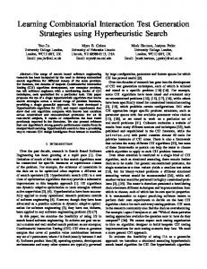
Fig. 1. The pCGMT9 vector construct and the process of scFv display on phage coat protein pIX.
To obtain a quality library, the individual steps were optimized to increase the antibody variable region gene diversity and the efficiency of scFv gene assembly and cloning. First, the primer design was optimized for amplification of variable region gene pools to maintain maximum diversity. Second, the efficiency of scFv gene assembly was increased by exploiting the presence of the DNA encoding the (G4S)3 peptide linker included at the end of the 3⬘-primer of the VH gene and 5⬘-primer of the VL gene. This design allowed us to assemble scFv genes from only two DNA fragments. Previously, scFv repertoires displayed by pIII were less efficiently assembled from three separate DNA fragments consisting of VH and VL gene repertoires and the linker DNA (28). Third, 25 extra nucleotides at the 5⬘-end of the SfiI cutting site were appended to ensure efficient digestion of amplified scFv gene fragments. The scFv library was constructed in two steps. First, the antibody variable region gene repertoires were amplified by PCR from the PBLs of 10 ‘‘normal’’ or asymptomatic human donors and a previously constructed scFv-phage library plasmid (28). To maximize diversity, VH, V, and V gene family specific primers were used individually for amplification. Then, scFv genes were assembled from VH, V, and V repertoires, digested with SfiI, and ligated into the phagemid vector pCGMT9 (Fig. 1). A human scFv library displayed on pIX and containing ⬇4.5 ⫻ 109 members was generated by only 20 electroporation procedures. To examine the integrity of the library, 40 clones were picked at random and all were found to contain scFv genes having the expected size. DNA sequencing of these clones revealed 40 unique sequences with a distribution in the length of the VH complementarity determining region 3 (CDR3) ranging from 5 to 19 residues. No obvious bias of V-gene usage was observed (data not shown). Panning and ELISA of scFv-Phage. The library was evaluated by affinity selection with six different purified protein antigens from various sources, including bovine, human, bacterial, and plant. In light of our interest in the generation of human 12614 兩 www.pnas.org兾cgi兾doi兾10.1073兾pnas.192467999
antibodies against potential biological warfare and bioterrorism agents (43), four of these proteins were toxins. SEB and CTB are bacterial enterotoxins (44–47), and RCA60 and RCA120 (‘‘ricin’’) are lectins from the castor-oil bean that act as potent inhibitors of protein biosynthesis (48–50). BSA was chosen as a well-known model antigen, and human TNF-␣ is a cytokine that has importance in cytotoxicity and as a proinflammatory substance (51– 53). Each round of panning selection comprised a cycle of scFv-phage binding to the immobilized antigen, washing away of unbound and nonspecifically bound scFv-phage, elution of the specifically bound scFv-phage, and propagation of enriched scFv-phage for entry into the next round of panning. By using this protocol, which is typical in our laboratory and that reported in the literature, we were gratified to find that the efficiency of enrichment of specific scFv-phage exceeded that which we generally observed by using the pIII-display format. Four rounds of panning with BSA and recombinant TNF-␣ were carried out for testing purposes, because this is our standard protocol, and excellent results were obtained. However, for the other proteins, panning was stopped after only three rounds with no decrease in performance. In all cases, we determined by ELISA that 90– 100% of the randomly selected and analyzed scFv-phage were positive in binding to their respective antigen (Table 1). From ⬇20 clones selected against each antigen, two to five distinct scFv sequences were found and each sequence showed strong binding by ELISA. Finally, testing of the anti-SEB scFvs against BSA, CTB, RCA60, and RCA120 indicated no detectable crossreactivity (data not shown). Fine Specificity of scFvs. As a stringent test of library diversity, phage-display characteristics, and panning performance, two protein antigens with a very high sequence homology were used in the selection process. RCA60 is a heterodimer (␣⬘) consisting of an A chain and B chain, and RCA120 is a tetramer (␣22) having two identical A chains and two identical B chains. Protein sequence alignments between RCA60 and RCA120 indicated a 93% and 83% sequence homology of the A chains and B chains, respectively. After three rounds of panning, single clones were randomly picked and the scFv-phages were rescued. From the RCA60 panning, 24 clones were represented by two different sequences, 23 of 24 clones were ranked as positive binders to RCA60 (one clone showed no detectable binding) by ELISA, and all 23 binders gave no detectable cross-reactivity with RCA120 or BSA. All clones obtained from the third round of panning against RCA120 were positive and showed no measurable crossreactivity with RCA60 or BSA (Table 1). Binding Affinity of scFvs. The Kd values were determined for some representative scFv- antigen pairings, focusing in particular on the toxin proteins. To obtain the Kd for each soluble scFv, the genes were subcloned into an expression vector (pETFlag) and purified from E. coli as described above. The kinetic parameters were measured by using BIAcore and the Kd calculated in each Gao et al.
SEB8 SEB10 SEB11 SEB13 SEB20 CTB12 RCA6028 RCA1208
kon, 105 M⫺1䡠s⫺1
koff, 10⫺3 s⫺1
Kd, 10⫺7 M
0.22 2.4 0.35 2.7 0.19 4.0 0.34 0.051
2.3 2.9 2.0 2.1 6.0 0.37 1.3 0.48
1.0 0.12 5.7 0.078 3.2 0.0093 0.43 0.94
Association (kon) and dissociation (koff) rate constants were measured for purified scFvs with BIAcore; Kd was calculated as (koff兾kon).
case which showed that several scFvs had high affinity (nanomolar) binding (Table 2). Discussion For more than a decade, phage-display technology has been an extremely practical way to isolate, engineer, and evolve combinatorial antibody libraries. In this regard, both pIII- and pVIIIdisplay formats have been the most instrumental in the construction of various libraries. Substantial information has been accumulated about the structural characteristics and the phagedisplay behavior of pIII and pVIII. However, a similar body of data concerning the proteins pVII and pIX remains to be acquired. Recently, we elucidated the orientation of pVII and pIX on the phage surface and, for the first time, showed that pVII and pIX could be used to individually display fusion proteins, notably the VH and VL of the antibody Fv fragment (33). Encouraged by these results, we explored the further application of pIX to display a library of human scFvs. In the library construction, individual steps were optimized to increase the efficiency of scFv gene assembly and cloning. A leader sequence was incorporated into the pIX-display vector that targeted the fusion proteins to the inner membrane and prevented accumulation in the cytoplasm. Directional cloning with a single, rarely occurring restriction site cut by the enzyme SfiI was also designed into the vector. The enzyme recognizes eight base pairs in sequences of 5⬘-GGCCNNNNNGGCC-3⬘ and cuts at the junctions of the degenerate region within the interrupted palindromic recognition site. Two different sticky ends allowed cloning of the scFv fragment in the correct orientation. Furthermore, the fact that SfiI always cuts two sites at once (54), and by ensuring efficient cutting by introducing 25 extra nucleotides at the 5⬘-end of the recognition site, facilitated construction of a large library containing ⬇4.5 ⫻ 109 members with minimal biases. In panning procedures against six different protein antigens, a ⬎104-fold enrichment in scFv-phage was obtained after only three rounds of panning, and more than 90% of the selected scFv-phages were positive binders. Moreover, specific, highaffinity scFvs were readily and consistently obtained without special panning protocols. For example, scFvs against SEB showed no cross-reactivity versus other antigens in the panel, and BIAcore analysis indicated a Kd ⫽ 7.8 nM for scFv SEB13, which was comparable to anti-SEB murine mAbs obtained from a secondary immune response requiring intrasplenic boosting (55). Also, a high-affinity scFv against CTB was obtained, with the Kd value the first reported to date, and the scFv affinity was estimated to exceed that of previously isolated murine mAbs from immunization (56, 57). The exquisite specificity of the scFvs between the highly homologous ricin agglutinins, RCA60 and RCA120, was also notable, and such scFvs should be useful for the detection and distinction of these toxins. The performance of the pIX format for scFv selection against protein targets was judged Gao et al.
as being broadly superior to the pIII-displayed scFv libraries used in our laboratory. The results from panning against hapten conjugates await to be determined. In general, pIX-display may overcome some disadvantages of the pIII-display format. It has been observed that scFv-phage fusions on pIII lost their viability dramatically during a commonly used high pH elution step during panning (10 mM triethylamine, 10 min), whereas wild-type helper phage were not effected (58). Similarly, other alternative phage recovery processes, such as low pH acid elution or DTT treatment, which also have no effect on wild-type phage, may have adverse effects on some fraction of the members of a particular library, a fact that cannot be predicted in advance. The N-terminal domain of pIII is required for phage infectivity by attachment to the tip of the F⬘ pilus of E. coli, whereas the C-terminal portion of pIII acts in combination with pVI to cap the trailing end of the filament during phage assembly, allowing its release from the cell (59). A potential advantage of pIXdisplay is that elution is not necessary to rescue phages that specifically bind to immobilized targets. Because the pIII proteins are freely exposed and unmodified, adding freshly prepared E. coli directly to the bound phage will produce infection. Hence, during selection, not only would one recover scFv-phage that might have lost infectivity in an elution step, but also other rare, very high-affinity binders that are difficult to obtain by most conventional elution procedures. Another important property of the N-terminal domain of pIII is its role in providing immunity to a cell already infected with a filamentous phage. This immunity function prevents superinfection by other phage (60). Most of the libraries displayed by using pIII are based on a phagemid that carries the pIII-fusion construct of interest and a helper phage that provides the proteins and enzymes required for phage packaging and replication. For phagemid systems using the entire pIII as the fusion partner, the phagemid-driven expression of the pIII-fusion protein must be shut down to allow superinfection by the helper phage, after which expression is induced to allow production and display of the fusion construct. However, it is very difficult to entirely shut down the background expression with the widely used lac promoter even with 2% glucose in the growth medium (61). The background leakage of the pIII-fusion protein before adding helper phage will cause some level of immunity for a fraction of the clonal population, and so one could expect a corresponding decrease in library diversity. To allow infection with helper phage, the N-terminal domain of the phagemid pIII-fusion construct may be deleted to avoid producing immunity to helper phage. Yet, both the intact and truncated pIII-display strategies are also subject to other possible disadvantages compared with pIX display that derive from the significant difference in the sizes of these coat proteins. The pIII itself is a large protein of 406 aa, and even the truncated C-terminal version is 150 aa in length (16). On the other hand, pIX is only a 32-aa peptide. In pIII-monodisplay formats, we suggest that displayed proteins are more sterically encumbered by neighboring wild-type pIII, as opposed to pIX displays, in which the protein encounters little steric interference from wild-type pIX. As a result, any pIXdisplayed fusion construct may be more accessible to all targets, and the differences could manifest significantly for some displays and some targets of interest. Regardless of which phage-display format is used, selection can suffer from the amplification of scFv-phage without specific binding activities, which increases the background and decreases enrichment efficiency. We suggest that the larger pIII proteins could play more of a role than pIX in this nonspecific binding through a greater likelihood of hydrophobic and兾or electrostatic interactions with the panned target antigen. Based on previous work from our laboratory with a truncated pIII-displayed scFv library, the background in the pIX system was at least five to 10-fold less by comparison of the PNAS 兩 October 1, 2002 兩 vol. 99 兩 no. 20 兩 12615
SCIENCES
scFv
APPLIED BIOLOGICAL
Table 2. Kinetic and binding parameters for scFvs
human scFv-antibody library derived from the PBLs of asymptomatic donors. Notably, we were able to efficiently construct the library and then readily select high affinity and exquisitely specific scFvs against six protein antigens of distinct sizes and structures, and from widely different sources. This pIXdisplayed human scFv-phage library may be a valuable source of human antibodies for numerous therapeutic and proteomic investigations.
output phage numbers from negative clones against the same antigen. The procurement of human antibodies specific for various endogenous factors and exogenous toxins is of particular interest for passive immunotherapy of disease and for protection. In addition, the progress in deciphering the human genome has given impetus to the application of engineered antibodies in proteomics. Thus, advances in antibody phage-display libraries will not only provide antibodies against newly discovered targets, but can help to define novel gene function and the measurement of abnormal protein expression in pathological states. Herein, we described a format using pIX for the display of a large, naı¨ve
We gratefully acknowledge the Skaggs Institute for Chemical Biology and support by the National Institutes of Health Program Project Grant P01CA27489, and a Louis R. Jabinson Fellowship (Louis R. Jabinson Investigatorship Fund for Graduate Education) (to C.G.).
1. Hoess, R. H. (2001) Chem. Rev. 101, 3205–3218. 2. Rodi, D. J. & Makowski, L. (1999) Curr. Opin. Biotechnol. 10, 87–93. 3. Vaughan, T. J., Osbourn, J. K. & Tempest, P. R. (1998) Nat. Biotechnol. 16, 535–539. 4. Griffiths, A. D. & Duncan, A. R. (1998) Curr. Opin. Biotechnol. 9, 102–108. 5. Zwick, M. B., Shen, J. & Scott, J. K. (1998) Curr. Opin. Biotechnol. 9, 427–436. 6. Dall’Acqua, W. & Carter, P. (1998) Curr. Opin. Struct. Biol. 8, 443–450. 7. Raag, R. & Whitlow, M. (1995) FASEB J. 9, 73–80. 8. Winter, G., Griffiths, A. D., Hawkins, R. E. & Hoogenboom, H. R. (1994) Annu. Rev. Immunol. 12, 433–455. 9. Marks, J. D., Hoogenboom, H. R., Griffiths, A. D. & Winter, G. (1992) J. Biol. Chem. 267, 16007–16010. 10. Barbas, C. F., Kang, A. S., Lerner, R. A. & Benkovic, S. J. (1991) Proc. Natl. Acad. Sci. USA 88, 7978–7982. 11. Kang, A. S., Barbas, C. F., Janda, K. D., Benkovic, S. J. & Lerner, R. A. (1991) Proc. Natl. Acad. Sci. USA 88, 4363–4366. 12. McCafferty, J., Griffiths, A. D., Winter, G. & Chiswell, D. J. (1990) Nature (London) 348, 552–554. 13. Huse, W. D., Sastry, L., Iverson, S. A., Kang, A. S., Alting-Mees, M., Burton, D. R., Benkovic, S. J. & Lerner, R. A. (1989) Science 246, 1273–1278. 14. Parmley, S. F. & Smith, G. P. (1988) Gene 73, 305–318. 15. Smith, G. P. (1985) Science 228, 1315–1317. 16. Webster, R. (2001) in Phage Display: A Laboratory Manual, eds. Barbas, C. F., Burton, D. R., Scott, J. K. & Silverman, G. J. (Cold Spring Harbor Lab. Press, Plainview, NY), pp. 1.1–1.37. 17. Berkowitz, S. A. & Day, L. A. (1976) J. Mol. Biol. 102, 531–547. 18. Lubkowski, J., Hennecke, F., Plu ¨ckthun, A. & Wlodawer, A. (1999) Structure (London) 7, 711–722. 19. Holliger, P., Riechmann, L. & Williams, R. L. (1999) J. Mol. Biol. 288, 649–657. 20. Holliger, P. & Riechmann, L. (1997) Structure (London) 5, 265–275. 21. Kishchenko, G., Batliwala, H. & Makowski, L. (1994) J. Mol. Biol. 241, 208–213. 22. Glucksman, M. J., Bhattacharjee, S. & Makowski, L. (1992) J. Mol. Biol. 226, 455–470. 23. Simons, G. F., Beintema, J., Duisterwinkel, F. J., Konings, R. N. & Schoenmakers, J. G. (1981) Prog. Clin. Biol. Res. 64, 401–411. 24. Houbiers, M. C., Spruijt, R. B., Wolfs, C. J. & Hemminga, M. A. (1999) Biochemistry 38, 1128–1135. 25. Cabilly, S. (1999) Mol. Biotechnol. 12, 143–148. 26. Hufton, S. E., Moerkerk, P. T., Meulemans, E. V., Bruı¨ne A. D., Arends, J.-W. & Hoogenboom, H. R. (1999) J. Immunol. Methods 231, 39–51. 27. Jespers, L. S., Messens, J. H., De Keyser, A., Eeckhout, D., Van Den Srande, I., Gansemans, Y. G., Lauwereys, M. J., Vlasuk, G. P. & Stanssens, P. E. (1995) Bio兾Technology 13, 378–382. 28. Gao, C., Bru ¨mmer, O., Mao, S. & Janda, K. D. (1999) J. Am. Chem. Soc. 121, 6517–6518. 29. de Haard, H. J., van Neer, N., Reurs, A., Hufton, S. E., Roovers, R. C., Henderikx, P., de Bruine, A. P., Arends, J. W. & Hoogenboom, H. R. (1999) J. Biol. Chem. 274, 18218–18230. 30. Sheets, M. D., Amersdorfer, P., Finnern, R., Sargent, P., Lindqvist, E., Schier, R., Hemingsen, G., Wong, C., Gerhart, J. C. & Marks, J. D. (1998) Proc. Natl. Acad. Sci. USA 95, 6157–6162. 31. Vaughan, T. J., Williams, A. J., Pritchard, K, Osbourn, J. K., Pope, A. R., Earnshaw, J. C., McCafferty, J., Hodits, R. A., Wilton, J. & Johnson, K. S. (1996) Nat. Biotechnol. 14, 309–314.
32. Griffiths, A. D., Williams, S. C., Hartley, O., Tomlinson, I. M., Waterhouse, P., Crosby, W. L., Kontermann, R. E., Jones, P. T., Low, N. M., Allison, T., et al.(1994) EMBO J. 13, 3245–3260. 33. Gao, C., Mao, S., Lo, C.-H. L., Wirsching, P., Lerner, R. A. & Janda, K. D. (1999) Proc. Natl. Acad. Sci. USA 96, 6025–6230. 34. Gao, C., Lin, C. H., Lo, C.-H. L., Mao, S., Wirsching, P., Lerner, R. A. & Janda, K. D. (1997) Proc. Natl. Acad. Sci. USA 94, 11777–11782. 35. Haidaris, C. G., Malone, J., Sherrill, L. A., Bliss, J. M., Gaspari, A. A., Insel, R. A. & Sullivan, M. A. (1999) J. Immunol. Methods 257, 185–202. 36. Welschof, M., Terness, P., Kolbinger, F., Zewe, M., Du ¨bel, S., Do ¨rsam, H., Hain, C., Finger, M., Jung, M., Moldenhauer, G., et al. (1995) J. Immunol. Methods 179, 203–214. 37. Marks, J. D., Tristem, M., Karpas, A. & Winter, G. (1991) Eur. J. Immunol. 21, 985–991. 38. Mao, S., Gao, C., Lo, C.-H. L., Wirsching, P., Wong, C.-H. & Janda, K. D. (1999) Proc. Natl. Acad. Sci. USA 96, 6953–6958. 39. Medaglia, M. V. & Fisher, R. J. (2002) Protein–Protein Interact. 255–272. 40. Roder, U. W. & Markey, F. (1998) Methods Mol. Med. 13, 531–554. 41. Malmborg, A.-C., Michae¨lsson, A., Ohlin, M., Jansson, B. & Borrebaeck, C. A. K. (1992) Scand. J. Immunol. 35, 643–650. 42. Jo ¨nsson, U., Fa¨gerstam, L., Ivarsson, B., Johnsson, B., Karlsson, R., Lundh, K., Lo ¨fås, S., Persson, B., Roos, H., Ro ¨nnberg, I., et al. (1991) BioTechniques 11, 620–627. 43. Zhou, B., Wirsching, P. & Janda, K. D. (2002) Proc. Natl. Acad. Sci. USA. 99, 5241–5246. 44. Jett, M., Ionin, B., Das, R. & Neill, R. (2002) Mol. Med. Microbiol. 2, 1089–1116. 45. Lencer, W. I., Hirst, T. R. & Holmes, R. K. (1999) Biochim. Biophys. Acta 1450, 177–190. 46. Spangler, B. D. (1992) Microbiol. Rev. 56, 622–647. 47. Gemmell, C. G. (1984) J. Med. Microbiol. 17, 217–235. 48. Olsnes, S. & Kozlov, J. V. (2001) Toxicon 39, 1723–1728. 49. Wellner, R. B., Hewetson, J. F. & Poli, M. A. (1995) J. Toxicol. Toxin Rev. 14, 483–522. 50. Nicolson, G. L., Blaustein, J. & Etzler, M. E. (1974) Biochemistry 13, 196–204. 51. Leong, K. G. & Karsan, A. (2000) Histol. Histopathol. 15, 1303–1325. 52. Luster, M. I., Simeonova, P. P., Gallucci, R. & Matheson, J. (1999) Crit. Rev. Toxicol. 29, 491–511. 53. Ruddle, N. H. (1992) Curr. Opin. Immunol. 4, 327–332. 54. Wentzell, L. M., Nobbs, T. J. & Halford, S. E. (1995) J. Mol. Biol. 248, 581–595. 55. Lapeyre, C., Kaveri, S. V., Janin, F. & Strosberg, A. D. (1987) Mol. Immunol. 24, 1243–1254. 56. Koike, N., Yabushita, Y., Yan, Z. D., Lida, T. & Honda, T. (1999) Microbiol. Immunol. 43, 1057–1060. 57. Honda, T., Ni, Y., Yoh, M. & Miwatani, T. (1989) Med. Microbiol. Immunol. 178, 245–253. 58. Johnsson, K. & Ge, L. (1999) Curr. Top. Microbiol. Immunol. 243, 87–105. 59. Rakonjac, J. & Model, P. (1998) J. Mol. Biol. 282, 25–41. 60. Scott, J. K. & Barbas, C. F. (2001) in Phage Display: A Laboratory Manual, eds. Barbas, C. F., Burton, D. R., Scott, J. K. & Silverman, G. J. (Cold Spring Harbor Lab. Press, Plainview, NY), pp. 2.1–2.19. 61. Krebber, A., Burmester, J. & Plu ¨ckthun, A. (1996) Gene 178, 71–74.
12616 兩 www.pnas.org兾cgi兾doi兾10.1073兾pnas.192467999
Gao et al.