Multimed Tools Appl (2009) 42:317–340 DOI 10.1007/s11042-008-0252-x
A mobile device application applied to low back disorders Juan C. Guerri & Ana Belén Antón & Ana Pajares & Manuel Monfort & Daniel Sánchez
Published online: 13 November 2008 # Springer Science + Business Media, LLC 2008
Abstract Many muscular function assessments require the monitoring of muscular activity in different environments. This can happen in the gymnasium during a training session or while practising a sport; at work, where movements and specific actions are executed; at school, where the child adopts an incorrect posture, or even while we walk. In this paper, we propose a system to allow the assessment of the muscular condition in any of these environments in a comfortable and simple way for the patient and using the advances in wireless communications. Just a wireless biomonitor (we have used the ME6000 biomonitor, a medical portable equipment of very small dimensions) and one PDA or mobile phone with wireless interface are needed. The medical device is configured from the mobile device. Then, wireless communications are used to transmit online the electromyographic signals registered by the medical equipment to a mobile device. The specific protocol used by the medical device is implemented to carry out the communication. There are two ways to configure the system: offline and online. In offline mode, once all the information is received, it is sent to a server using a connection to the Internet instead of the online way, where this information is sent simultaneously. The server includes a J. C. Guerri (*) : A. B. Antón : A. Pajares Institute of Telecommunications and Multimedia Applications (iTEAM), Universidad Politécnica de Valencia, Camino de Vera, 46071 Valencia, Spain e-mail:
[email protected] A. B. Antón e-mail:
[email protected] A. Pajares e-mail:
[email protected] M. Monfort Departamento de Didáctica de la Expresión Corporal, Unidad docente de Educación Física, Universidad de Valencia, Alcalde Reig 8, Valencia, Spain e-mail:
[email protected] D. Sánchez Departamento de Anatomía y Embriología Humana, Universidad de Valencia, Blasco Ibáñez 15, 46010 Valencia, Spain e-mail:
[email protected]
318
Multimed Tools Appl (2009) 42:317–340
background application to process the information in real-time with the aim to evaluate the function of the muscle during the exercise and to establish healthy behaviours for the patient. Then, this information can be consulted by the specialist and also by the client using the PDA or mobile phone to show the report. A prototype of this system has been developed and implemented. The system has been evaluated by a preliminary usability, reliability, feasibility and communication performance study. Keywords Wireless telemedicine . Handheld devices . Multimedia . Health monitoring . Electromyography . Usability
1 Introduction Low back disorders are a major health problem in western countries. It is estimated that 58–84% of the population will suffer back pain at some point of their lives. An annual incidence of 28 low back pain episodes per 1,000 people has been reported, with the highest incidence seen in those aged 25–64 (this is to say, in people of working age) [1]. That is the reason why these disorders have an enormous socioeconomic impact in industrialized countries. In order to deal with this major issue, health systems are actively developing new technologies such as telemedicine. The main goal of telemedicine is to “contribute to the improvement of the quality of health care through the integration of medical information and new technologies”. A generic telemedicine system is formed by three components: 1) the infrastructure of telemedicine, which consists on the equipments and processes to acquire and to present the clinical information; 2) the infrastructure of telecommunications, which provides the resources to transfer information using voice, video and data technologies; and 3) the applications of telemedicine: a collection of processes and interfaces for the benefit of all the actors who take part in the sanitary system [2]. On the other hand, wireless technologies are widely used nowadays, even for telemedicine applications. Concretely, the evolution of 3G wireless communications (UMTS) will be a very important aspect for future developments in telemedicine. Also, in recent years other wireless technologies such as WLAN (WiFI) and WPAN (Bluetooth, Ultrawideband) have been implemented as an alternative to make the communications more flexible and powerful. In this paper, we have tried to take advantage of wireless technology in benefit of health care through the use of electromyography techniques. Electromyography is the study of muscle function through the recording of the electrical activity associated with functioning muscles [3]. It describes and evaluates the neuromuscular response of the muscular contraction in subjects with and without pathologies of the musculoskeletal system from the bioelectrical information recorded during specific activities. Specifically, surface electromyography (sEMG) is a non-invasive kinesiology technique (surface electrodes are mounted directly on the skin) used for studying body movements [4]. In most cases patients have to obtain signals data at regular intervals or several times a day. In these situations, they have to visit the health care centre or take the signals at home using complex and expensive devices. The aim of the system proposed in this paper is to avoid such displacement using wireless technologies, to widen the usability of the telemedicine applications and to generate automatic feedback reports. With this objective, the data captured by the electromyograph are sent online and using wireless technology to a handheld device (PDA or mobile phone). Later, the data are relayed to a server placed in the sanitary centre accessible by Internet. A background application processes the information
Multimed Tools Appl (2009) 42:317–340
319
in real-time and it generates a feedback report which can be consulted and modified by the specialist and downloaded by the client using the PDA or mobile phone to visualize it. The rest of this paper is organized as follows. In section 2 the theoretical background of this work is shown. Section 3 describes the complete system and its elements. In section 4, the system implementation is explained. In this section, an example of the results and the expert system are presented. Section 5 shows the usability feasibility, reliability and communication performance of the system, Finally, in section 6, the different conclusions which can be extracted and future lines of work are established.
2 Background There have been important advances in wireless communications and network technologies in the last decades. These advances have had a significant impact on m-Health (Mobile Health) systems, defined as “mobile computing, medical sensor and communications technologies for health care” [5]. This impact is mainly due to an important growth in the use of handheld devices (PDAs and mobile phones). On the other hand, the use of wireless technologies represents a natural evolution of traditional telemedical systems. Among wireless medical applications, those related to home monitoring [6–8] and computer-assisted rehabilitation and therapy [9] should be highlighted. Both involve the monitoring of different parameters like heart rate, blood pressure or movement (for example, fall detection) [10]. There are several previous works which use PDAs for different aims: to have access to the patient’s information; data acquisition, processing and communication [11–15]; a proxy for medical devices [16]; data integration from multiple sources [17]; and a useful tool in high mobility environments [18, 19]. The most important benefits of using handheld devices are the possibility of having access to information from anywhere, sending immediate clinical orders to patients and being able to consult medical experts at any moment. Part of the content of the paper is focused on the aspects related to problems in the integration and implementation of mobile and medical devices and also mobile networks. In our opinion, the most interesting and motivating point of the paper is, in fact, demonstrating that the objective of m-Health can be achieved; being this objective “to utilize health care application written on mobile devices that are connected through the wireless networking and communication technology to improve healthcare safety and outcomes, whereas reducing costs”. Most of the studies related to m-health consider the following challenges:
&
& &
Healhcare professionals: most of healcare professionals haven’t experienced the new technologies and are even reluctant to their use if that usage implies a hard and long learning process. In this context, aspects such as user-friendliness are essential. Lightness and functionality in application design, even at the cost of realizing less attractive user interfaces. Integration into clinician’s workflow and work methodology: the success of the applications based on new technologies depends on their integration in the proceses and methodology used by health professionals. Technology: the evolution of communication networks makes us think about the need of developing applications which allow the use of different technologies: internet, WiFi, Bluetooth and cellular networks (2.5G, 3G and future 4G) and evaluating their performance to support the m-health services. The development of
320
&
Multimed Tools Appl (2009) 42:317–340
the applications should also allow the user to select among the different connection alternatives available. Mobile devices: nowadays there is a great variety of mobile devieces available in the market (PDAs, smart phones, tablet PCs, laptops, etc.). Different studies consider PDAs and mobile phones as medical specialists favourite mobile devices. As computing standards and data format standards for m-health do not exist, the interoperability between systems is even more complicated.
Our previous works [20–22] were focused on using local area networks and the Internet as communication network. The present work also represents an evolution towards using wireless technologies to improve the quality of health care. However, there are some differences between our work and other authors’ works mentioned before:
&
&
&
&
&
Handheld devices: the developed software can be used both in PDAs and mobile phones. The possibility of using it in a mobile phone has greatly increased its usability. The system has been developed taking into account aspects as usability, reliability and feasibility. Open interface: most health care systems have their own medical devices. Most previous works have not taken into account the fact that these devices use their own proprietary configuration protocols. Our system has been developed to use commercially available hardware and software components. We have designed a scalable and modular software which allows us to include new technologies as they appear in the market. Configuration: in the proposed system, the handheld device (PDA or mobile phone) is not only used as a data acquisition and communication platform. The handheld device is also used as a configuration interface of the medical device. Through a user friendly graphical interface and an emulation of the proprietary configuration protocol, it provides a straightforward interface for users, regardless of their degree of technical training. Gateway: in our system, the handheld device can act as a gateway between multiple devices (EMG, ECG, video, audio, etc….) and the Internet. The fact that medical devices do not need to have a direct access to the Internet has some advantages. For example, medical devices just need to implement wireless technologies with short ranges, saving power and costs. In addition, medical devices are manufactured by different vendors and use incompatible configuration protocols, but our system can translate proprietary protocols and integrate data from heterogeneous sources. OffLine and OnLine mode: the system can be configured in either offline or online way. If the online mode is chosen, the measurements are transmitted in real time. Therefore a permanent internet connection is necessary for the whole exercise length. However the offline mode allows the movement assessment regardless of whether the user has or not a permanent connectivity. With the aim of checking that 3G network is suitable to support our m-health service we analyse the impact of the wireless network in the communication between the mobile device and the server.
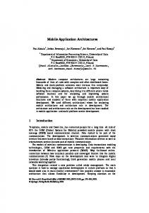
3 Description of the system The generic structure of the proposed system is shown in Fig. 1. The part of the user (patient) and the part of the server and specialist (sanitary professional, trainer, etc) are
Multimed Tools Appl (2009) 42:317–340
321
SERVER
AdHoc Medical Network
WI-FI or UMTS
Internet
WI-FI MEDICAL DEVICE
MOBILE DEVICE USER
SPECIALIST
Fig. 1 This is the structure of the complete system. Two parts are distinguished: user or patient and specialist or sanitary professional. Wi-Fi is the technology used for the connection between the medical and mobile devices. Wi-Fi or UMTS access is used for the connection between mobile device and server
distinguished. The user must have a mobile device with wireless capability WLAN Wi-Fi 802.11b or UMTS interface, and an electromyograph, which registers the electrical signals (EMG). Both devices (medical and mobile) communicate across a wireless net that has been named ME6000 Network, using Wi-Fi technology. The specialist must have access to the server where the web page, which will be used for the interchange of information between patient and sanitary professional, is located. The medical device used is the ME6000 Biomonitor System (Mega Electronics Ltd. www.meltd.fi) (Fig. 2). It is an 8-channel electromyograph which carries out surface measurements of electrical activity on 8 muscles simultaneously. It needs a precise amplification because the electrical impulses caused by the muscular fibers are very small, with minimal amplitudes of approximately 1 μV. This electromyograph can be used both in laboratory and field tests because it is small, light and portable (it only weighs 344 g approximately). Besides, it allows the use of other types of sensors, both physical and environmental (goniometers, meters of cardiac frequency, etc.). It has trigger in/out connections which facilitate a simple synchronization with other medical equipments. It allows online records using USB cable or telemetry to a PC with a proprietary application and offline without a computer, using a compact flash memory card (256 MB extendable). The electromyograph uses an adapter PCMCIA Cardbus D-Link AirPlus to 2.4 GHz. It establishes one WLAN interface using IEEE 802.11b technology. Using the system proposed in this paper we can remote monitoring the muscular activity during the patient’s daily life. This remote monitoring of patients can reduce the expensive health care costs and increase the quality of patient’s life. The mobile device can be a PDA or a mobile phone. We have worked with both of them, a PDA Hewlett Packard IPAQ HX4700 Pocket PC and a mobile phone Nokia E61. It
322
Multimed Tools Appl (2009) 42:317–340
Fig. 2 ME6000 Biomonitor
requires two necessary features: WLAN WI-FI 802.11b wireless standard (or UMTS interface) and JAVA MIDP 2.0 profile with CLDC 1.1 configuration [23]. The connection between the electromyograph and the mobile device is a connection in ad-hoc mode [24]. While traditional networks have fixed nodes with wired connections (either optical fibers or copper lines), ad-hoc wireless networks can, in general, be described as multi-hop wireless networks with mobile nodes. However, the mobility condition can be relaxed and an ad-hoc wireless network can be identified as a network where all the nodes are connected through wireless links and where there is no central or dominant node (as opposed to the case of cellular wireless networks, for example, where a base station (BS) exists in each cell). On the other hand, in medical scenario it is very important to prevent non-authorized access to patients’ data. It is particularly easy to obtain this information in wireless applications where data are transmitted via radio interface, using different kinds of software, such as a sniffer. The 802.11b WLAN standard incorporates WEP as mechanism to provide security. WEP is based on encryption and authentication. A longer key length makes more difficult for hackers to access the data: therefore, an extended 128-bit WEP protocol with a 104-bit key size is used. When the application developed for the mobile device runs and its profile and configuration have been installed, the .jar and .jad files have to be downloaded. A Java Application Descriptor (JAD) file contains a predefined set of attributes which allow application management software to identify, retrieve, and install the MIDlets. A MIDlet is an application written for a Mobile Information Device (MID) Profile, which requires a device that implements Java ME to run. All attributes appearing in the JAD file are made available to the MIDlets. A Java Archive (JAR) file contains: Java classes for each MIDlet in the suite (a group of MIDlets that can share resources at runtime), Java classes shared between MIDlets, resource files used by the MIDlets (for example, image files) and a manifest file describing the JAR contents and specifying attributes used by application management software to identify and install the MIDlet suite.
Multimed Tools Appl (2009) 42:317–340
323
A computer system (server) operates continuously on the network and awaits requests for services from other devices on the network (in this case an application running in a PDA or mobile phone). The continuously running application which receives the medical data is in charge of the data processing and its storage. The server is also used to host the web site. The web site allows the user to know how to use the Biomonitor System and to locate the electrodes, among other information. It also allows the specialist to access the medical data sent by the mobile device and the patient to access the feedback report generated by the system.
4 Implementation of the system Two main applications have been developed using Java: one to be running in the server and another to be executed in the mobile device. Object-oriented features have been used in the programming process. Special mention has to be made to the use of Java 2 Micro Edition (J2ME) in the application developed for the mobile device (PDA or mobile phone). J2ME is a specification of the Java platform oriented to the programming of applications (MIDlets) for electronic devices with very small computational and graphic capacities, like cell phones, PDAs or intelligent electric appliances. This edition uses a virtual machine called KVM (Kilo Virtual Machine) given that it needs only a few kilobytes of memory to work, instead of the use of the classic JVM (Java Virtual Machine). Besides these two main applications, other developed programs provide the necessary resources to carry out the proposed system of telemedicine. Figure 3 and Table 1 describe the sequence of actions to be carried out by the participant actors in the proposed system and the function of the developed applications. The full execution of the system can be divided into three big phases (Table 1) explained in the next sections.
Action 3. Medical deviceremote configuration and capture Action 2. Download work plan
Action 5. Real-time analysis and feedback reports generation
Server Medical device
PDA or mobile phone
Action 4. Send data (offline or online)
Action 6. Download report
Action 7. Download captured data and report
Specialist Fig. 3 Overview of the system components and actions
Action 1. WorkPlan (text, videos, images, etc.)
4. To send the data (EMG measurements) to the server.
User: patients
PHASE 3
PHASE 2
5. To generate the feedback reports in real-time.
6. To download the reports.
7. To download the recorded data.
Server: Expert System
User: patients
Specialist: trainer, health worker
User: patients
User: patients
1. To generate the work plan and the auxiliary information about the execution of the movement to evaluate. 2. To look up the proposed plan and the instructive information. 3. To turn on the electromyograph and to place the surface electrodes. To execute the application in the mobile device (PDA or mobile phone) and to begin with the activity to assess.
Specialist: trainer, health worker
PHASE 1
ACTION
ACTOR
?
The reports are accessed by the user via the web page. Previous authentication is necessary. The information sent by the mobile device and the reports generated by the expert system (stored in the server) are accessed via the web page. Previous authentication is necessary.
The mobile device sends the parameters of the measurement protocol, selected by the user, to configure the ME6000. The EMG signals are captured by the electromyograph while the user performs the exercise. They are sent online to the mobile device which stores them or sends them online to the server. The information stored (in offline mode) in the mobile device is translated into EMG values. They are sent via Internet to the server.
The web page is accessed via the mobile device.
Text, videos and images are created and stored in the server.
HOW?
Table 1 This table enumerates the different actors, phases, actions and applications which are in the use of the proposed system
The server receives the medical data. An ASCII file is created with a specific format. The server includes an expert system that processes de EMG raw data (wavelet filter, EMG percentages, parameters) and generate reports for the user.
324 Multimed Tools Appl (2009) 42:317–340
Multimed Tools Appl (2009) 42:317–340
325
4.1 The first phase: groundwork of the exercise A web site has been developed to include the information that the professional considers necessary about the correct execution of a specific movement to evaluate or about the right collocation of the electrodes on the skin. The information can be presented as a text, as images or as demonstrative videos. They can be displayed in a PC screen or in screens with graphical limitations, such as a PDA or mobile phone screen, given that the web site has been designed to be accessed by handheld devices. Figure 4 shows an example of this kind of information. 4.2 The second phase: execution of the muscular movement and acquisition of EMG values The application to run in the mobile device (named EMG-MIDlet), developed in J2ME, provides the user with four possible actions. The first action (OnLineCommunication or OffLineCommunication, in Fig. 5) is to select the communication mode. With the second one (Start, in Fig. 5), the user begins the collection of EMG measurements once the connection has been successful, while the user is executing the proposed movement. This information is sent online by the electromyograph to the mobile device, which either stores the raw data using the RMS (Record Management System) [25] (in OffLine mode) or it sends it directly to the server (in OnLine mode). The application allows selecting between three different protocols whose parameters of configuration have been defined and stored previously. The most important parameters are the duration of the exercise and the sampling frequency. The number of measurements is
Fig. 4 Views of the different formats of information which are present in the web page (text, image or video)
326
Multimed Tools Appl (2009) 42:317–340
OnLine Communication OffLine Communication Exit Fig. 5 Screenshot with the main menu of the mobile device application
calculated with both parameters. So, when all values have been received, the mobile device sends a message of end of connection. Then, the electromyograph stops the measurement and sends its last message. Therefore, the communication between the medical and mobile devices comprises five specific steps: Connection, to establish the wireless connection; Medical Device Configuration, to define the kind of signal, number of active channels, etc; Registered Measurements, to send the packets with the EMG values; End, to mark the end of the recording; and Disconnection, to finish the wireless connection (Fig. 6). The wireless connection established between the medical and mobile devices works through an ad-hoc network. The third action (Update, in Fig. 5) is the transmission of the information stored in the record stores (in OfflLine mode) to the server, using a simple wireless connection by Internet. Among the information sent are the username of the patient, the patient profile and the protocol name. These data are necessary for the storage and creation of the file with the results and feedback report. MOBILE DEV.
MEDICAL DEV.
Wireless Conn
Data Res (seq = x )
…
Wireless Conn
Data Res (seq= x + N)
Conf Req 1 Conf Res 1
End Req End
Conf Req 2
End Res
Conf Res n
Disconnection
Conf Res 2
…
Medical Dev. Configuration
MOBILE DEV.
Data Req (seq= x) Registered Measurements
Connection
MEDICAL DEV.
Fig. 6 Communication between the medical and mobile devices
Wireless Disconn Wireless Disconn
Multimed Tools Appl (2009) 42:317–340
327
Finally, the fourth action (Exit, in Fig. 5) allows aborting or exiting the application when the measurement is finished; therefore, the MIDlet which was being executed in the mobile device is destroyed. Figure 7 describes the main processes of the mobile device application developed. Since two alternatives are provided to the user, OnLine and OffLine communication with the server, two main processes are described. They are very similar regarding the process definition; the main difference is the use of the Record Management System (RMS) which provides a mechanism through which MIDlets can persistently store data and retrieve it later. In the Offline communication strategy three processes are available. When communication is established between electromyograph and mobile device the StartMeasurements process is executed. UDP datagram connections are opened and the sender is created with the data necessary for configuring the electromyograph. Conf_Req provides the information necessary for this configuration according to the protocol chosen. After the configuration process, the sender provides Data_Req which are the ACKs (see Fig. 7) for the maintenance of the communication. In fact, the sender message is the datagram which is sent in the communication process. Each time information is received from the medical device, the receiver stores this information using the RMS. This record-oriented approach comprises multiple record stores. Each record store can be seen as a collection of records and each record is an array of bytes and it has a unique identifier. In updating measurements stage a socket connection is opened and an output stream is used for writing the measurements in the file stored in the server. The bytes stored in the RMS are read in order to define the filename, which consists of the username and also some configuration data such as the protocol name and the date, and to transform the measurements in EMG values. Finally, the process to destroy the application is briefly remarked. In the Online communication strategy the processes are the same, however the RMS is not used and the measurements are sent while they are being captured and transformed to human readable EMG values. UDP Datagram Connection is used for the communication with the medical device, too, and a socket connection is opened for sending the user information and the measurements registered to the socket in the server side. In this description, the server application processes have also been added in order to give an overview of the online EMG_MIDlet
EMG_OffLine_MIDlet START START
UPDATE UPDATE
EXIT EXIT
UpdateMeasurements(this, protocol, username);
StartMeasurements(this, protocol); StartMeasurements
EXIT EXIT
destroyApp(true); notifyDestroyed(); ReceiveMeasurements
UpdateMeasurements
UDPDatagramConnection udc
Application Server
EMG_OnLine_MIDlet START
SocketConnection
SocketConnection Sender(udc) Sender
sender Configuration ACKs
send(addr,conf)
Sender(dc) send(addr,msg)
send(addr,ack)
udc.newdatagram() receiver
OutputStream writeName()
readName()
Filename
RMS Measurements
writeRegister()
InputStream receiveUserInfo()
sendUserInfo()
UserInfo
Filename Measurements
readRegister()
Internet sendMeasurements()
Conf_Req Conf_Req(protocol) getData (id) getDuration() getNChannel() getSamplingFreq()
Fig. 7 Process diagram of the EMG_MIDlet
receiveMeasurements() EMG_values
EMG_values
Data_Req Data_Req() getData (id)
Measurements
OnLine Mode OffLine Mode Common actions
328
Multimed Tools Appl (2009) 42:317–340
communication behaviour. Note that the server application is the same in both approaches. This is very simple; it consists of an opening socket connection and an input stream for receiving the information which is written in an ASCII document (Fig. 8) taking into account the specific configuration information (Conf_Req). This allows establishing configuration information such as the sampling rate, the name of the monitored muscles, the date of the measurement, etc. The remaining data are the recorded EMG values. The name (or username) of the patient is sent too, because the measurements are stored according to this identification. 4.3 The third phase: data processing and evaluation of results This section presents an example of the results obtained during a programmed session, and how these data were analysed. Two young male adults participated. Both were physically fit, with similar weight, height and age. The first subject (control) had never suffered from low back pain or any other kind of low back disorders. The second subject (patient) showed repeated episodes of pain on the lumbar region during the last month, with time off work due to low back pain, and a diagnosis of L4-L5 lumbar disc herniation given by a medical specialist (clinical history, magnetic resonance evidence and radiculopathy symptoms confirmed by physical exploration) [26]. Written consent to participate in the investigation was obtained from the subjects after they had been informed about the study. All the procedures were conducted in accordance with the principles of the World Medical Association’s Declaration of Helsinki. The chosen protocol was the one called “Flexion-Extension Slow”. The instructions for the correct execution of this exercise included a demonstrative video. The user looked up in the web page, by means of their PDAs or mobile phone, how to place the electrodes. They were attached bilaterally on the upper and lower rectus abdominis, erector spinae, and biceps femoris muscles. Fig. 8 Example of ASCII file format. It is a format compatible with EMG software. Information about configuration parameters or name of muscles is distinguished. The EMG values are distributed in columns according to the channel which they belong to
MegaWin ASCII file [DEFINITIONS] Version=1 ChannelCount=4 DataType=1 SamplingFreq=1000.000000 FirstSampleTime=0.001000 PhaseName=Raw Free 02 Nov 2006 12:18:19 [COMMENT] [SOURCE NAMES] Upper Erector spinae muscle Lower Erector Spinae Muscle Upper Erector spinae muscle Lower Erector Spinae Muscle [SIDE INFO] R R L L
[UNITS] uV uV uV uV [DATA] 16 -1 -3 -5 12 0 -2 2 -5 2 -2 3 -31 10 -3 0 -35 13 -3 -3 -23 12 -3 0 -17 16 -3 2 ............... -14 -6 0 3 -12 -25 -1 3 -10 -22 -2 3 -14 -14 -2 0 -18 -18 -2 2 -4 -16 -2 0 6 -11 -2 0
Multimed Tools Appl (2009) 42:317–340
329
Five repetitions of the trunk flexion-extension cycle were performed. Both trunk flexion and extension took 4 s each, resting for a second at maximum flexion. Each cycle was distributed as follows: seconds 1-2, neck and trunk flexion, until the hands reach the knees; seconds 3-4: further trunk flexion, until second 5, when the subject kept a position of maximum non-forced flexion. Extension was performed inversely during seconds 6 to 9. The full duration of the exercise was 55 s, given that every protocol includes three seconds of relaxation in the standing position at the beginning and at the end of the protocol and one second between each cycle. Given that the sampling rate is 1000 Hz and 8 measurement channels are used, the application expected to receive 440,000 samples (8 channels x 1000 samples/s x 55 s). The medical data were sent to the server using the developed application and when all such values were received, the application sent a last message ending the communication. The expert system (Fig. 9) analyses the received data and provides real-time feedback information. The input information are the raw EMG data and the items of the patient profile (age, sex, height, weight, clinical diagnosis, results of auto-applied disability questionnaires).
RAW EMG DATA
PATIENT PROFILE
WAVELET FILTER
70,00
FLEXION
PAUSE
EXTENSION
60,00
% EMG
50,00
AVERAGE EMG PERCENTAGES
40,00
30,00
20,00
QUERY (profile, thresholds)
10,00
Erector Spinae 0,00 1
2
3
4
5
6
7
8
9
10
11
12
13
14
15
16
17
18
Time Periods
EMG Parameter1: ES RELAXATION DURATION
EMG Parameter2: FLEXION-EXTENSION INDEX
EMG Parameter3: RELAXATION INDEX
BBDD _ or >
>
(thresholds)
FEEDBACK RESULTS (xml)
Fig. 9 Expert system diagram
330
Multimed Tools Appl (2009) 42:317–340
The first step of the process is the elimination from the captured signal of interferences due to movement artefacts, by means of Wavelet filters [27]. The automatic filtering of interferences allows us to avoid subsequent acquisitions. This would be particularly useful when the aim of the tests, for example, is the analysis of muscle fatigue: fatigue analysis requires a long recovery period between recordings, which makes difficult to repeat the testing if the signal cannot be used due to movement interferences. In addition, the possibility of a digital processing of the signal and a server-based interference removal process could allow relaxing the specifications of the acquisition devices and the use of lower cost equipment. The second step is the calculation of the average EMG percentages in the five trunk flexion-extension cycles. To this end, the raw EMG data were full-wave rectified, averaged over 500 samples (Root Mean Square) and normalized as a percentage of their maximum value to make the interpretation and evaluation of the results easier. The duration of each cycle of movement was divided into 18 equal time intervals. Then, the mean of the five repetitions was calculated, getting only one EMG pattern of trunk flexion-extension movement (Fig. 10). The results showed that the muscles stimulation of both sides is coordinated in time and amplitude. Accordingly, the graphs show only the EMG of one side and the upper rectus abdominis. Figure 10 shows that the first activation peak of the erector spinae starts with the beginning of the flexion movement and lasts until the second half of flexion time, when an abrupt decrease of the electrical activity takes place. Muscle activity shows values below the initial baseline (myolectrical silence) until the beginning of extension time, when the
70,00
FLEXION
PAUSE
EXTENSION
60,00
% EMG
50,00 40,00 30,00 20,00 10,00 0,00 0, 1
2
3
4
5
6
7
8
9
10 1
11
12
13
14
15 1
Ti Time Periods Erector Spinae
Rectus Abdominis
Biceps Femoris
Fig. 10 Average EMG percentages in the five trunk flexion-extension cycles
16
17 1
18
Multimed Tools Appl (2009) 42:317–340
331
second activation peak starts, and lasts until the end of extension. This is the normal erector spinae activation pattern in pain-free subjects [28, 29]. Moreover, the subject shows a very good pattern of abdominal muscles activation in order to stabilize the trunk in the periods with absence of activation of trunk posterior muscles. Such pattern is shown by the increase of the activity in the rectus abdominis during the period of erector spinae myolectric silence. Figure 11 shows the patterns of activity of the right erector spinae muscles of both subjects (pain-free and low back pain). In the patient, erector spinae muscles kept a high level of activation during the whole flexion—extension cycle: this is to say, in the subject with low back pain myolectrical silence of the erector spinae was absent. This is a wellknown sign of low back pain [28, 29], which is clearly shown by our application. Using these data, several useful EMG parameters could be easily calculated in real-time: 1) Duration of ES relaxation. The start of ES relaxation would be considered to occur at the first time period during flexion at which electrical activity is lower than the baseline value plus two standard deviations. The interval after the start of ES relaxation and before the maximum EMG activity peak during extension, at which activity reaches a value higher than the baseline value plus two standard deviations, would be considered the ‘end’ of ES relaxation. The baseline EMG activity value would be the average of the five intervals of lowest activity in the relaxation phase. The level of ES EMG activity during myolectrical silence has been found to offer a promising method for individualizing rehabilitation treatment and assessing impairment rating validity. 2) EMG activity ratios. In order to quantify muscle activity during flexion and extension, it is used a flexion-extension index, a
FLEXION
80,00
PAUSE
EXTENSION
70,00
% EMG
60,00 50,00 40,00 30,00 20,00 10,00 0,00 0, 1
2
3
4
5
6
7
8
9
10
11
12
13
14
15
16
17
18
Time Periods Erector Spinae
Pain Free
Erector ecto cto Spinae in ina Low ow Back B Pain ain
Fig. 11 Average EMG percentages from the five trunk flexion-extension cycles in a pain-free subject and a low back patient
332
Multimed Tools Appl (2009) 42:317–340
ratio between the maximum EMG activity during flexion and extension. A similar index was developed in previous works to quantify the relaxation level in the intervals of maximum trunk flexion: the relaxation index, calculated as a ratio between the mean ES EMG activity at the end of flexion (85–100% of the flexion cycle) and the activity recorded at mid-flexion (45%–60% of the flexion cycle). The system would calculate these parameters and compare them to the normal threshold values stored in the server’s database. The newly obtained data would be also stored in the database. Finally, an xml document is also generated to provide the patient with feedback information, which can be downloaded to the patient’s mobile device: such information could include warnings about possible lesions, reports of rehabilitation prognosis, etc. Thus, starting from the real-time information received in the server, the system could send support instructions directly to the patients’ mobile device, with the subsequent improvement in their care.
5 Performance evaluation It is clear that the ubiquity of mobile devices, their increasing computational capabilities and the advances of wireless networks offer an opportunity for health monitoring and assessment applications. However, the success of these applications greatly depends on their usability, feasibility, and performance of transport services offered by the wireless network. The characteristics of mobile technology are very different from traditional webbased systems. In the literature there are several works about requirements for designing handheld computers systems [30], ubiquitous computing evaluation [31] and more specifically, about usability studies for mobile applications [32, 33] and [11]. On the other hand, also there are works focused on experimental performance evaluation of wireless networks (i.e. 3G technology) as support offering mobile health services [34–37]. 5.1 Feasibility, reliability and usability evaluation Using [11] as reference, a two-phase preliminary study was carried out in a laboratory environment. The aim of the first phase was to know the reliability and feasibility of the medical protocol; and the aim of the second phase to assess the usability of the system. 5.1.1 Phase 1. Feasibility and reliability assessment For the study, 12 healthy volunteers participated in this phase of the assessment (Table 2). The exercise protocol was the same as in Section 5. Prior to the test, the subjects read the
Table 2 Age and anthropometric measurements of the participants in the feasibility and reliability study. Protocol learning assessment
Age (years) Weight (kg) Height (cm) Time used in Medical protocol (m) Time used in Learning (m)
Mean
Sd
21.3 57.3 168.8 24.3 5.1
1.1 11.5 8.0 2.4 0.4
Multimed Tools Appl (2009) 42:317–340
333
protocol instructions and practised the trunk flexion-extension movements. The quality and adequacy of the information for the learning of the protocol was assessed by means of a questionnaire which included items about the time devoted to the reading and learning of the protocol (Table 2), the correct placement of the electrodes and the correct performance of the movements (Table 3). The results showed that little time was spent learning the recording protocol and that both, the placement of the electrodes and the performance of the movements, were correct. An experienced observer (expert) performed two recordings in each subject. Then each subject (user) performed his/her own two recordings, under the supervision of the expert. In order to test the reliability of both the expert and user measurements, the intra-class correlation coefficient (ICC) [38–40] was calculated for the average levels of erector spinae EMG activity during both flexion and extension, between the two measurements of the expert (intra-observer reliability of the expert), the two measurements of the users (intra-observer reliability of the users) and the second measurement of the user and the second of the expert (inter-observer reliability). The results of the ICC show in all cases a good reliability for the measurements with values ranging from 0.73 to 0.93 [40, 41] (Table 4). 5.1.2 Phase 2. Usability assessment Usability data were collected using questionnaires at the end of the trial. The tests were completed by seven experienced users in communications technology (with knowledge about the usability concept). The questionnaire was based on [11] and [30], and included such items as attention, trust, conceptual model, interaction, invisibility and impact. The answers were either yes/no or chosen from a Likert scale, ranging from 1 to 5 (lower is better) (Table 5). The results showed a good satisfaction about the usability of the application, especially with the functionality and interaction. 5.2 Communication evaluation Regard the communication performance, Fig. 12 shows the different elements of the testbed system (mobile operator network, Internet, corporate network) and the delays they cause. Since communication between medical device and mobile device is held via a wireless connection in a local network, its influence has not been taken into account in the evaluation. When the mobile device receives the measurements they are relayed to the server. At the aforementioned communication system, the data can be sent using a mobile operator network (i.e. UMTS). This network operator is in charge of the data transport to internet and finally the data is stored and processed in the server (located in the specialists’ corporate network). The transport system used is based on the TCP protocol and provides a reliable service. Each element in the system provides an additional delay at the end to end delay. Due to it is Table 3 Expert assessment of the use of electromyography. Likert scale: from 1 (Very Correct) to 5 (Very Incorrect) Ab
Mean
Sd
Use of electrodes and electromyograph Movement Performance
1 1.04
0 0.1
334
Multimed Tools Appl (2009) 42:317–340
Table 4 Reliability. Average levels of erector spinae EMG activity during both flexion and extension in 12 pain-free subjects (% of maximum EMG activity during the test ± standard deviation). Intra-class Correlation Coefficient (ICC)
Trial 1 (expert) Trial 2 (expert) Trial 1 (user) Trial 2 (user) ICC intra-observer (trials 1 and 2, expert) ICC intra-observer (trials 1 and 2, user) ICC inter-observer (trial 2, expert and user)
Average EMG level Flexion
Extension
26.5± 28.3± 26.2± 24.6± 0.88 0.93 0.81
51.8± 50.8± 50.3± 47.8± 0.79 0.87 0.73
7.4 6.3 6.2 5.9
8.6 5.3 5.7 8.1
not possible to evaluate the intermediate points in the path, the study has been based in the delay suffered by the users. This delay consists of the time spent on the packet transmission between the mobile device and the server via TCP (t1-t0); the time spent by the expert system on the data processing, with a range of several seconds; and the time spent on the packet transmission during the feedback report downloading (t3-t2). Since the system can be used by patients with critical data, the performance of the transport service is very important to encourage the success of the services provided. In order to configurate the transmission parameters (size of the packages sent in the uplink and downlink stages), several experiments have been developed for measuring the time on a TCP packet relay from the mobile device to the server (t1-t0) and from the server to the mobile device (t3-t2) via UMTS as mobile network. Note that the uplink has different capacities: 0–16 kbps (common bearer) and 16–64 kbps (dedicated bearer); and the downlink has: 0–16 kbps (common bearer), 16–64 kbps (dedicated bearer 1), 64–128 kbps (dedicated bearer 2) and 128–384 kbps (dedicated bearer 3). Following [34], packages of different sizes have been evaluated for the uplink (uplink message size) and for the downlink (downlink message size). However, in our evaluation, in spite of [34] that evaluates the best case scenario, we have considered real features such as the network Table 5 Usability evaluation of the application. Likert scale: from 1 (Very Correct) to 5 (Very Incorrect) Variable Attention Time spent using the application interface (m) , mean (sd) Trust Feel info was private (1–5), mean (sd) Conceptual model Function as expected (1–5), mean (sd) Interaction Disrupting (1–5), mean (sd) Frustrating (1–5, higher is better), mean (sd) Invisibility Understand key functions (1–5), mean (sd) Guidance using the application, (1–5), mean (sd) Impact Motivating (1–5), mean (sd)
Mean
Sd
12
1.8
2.28
0.7
1.28
0.5
1.85 4.57
0.4 0.5
1.71 2
0.5 0.8
2
0.8
Multimed Tools Appl (2009) 42:317–340
335 Internet Gateway
Server
Corporate Router
PDA or mobile phone
Mobile Operator Network
Corporate Network
Wireless Connection TCP Packet transmission (EMG Data -->, ACK