A Model for Predicting Magnetic Particle Capture in a Microfluidic Bioseparator
a
a
a
b
c
E. P. Furlani *, Y. Sahoo , K. C. Ng , J. C. Wortman , and T. E. Monk
a
Institute for Lasers, Photonics and Biophotonics,
University at Buffalo (SUNY), Buffalo, NY, 14260 b
Department of Physics, Harvey Mudd College, Claremont CA, 91711
c
Department of Physics, Truman State University, Kirksiville MO, 63501
*
E. P. Furlani is the corresponding author: email
[email protected],
Abstract
A model is presented for predicting the capture of magnetic micro/nano-particles in a bioseparation microsystem. This bioseparator consists of an array of conductive elements embedded beneath a rectangular microfluidic channel. The magnetic particles are introduced into the microchannel in solution, and are attracted and held by the magnetic force produced by the energized elements. Analytical expressions are obtained for the dominant magnetic and fluidic forces on the particles as they move through the microchannel. These expressions are included in the equations of motion, which are solved numerically to predict particle trajectories and capture time. This model is wellsuited for parametric analysis of particle capture taking into account variations in particle size, material properties, applied current, microchannel dimensions, fluid properties, and flow velocity.
Keywords: Bioseparation, Magnetic–based bioseparation, Microfluidic bioseparation, Magnetophoretic bioseparation, High gradient magnetic separation, Micro total assay system
1. Introduction In molecular biology the ability to separate biomaterials such as cells, enzymes, antigens, or DNA from their native environment is fundamental to the detection and analysis of such entities (Pankhurst et al., 2003; Molday et al., 1977). Broadly speaking, the primary function of a bioseparator is to separate a target biomaterial from a low concentration solution, and re-release it in sufficiently high concentration to enable a desired analysis. Magnetic bioseparation is a versatile and well-established method to achieve this. It involves the use of magnetic micro/nano-particles with surface treatments (immobilized affinity ligand) that are designed to bind with a target biomaterial. Magnetic separation is usually implemented using either a direct or indirect approach (Safarýk and Safarýkova, 1977). In the more common direct approach surfacetreated particles are mixed with a solution containing the target biomaterial. The mixture is allowed to incubate until a sufficient amount of biomaterial binds to the particles (bound biomaterial is said to be magnetically tagged or labeled). The labeled biomaterial is separated from the solution using a magnetic separation system, and then re-released in higher concentration for further processing. In the indirect approach, the target biomaterial is first incubated in solution with an affinity ligand (primary antibody), which is added in free form. After a sufficient amount of biomaterial binds to the primary antibody, magnetic particles with surfacebound secondary antibodies (antibodies against the primary antibodies) are introduced, and the mixture is allowed to incubate until a sufficient amount of the target biomaterial
becomes magnetically tagged. This material is then separated and re-released in higher concentration for further processing. The use of magnetic separation in molecular biology has enjoyed a resurgence of interest over the last decade (Zborowski et al., 1995). It has advantages over competing techniques in that it is significantly faster than other methods, and enables the target biomaterial to be isolated directly from crude samples such as blood, bone marrow, and tissue homogenates. Furthermore, the relatively low permeability of the aqueous medium enables efficient coupling between the applied field and the magnetically labeled material. Moreover, since biomaterials have a relatively low intrinsic magnetic susceptibility, there is substantial contrast between tagged and untagged material, which enables a high degree of selectivity. In
conventional
magnetic
separation
systems
rare-earth
magnets
or
electromagnets are used to produce a non-uniform field distribution throughout the separation region. When magnetic particles enter this region they experience a force that moves them towards areas of high field gradient where they can be captured. The particles have a high susceptibility and acquire a dipole moment in an external field, but quickly relax back to an unmagnetized state once the field is removed. Thus, when the field is removed the separated particles re-disperse in solution, thereby enabling the primary function of the separator, the enhancement of target material concentration. Conventional magnetic separation systems have drawbacks in that they tend to be relatively large, costly, and complex, requiring significant energy to operate. Moreover, the accurate manipulation of microscopic particles in small sample volumes is awkward and time consuming in such systems, and the ability to precisely monitor the separation
process
is
limited.
However, advances in microsystem technology have
led
to
the
development of novel integrated
magnetic
bioseparation microsystems that are energy
efficient
and
ideal for the analysis and monitoring of small samples
(Gijs,
2004,
Han et al., 2004). Such
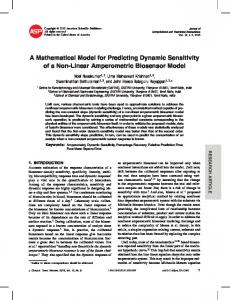
Fig.1. Microfluidic Bioseparator: (A) Perspective view with reference frame, (B) Cross-section of microfluidic bioseparator illustrating bioseparation sequence: (a) magnetic particles with surface-bound antibodies enter the microchannel, (b) energized conductive elements capture the particles, (c) target antigens are introduced into the microchannel, (d) immobilization of target antigens on magnetic particles, (e) separated material is released for further processing and analysis.
a system has been fabricated and characterized by Choi (Choi et al., 2000, 2001). This microsystem consists of a parallel array of rectangular conductive elements, which are semi-encapsulated in permalloy (not shown), and embedded beneath a rectangular microfluidic channel (Fig. 1A). The microsystem is small, occupying a volume of only a few cubic millimeters. A hypothetical separation sequence for this system is depicted in a cross-sectional view in Fig. 1B. First, magnetic particles with surface-bound antibodies enter the microchannel and are captured by the energized conductive elements. Next, target antigens are introduced into the microchannel and bind to the antibodies on the captured particles, thereby becoming immobilized. Lastly, the conductive elements are de-
energized and the separated material is re-released in high concentration for further processing. In this article we develop a model for predicting the transport and capture of magnetic particles in a bioseparation microsystem similar to that developed by Choi (Choi et al., 2000, 2001). We obtain analytical expressions for the dominant magnetic and fluidic forces on the particles, and test the magnetic force expression using finite element analysis (FEA). We include the magnetic, fluidic, and gravitational forces in the equations of particle motion, and solve these equations to study particle movement within the bioseparator. Specifically, we compute particle trajectories and capture times, and show that efficient separation can be achieved in a few minutes with modest power dissipation. The model takes into account key variables including the size and magnetic properties of the particles, the dimensions and spacing of the conductive elements, the magnitude of the applied current, the dimensions of the microchannel, and the viscosity and flow rate of the fluid. An important advantage of this approach is that it is based on analytical analysis, which provides insight into the basic physics and dominant factors governing particle capture, and enables rapid parametric analysis of system performance. This is in contrast to a purely numerical approach (e.g., finite-element analysis) that tends to be awkward for parametric analysis. The model is demonstrated via application to a practical microsystem design, and our analysis indicates that efficient particle capture can be achieved in a few minutes.
2. Theory In this section we derive a model for predicting the motion of a spherical magnetic particle of density ρp , radius R p , volume Vp =
4 πR 3p , and mass m p = ρp Vp in the 3
microfluidic bioseparator shown in Fig. 1. The trajectory of the particle is governed by several forces including, (a) the magnetic force due to all field sources, (b) fluidic drag, (c) particle/fluid interactions (perturbations to the flow field), (d) inertia, (e) buoyancy, (f) gravity, (g) thermal kinetics (Brownian motion), and (h) interparticle effects that include magnetic dipole interactions. We are interested in the behavior of magnetic particles in low concentration and slow flow regimes where the magnetic and viscous drag forces dominate. Therefore, we neglect particle/fluid interactions and interparticle effects. However, we include the gravitational force, which while of second order relative to the dominant forces, is not negligible. We use classical Newtonian dynamics to study particle motion, mp
dv p dt
= Fm + Ff + Fg ,
(1)
where v p is the velocity of the particle, and Fm , Ff , and Fg are the magnetic, fluidic, and gravitational forces, respectively. The magnetic force is obtained using an “effective” dipole moment approach where the magnetized particle is replaced by an “equivalent” point dipole with a moment m p,eff (Furlani and Ng, 2006). The force on the dipole (and hence on the particle) is given is given by: Fm = µ f ( m p,eff • ∇ ) H a ,
(2)
where µ f is the permeability of the transport fluid, m p,eff is the “effective” dipole moment of the particle, and H a is the (externally) applied magnetic field intensity at the center of the particle, were the equivalent point dipole is located. If the particle is in freespace, m p,eff = Vp M p and Eq. (2) reduces to the usual form Fm = µ0 Vp ( M p • ∇ ) H a , where Vp and M p are the volume and magnetization of the particle, and µ0 = 4π ×10−7 H/m is the permeability of free space. The fluidic force is predicted using the Stokes’ law for the drag on a sphere in uniform flow, Ff = −6πη R p ( v p - v f ),
(3)
where η and v f are the viscosity and the velocity of the fluid, respectively. For the bioseparation applications of interest here, the flow in the microchannel can be considered to be laminar with a velocity profile that varies in a parabolic fashion along the height of the channel. Since the particle diameter is much smaller than the channel height, the fluid velocity is relatively constant across the particle. As such, we use Eq. (3) to estimate the drag force at a given time using the particle velocity at that time, and the fluid velocity at the position of the particle at that time. It should be noted that a rigorous analysis of the fluidic force for this application is complicated and beyond the scope of this study. The gravitational force is given by Fg = −Vp ( ρ p - ρf )g yˆ ,
(4)
where ρ p and ρ f are the densities of the particle and fluid, respectively, and g = 9.8 m/s 2 is the acceleration due to gravity. The gravitation force acts in the -y direction. It is worth noting that the gravitational force is often ignored when analyzing the magnetophoretic motion of submicron particles, as it is usually much weaker than the magnetic force (Furlani, 2006; Furlani and Sahoo, 2006). However, in this application the magnetic force is relatively weak and the particles can be micron-sized. Therefore, we include the gravitational force in the analysis. We digress briefly to discuss the limitations of the Newtonian approach. As noted above, Eq. (1) does not take into account Brownian motion, which can influence particle capture when the particle diameter D p is sufficiently small. Gerber et al. have developed the following criterion to estimate this diameter (Gerber et al., 1983) F D p ≤ kT ,
(5)
where F is the magnitude of the total force acting on the particle, k is the Boltzmann constant, and T is the absolute temperature. In order to apply Eq. (5), one needs to estimate F . If the magnetic field source is specified, one can estimate F for a given particle by taking a spatial average of the force on the particle over the region of interest. Gerber et al. have studied the capture of Fe3O4 particles in water using a single magnetic wire, and have estimated the critical particle diameter for their application to be Dc , p ≡ kT / F = 40 nm, i.e., F = 0.1 pN (Gerber et al., 1983). For particles with a diameter below Dc , p (which is application dependent) one solves an advection-diffusion equation for the particle number density n(r, t ) , rather than the Newtonian equation for
the trajectory of a single particle. Specifically, n(r, t ) is governed by the following equation (Gerber et al., 1983; Fletcher, 1991),
where J = J D + J A
∂n(r, t ) +∇•J = 0, (6) ∂t is the total flux of particles, which includes a contribution
J D = − D∇n(r, t ) due to diffusion, and a contribution J A = v n(r, t ) due to advection of
particles under the influence of applied forces. Equation (6) is often written in terms of the particle volume concentration c(r, t ) , which is related to the number density, c(r, t ) = 4π R 3p n(r, t ) / 3 . The diffusion coefficient D is given by the Nernst-Einstein
relation D = µ kT , where µ = 1/(6πη R p ) is the mobility of a particle of radius R p in a fluid with viscosity η (Stokes’ approximation). The particle drift velocity v in J A is obtained from Eq. (1) in the limit of negligible inertia ( m p
dv p dt
→ 0 ), i.e. by setting
Fm + Ff + Fg = 0. Specifically, from Eqs. (1)-(3) we find that v (r ) = µ F(r ) , where F(r ) = 6πη R p v f (r ) + Fm (r ) + Fg (r ) . Note that if the Stokes’ drag is the only force, then v = v f . Equation (6) can be rewritten in the form, ∂n(r, t ) kT 1 = ∇ 2 n(r, t ) − ∇i( F(r )n(r, t ) ) . ∂t ( 6πη Rp ) ( 6πη Rp )
(7)
In order to solve either Newton’s equation (1) or the advection-diffusion equation (7), one needs an expression for Ff and Fm , which we derive below. 2.1. Magnetic Force
Consider a spherical magnetic particle in the presence of an applied magnetic field H a . Assume that particle is uniformly magnetized, and that the magnetization is a
linear function of the field intensity up to saturation, at which point it remains constant at a value M s . Specifically, below saturation, M p = χ p H in ,
(8)
where χ p = µp / µ0 − 1 is the susceptibility of the particle, µp is its permeability, and H in = H a − H demag . H demag is the self-demagnetization field that accounts for the magnetization of the particle, i.e. its magnetic “surface charge”. It is well known that H demag = M / 3 for a uniformly polarized spherical particle with magnetization M in freespace (Furlani, 2001). If the particle is suspended in a magnetically linear fluid of permeability µf , the force on it in an applied field H a is (Furlani and Ng, 2006), Fm = µf Vp
3( χp − χf )
( χ p − χ f ) + 3 ( χ f + 1)
( H a i∇ ) H a .
(9)
We assume that χ f w) ( x = w) ( x < w)
( x > − w) ( x = − w) , ( x < − w)
−1 y + h tan x + w θ 2 ( x, y ) = π /2 y+h π + tan −1 x +w
−1 y − h tan x − w θ 4 ( x, y ) = π /2 y−h π + tan −1 x −w
( x > − w) ( x = − w) ,
(22)
( x < − w)
( x > w) ( x = w).
(23)
( x < w)
Eqs. (19) and (20) are given by Binns (Binns et al., 1992), but the expression there for Eq. (20) is missing a minus sign, which has been corrected here. We substitute Eqs. (19) and (20) into Eqs. (16) and (17), and obtain the magnetic force components for a single conductor, (0) Fmx ( x, y ) =
µ0 Vp f (H a )I 2 r2 r1 ( y + h )(θ1 − θ 2 ) + ( x + w ) ln − ( y − h )(θ 4 − θ3 ) − ( x − w ) ln 2 (8π wh ) r4 r3
2 2 2 2 2 2 2 2 r r 2r −r 2r −r 2r −r 2r −r × ( y + h ) 1 2 22 − ( y − h ) 4 2 23 + ( x + w ) 3 2 22 − ( x − w ) 4 2 21 + ln 2 4 r1 r2 r1 r4 r1r3 r3 r4 r2 r3 r r − ( x + w )(θ 2 − θ3 ) + ( y + h ) ln 2 − ( x − w )(θ1 − θ 4 ) − ( y − h ) ln 3 (θ1 − θ 2 ) − (θ 4 − θ 3 ) , r1 r4
(24)
and
(0) Fmy ( x, y ) =
µ0 Vp f (H a )I2
( 8π wh )
2
( y + h )(θ1 − θ 2 ) +
r2 − ( y − h )(θ 4 − θ 3 ) − r3
( x + w ) ln
r1 r4
( x − w ) ln
× (θ 2 − θ 3 ) ) − (θ1 − θ 4 ) − ( x + w )(θ 2 − θ3 ) +
r2 − ( x − w )(θ1 − θ 4 ) − r1
( y + h ) ln
(25)
r3 r4
( y − h ) ln
2 2 2 2 2 2 2 2 r r 2r −r 2r −r 2r −r 2r −r × ( y + h ) 1 2 22 − ( y − h ) 4 2 23 + ( x + w ) 3 2 22 − ( x − w ) 4 2 21 + ln 2 4 . r1 r2 r1 r4 r3 r4 r2 r3 r1 r3
Next, consider an array of Nc conductors with the fist conductor centered with respect to the origin in the x-y plane, and all other conductors positioned along the x-axis as shown in Fig. 2B. The direction of current is opposite in adjacent conductors, i.e., into the page and then out of the page as shown. We identify the conductors using the index n = (0,1,2,3,4, …, Nc-1). The field components due to the first conductor (n = 0) are given by Eqs. (19) and (20). The n’th conductor is centered at x = sn and its field components can be written in terms of Eqs. (19) and (20) as follows, H ax( n ) ( x, y ) = (−1) n H ax(0) ( x − sn , y )
H ay( n ) ( x, y ) = (−1) n H ay(0) ( x − sn , y )
(n = 1, 2,3,…) (26)
The coefficient (−1)n takes into account the alternating direction of current through adjacent elements. Finally, the total field of the Nc element array is obtained by summing the contributions from all the conductors, H ax ( x, y ) =
N c −1
∑ (−1) n=0
n
H ax(0) ( x − sn , y ),
(27)
H ay ( x, y ) =
N c −1
∑ (−1) n=0
n
H ay(0) ( x − sn , y ),
(28)
We substitute Eqs. (27) and (28) into Eqs. (16) and (17) and obtain the force components Nc −1 Nc −1 ∂H ax(0) ( x − sn , y ) Fmx ( x, y ) = µ 0 Vp f (H a ) ∑ (−1) n H ax(0) ( x − sn , y ) ∑ ( −1) n ∂x n=0 n = 0 ∂H + ∑ ( −1) n H ay(0) ( x − sn , y ) ∑ ( −1) n n =0 n=0 N c −1
N c −1
(0) ax
(29)
( x − sn , y ) , ∂y
and Nc −1 ∂H ay(0) ( x − sn , y ) Nc −1 Fmy ( x, y ) = µ0 Vp f (H a ) ∑ (−1) n H ax(0) ( x − sn , y ) ∑ (−1) n ∂x n =0 n =0 ∂H + ∑ (−1) n H ay(0) ( x − sn , y ) ∑ (−1) n n=0 n=0 N c −1
N c −1
(0) ay
(30)
( x − sn , y ) , ∂y
Equations (29) and (30) are used in the equations of motion below. 2.2. Fluidic Force
As noted above, we use Stokes’ law to predict the fluidic drag force on the particle. Specifically, to obtain the drag force at a given time t, we substitute the particle velocity at that time v p (t) , and the fluid velocity at the position of the particle at that time
v f (x p (t)) , into Eq. (3), Ff = −6πη R p v p (t) - v f (x p (t)) ,
(31)
To evaluate Eq. (31) we need an expression for fluid velocity v f in the microchannel. Let L denote the length of the channel and hc and wc denote the half-height and half-width of its rectangular cross section (Fig. 1A). The nature of the flow, laminar or turbulent, is estimated from the Reynolds number Re = vf D ρ / η , where vf is the average fluid velocity , D is the characteristic length of the channel (the hydraulic diameter), and ρ
and η are the density and viscosity of the fluid, respectively. In bioseparation applications vf < 1 m/s, D ≈ 100 µm, ρ ≈ 1000 kg/m3 , and η ≈ 0.001 Ns/m 2 . Therefore, Re < 100 which indicates laminar flow (i.e., Re < 2300 ). We assume fully developed
laminar flow with the flow velocity parallel to the x-axis, and varying across the cross section, ˆ v f = vf ( y′, z′) x.
(32)
It is convenient to use coordinates y′ and z′ centered with respect to the cross section of the channel, and it is understood that these differ from the coordinate system used for the magnetic analysis (Fig. 2B). Here, z ' spans the width of the channel. The velocity profile for fully developed laminar flow is
vf ( y′, z′) =
16hc2 ∆P ∞ (−1) n cosh((2n + 1)π z '/ 2hc ) 1− cos((2n + 1)π y '/ 2hc ), ∑ ηπ 3 L n = 0 (2n + 1)3 cosh((2n + 1)π wc / 2hc )
(33)
where ∆P is the change in pressure across the length L of the channel (Ichikawa et al.,
2004). The volume flow rate Q through the channel is
Q = Avf ,
(34)
where A = 4h c wc is the cross-sectional area. If the channel is short relative to its width ( hc / wc
1 ), which is typically the case, and if we ignore the variation in velocity along
the width of the channel (i.e. along the z ' -axis), then the velocity profile reduces to, 2 3 vf y ′ 1 − . v f ( y′) = 2 hc
(35)
In order to include this expression in our analysis we rewrite it in terms of the coordinate
y of Fig. 2B in which y′ = y − (h + hc + tb ) , where tb is the thickness of the base of the channel (i.e., the distance from the top of a conductive element to the lower edge of the fluid). This gives, 2 3 vf y − (h + hc + tb ) 1 − vf ( y) = . 2 hc
(36)
Finally, we substitute Eq. (36) into Eq. (3) and obtain the fluidic force components on a particle with velocity v p = v p,x xˆ + v p,y yˆ at a position x p = x p xˆ + y p yˆ , 3v Ffx = −6πη R p v p,x − f 2
y p − (h + hc + tb ) 2 1 − , hc
(37)
and Ffy = −6πη R p v p,y .
(38)
In these expressions, v x and v y are the components of the particle velocity. We use these in the equations of motion.
2.3. Equations of Motion
The equations of motion for a magnetic particle traveling through the bioseparator can be written in component form by substituting Eqs. (29), (30), (37) and (38) into Eq. (1), mp
3v = Fmx (x p , y p ) − 6πηR p v p,x − f dt 2
dv p,x
mp
dv p,y dt
y p − (h + h c + t b ) 2 1 − , hc
= Fmy (x p , y p ) − 6πηR p v p,y − Vp (ρp - ρf )g ,
(39)
(40)
v p,x (t ) =
dx p dt
v p,y (t ) =
,
dy p dt
.
(41)
Equations (39) - (41) constitute a coupled system of first-order ordinary differential equations (ODEs) that are solved subject to initial conditions for the position x p (0) , y p (0) , and velocity v p , x (0) , and v p , y (0) of the particle. We solve these equations numerically using the Runge Kutta method. Equations (39) - (41) predict the motion of a magnetic particle in a moving fluid that is permeated by a magnetic field. This applies to bioseparation in which the bound biomaterial is much smaller than the magnetic particle and does not appreciably influence particle motion.
However, in many applications the biomaterial is much larger than a
single particle. Some nominal sizes for various biomaterial are as follows (Pankhurst et al, 2003): cells (10-100µm), viruses (20-450 nm), proteins (3-50 nm), and genes (10 nm wide and 10-100 nm long). Thus, for example, if a cell is 20 microns in diameter, several micron-sized magnetic particles must bind to its surface to implement effective separation. If there are Np magnetic particles bound to a cell, the mass, volume, radius, and density of the combined cell/particle structure can be estimated using (Safarýk and Safarýkova, 1977) m cp = m cell + N p m p = ρ cell Vcell + N p ρ p Vp ,
Vcp = Vcell + N p Vp ,
(42)
(43)
1/ 3
3 Rcp = Vcp , 4
(44)
and ρ cp =
ρ cell Vcell + N p ρ p Vp Vcell + N p Vp
.
(45)
The equations of motion (39) - (41) need to be modified to account these relationships. To simplify the analysis, we assume that that the magnetic force acts only on the bound magnetic particles, whereas the fluidic force acts on the entire structure, which to first order has an effective radius Rcp . Let x p,k , y p,k denote the coordinates of the k’th magnetic particle on the surface of the cell, and let xcp , ycp and vcp,x , vcp,y denote the coordinates and velocity components of the center of mass of the structure, respectively. We adapt the Eqs. (39) - (41) to the cell/particle structure and obtain, m cp
dvcp,x dt
2 3 vf y cp − (h + h c + t b ) 1 − = ∑ Fmx (x p,k , y p,k ) − 6πηR cp vcp,x − , 2 hc k =1
m cp
Np
dvcp,y dt
(46)
Np
= ∑ Fmy (x p,k , y p,k ) − 6πηR cp vcp,y − Vcp (ρcp - ρf )g,
(47)
k =1
vcp,x (t ) =
dxcp dt
,
vcp,y (t ) =
dycp dt
.
(48)
In Eqs. (46) and (47) the magnetic force components Fmx (x p,k , y p,k ) and Fmy (x p,k , y p,k ) on the k’th particle are computed using the volume and magnetic properties for that particle. In this analysis, we have assumed that the cell/particle structure is rigid, and have ignored its rotation. A more complete analysis would include the effects of rotation and structural deformation, as well as the influence of motion on the local fluid flow (i.e. the coupled structure/fluid interaction). However, these effects are beyond the scope of the present work.
3. Results We use Eqs. (27)-(30) and (39)-(41) to study the bioseparator shown in Fig. 1A. We assume that the transport fluid is nonmagnetic ( χ f = 0 ), and has a viscosity and density equal to that of water, η = 0.001 Ns/m2 and ρ f = 1000 kg/m3. The force calculation requires a choice of particle size and material properties. We choose a commercially available particle that is commonly used for bioapplications, the MyOne particle from Dynal Biotech (www.dynabead.com). The properties of this particle as obtained from Dynal Biotech are as follows: radius R p = 0.5 µm, density
ρ p = 1800 kg/m3 , and saturation magnetization M s = 4.3 ×10⁴ A/m. The intrinsic susceptibility χ p is not available. Instead, Dynal Biotech specifies an “effective” susceptibility of χ p,e = 1.4 . The values of χ p and χ p,e are measured with respect to the internal and applied field, respectively, and for a given sample the two are related as follows,
χ p,e =
χp 1 + Nχ p
,
(49)
where N is the shape dependent demagnetization factor of the sample (e.g., N = 1/3 for a sphere). We modify our model and use χ p,e instead of χ p , which amounts to replacing Eq. (14) by
χ p,e f (H a ) = M s / H a
Ha