BY Sanford M. Meyers MD,* Mikhail A. Ostrovsky PhD DSc, AND Robert F. Bonner PhD. ABSTRACT. Background/Purpose: Cumulative sunlight exposure and ...
A MODEL OF SPECTRAL FILTERING TO REDUCE PHOTOCHEMICAL DAMAGE IN AGE-RELATED MACULAR DEGENERATION BY
Sanford M. Meyers MD,* Mikhail A. Ostrovsky PhD DSc, AND Robert F. Bonner PhD
ABSTRACT
Background/Purpose: Cumulative sunlight exposure and cataract surgery are reported risk factors for advanced age-related macular degeneration (AMD). Laboratory studies suggest that accumulation and photochemical reactions of A2E (N-retinylidene-N-retinylethanolamine) and its epoxides, components of lipofuscin, are important in AMD. To relate this data to the clinical setting, we modeled the effects of macular irradiance and spectral filtering on production of A2E and reactive oxygen intermediates (ROIs) in pseudophakic eyes with a clear or “yellow” intraocular lens (IOL) and in phakic eyes. Methods: We calculated relative changes of macular irradiance as a function of light (390 to 700 nm) intensity, pupil size, age, and lens status, and modeled resulting all-trans-retinal concentration and rates of production of A2E-related photochemicals and photon-induced ROIs in rods and retinal pigment epithelium (RPE). We compared these photoproducts following cataract surgery and IOL implantation with and without spectral sunglasses to normal age-related nuclear sclerotic lens changes. Results: Following cataract and IOL surgery, all-trans-retinal and lipofuscin photochemistry would theoretically increase average generation of 1) A2E-related photochemicals, 2) ROI in rods and 3) ROI in RPE, respectively, 2.6-, 15- and 6.6fold with a clear IOL, and 2.1-, 4.1- and 2.6 fold with a yellow IOL, but decrease approximately 30-, approximately 20and 4-fold with a vermillion filter sunglass and clear IOL compared to an average 70 year old phakic eye. Conclusion: Sunglasses that strongly decrease both deep blue light and rod photobleaching, while preserving photopic sensitivity and color perception, would provide upstream protection from potential photochemical damage in subjects at risk for AMD progression after cataract surgery. Trans Am Ophthalmol Soc 2004;102:83-95
INTRODUCTION
The late stages of age-related macular degeneration (AMD), neovascularization and geographic atrophy, are important causes of severe visual loss and legal blindness in persons over 60 years of age in the United States.1-3 Clinical and epidemiological studies that report a significant association between both prior cataract surgery and cumulative exposure to sunlight and late-stage AMD lend support to the hypothetical role of photochemical reactions in the pathogenesis of AMD.4-10 In 1920, van der Hoeve11 observed that AMD was less common in eyes with cataracts and proposed that opacity of the lens diminished the severity of AMD; in 1925,
From Retina Consultants, Des Plaines, Illinois (Dr Meyers); the Russian Academy of Sciences, Moscow (Dr Ostrovsky); and the National Institute of Child Health and Human Development, National Institutes of Health, Bethesda, Maryland (Dr Bonner). *Presenter. Bold type indicates AOS member.
Trans Am Ophthalmol Soc / Vol 102 / 2004
Gjessing12 reported an inverse relation between lens opacity and AMD. Recent epidemiological studies have reported conflicting data on AMD and lens opacities but consistently suggest that prior cataract surgery, aphakia, and pseudophakia are risk factors for late-stage AMD.5,6,13,14 The Chesapeake Bay Waterman Study and the Beaver Dam Eye Study reported a significant association between AMD and cumulative sunlight exposure, 400 to 700 nm, the former with late-stage AMD and the latter with early AMD.7,8 Neither of these studies reported an association between AMD and exposure to ultraviolet A (320 to 400 nm) or B (290 to 320 nm) light. Laboratory studies on acute photochemical injury to mammalian rod photoreceptors and retinal pigment epithelium (RPE) cells document two well-defined action spectra: (1) Ham-type photochemical injury to the RPE caused by intense blue and ultraviolet light with peak sensitivity at approximately 350 nm in aphakic monkeys and (2) rod damage associated with the rhodopsin absorption spectrum and enhanced by blue light.15,16 Recent in
83
Meyers et al vitro studies have demonstrated that RPE lipofuscin granules generate oxygen free radicals, singlet oxygen, and other reactive oxygen intermediates (ROIs), with an action spectrum very similar to that in the Ham-type in vivo study.17-19 Additionally, lipofuscin granules contain a number of different fluorescent species that (1) originate largely from photochemical reactions involving all-trans-retinal and include A2E (N-retinylidene-Nretinylethanolamine) and its epoxides and (2) are capable of acting as photosensitizers of singlet oxygen.18,19 A2Ederived fluorophores in lipofuscin appear to be produced during periods of photopic vision associated with significant rhodopsin bleaching and high levels of all-trans-retinal in the rod outer segment (ROS) disks. All-trans-retinal in the ROS disks can absorb short-wavelength light with a 380-nm peak and known wavelength dependence (or action spectrum) leading to a long-lived triplet state that acts as a photosensitizer of oxidative damage.20 A2Ederived fluorophores that accumulate in the RPE lipofuscin are potent photosensitizers of oxidative damage with a known aggregate action spectrum.21 To relate the epidemiological and laboratory data to the clinical setting, we modeled the effects of retinal irradiance and spectral filtering on the relative rates of alltrans-retinal photosensitization, production of A2E and A2E-derived lipofuscin fluorophores, and 1O2 generation due to RPE lipofuscin photosensitization in phakic eyes and in eyes with a clear or “yellow” intraocular lens (IOL) with and without external spectrally selective filters. We discuss this model in relation to hypothetical pathogenic pathways in AMD and describe a possible preventive strategy for decreasing the potential risk of photochemical damage in AMD. METHODS
We have developed a model relating retinal spectral irradiance as a function of age and lens status to the relative rates of rod bleaching, steady-state concentration of alltrans-retinal, its photosensitization of oxidative damage in the rod outer segments, or alternatively its reactions to form A2E-related species. In RPE cell culture experiments, A2E is rapidly ingested and concentrated through lysosomal processing into prototypical nascent lipofuscin granules.19 On irradiation, these granules form a complex mixture of oxidized A2E-related fluorophores that are potent photosensitizers of singlet oxygen generation and spectroscopically comparable to mature lipofuscin granules harvested from aged human retinas (R. Bonner, unpublished data, 2004). In our model, we assume that averaged over long time periods (1 year), the relative rate of “mature” lipofuscin accumulation within the RPE is proportional to the rate of production of A2E precursors
84
in the rods. From the literature, we applied a variety of values describing the normal age dependence of critical ocular parameters to standard formulas in order to determine average retinal spectral irradiance as a function of age. We then used our model and published action spectra to estimate the normal average age dependence of these potential causes of chronic light injury at 7 to 11 degrees from the center of the fovea, a region of high lipofuscin accumulation and rod loss.22 Finally, we compared the estimates for the normally aging phakic subject following replacement of the “yellowed” aged lens with two different commercial IOLs (Alcon clear AcrySoft MA60BM UV-absorbing and light yellow AcrySoft Natural) with or without rod-sparing or blue-blocking spectrally selective sunglasses. We calculated relative changes of macular irradiance as a function of light spectral (390 to 700 nm) intensity, pupil size, age, and lens status. The solar radiance has direct, diffuse (scattered light from the atmosphere), and ground-reflected components and varies with the day of year, time of day, altitude, and latitude. For our calculations we used the following formula and daylight radiances at the cornea between 9 and 4,400 candelas[cd]/m2 for the solar spectrum through air mass 1.2: 700
HR = (A n2/f 2) ∫390
HP(λ) t(λ) dλ
(1)
where HR is the retinal irradiance where the specific photochemical reaction occurs; Hpupil = Hp≅ is the “effective” solar radiance at anterior corneal surface, Hcornea ; A is the area of pupil; f is the distance from pupil to macula; n is the index of refraction of ocular media; t is the transmission of the ocular media; and λ is the wavelength of light. For diffuse sources, the solid angle of the retinal image determined by the pupil area (A n2/f 2) greatly diminishes retinal irradiance. For diffusely reflected sunlight, the surface reflectance or albedo of viewed objects further diminishes corneal irradiance so that at 500 nm the spectral irradiance of the sun hitting the ground might be approximately 1 mW/cm2/10 nm, but the retinal irradiance only a few microwatts per cm2/10 nm. Acute phototoxicity experiments in animals or cell cultures capable of inducing apoptosis generally use light intensities hundreds or thousands times greater than normal retinal irradiance in daylight. We assumed n was 1.33 and f was 21.5 mm. This formula and the assumptions we made are similar to those used in studies to calculate retinal irradiance at the surface of the retina from indirect ophthalmoscopes, slit lamps, and surgical lamps.23 In the visible spectrum, ocular spectral transmission t(λ) is determined mainly by absorption of light by the
A Model of Spectral Filtering to Reduce Photochemical Damage in Age-Related Macular Degeneration lens(tL), the macular pigment(tMP), the photoreceptor visual pigments(tPH), and melanin in the RPE (tM) in the macula. Each of these components is a function of wavelength: t(λ)= tL(λ) tMP(λ,r) tPH(λ,r,z) tM(λ,r,z)
(2)
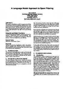
Spectral irradiance at the surface of the macula is determined by (1) the source spectral irradiance at the cornea, (2) the pupil diameter, and (3) the transmission of the lens (and cornea). The cornea absorbs virtually all light below 300 nm. The lens transmits virtually 100% of light over 660 nm. Because the lens and UV-absorbing IOLs transmit less than 1% light below 390 nm, we considered light below 390 nm to have an insignificant effect on our calculations. We are principally interested in the role of photoprotection of the lens as it ages (“yellows”) compared to IOLs used to replace the lens following cataract surgery. We used published data on lens transmittance as a function of age in normals (due to nuclear sclerosis) and data on the transmittance of the Alcon clear AcrySoft UVabsorbing and light yellow AcrySoft Natural IOLs and a similar yellow Spectrum IOL developed and used in Russia beginning in the mid-1980s.24-26 We also examined the effects of external spectrally selective sunglasses after cataract surgery (a vermilion, a yellow or 480-nm longpass, and a deep red or 570-nm long-pass; see Figure 1). The selected vermilion filter allows (1) little attenuation of
FIGURE 1 Spectral transmission of ocular filters analyzed with our model. Numbered from left to right (1) Acrysoft clear UV-absorbing IOL, (2) Russian Spectrum (light yellow) IOL, (3) Acrysoft light yellow IOL, (4) 480-nm long-pass filter, (5) 70-year-old lens, (6) vermilion filter, and (7) 570-nm long-pass filter.
long wavelengths and photopic sensitivity, (2) sufficient midrange blue transmission to provide good color perception, and (3) blockage of wavelengths efficiently absorbed by rod rhodopsin and short-wavelength blue light. Retinal irradiance is a strong function of pupil diameter and corneal irradiance. We used published average normal data on pupil size as a function of age and after cataract removal and IOL implantation.27,28 The lightfiltering effects of macular and photoreceptor pigments and RPE melanin are complicated functions of their radial and depth (z) distributions.29,30 Although we have used literature values for the radial (r) and depth (z) dependence, we present here only our model predictions for the photochemistry and spectral irradiance at 7 to 11 degrees from the center of the fovea. The effect of the macular pigment on the spectral irradiance of the underlying macular photoreceptors and RPE is highest in the central 1 degree of the fovea, rapidly decreases from 1 to 5 degrees from the center of the fovea, and is considered negligible beyond approximately 7 degrees.29 Rod loss and lipofuscin accumulation occur predominantly outside the region of significant macular pigment absorption, and therefore its possible photoprotective role does not affect our calculations for 7 to 11 degrees. Our calculations of the “internal filter” photoprotection of rhodopsin and melanin absorption have been estimated assuming age-independent concentrations of rhodopsin and melanin at 7 to 11 degrees from the fovea.30 We modeled only the average age-dependent effects of macular spectral irradiance in a typical subject before and after cataract surgery. In eyes with early AMD, pigmentary and drusenoid changes may critically affect the potential for local photodamage and not be reflected in the reported average values (such as rod dark-adaptation time constant) that we used. Such complex interactions with spectral irradiance could not be reliably predicted and are beyond the scope of our model. In order to estimate the average age-dependent rates of all-trans-retinal photosensitization of ROI production in the rods or of its reactions to form A2E and related molecules, we related the macular irradiance to the corresponding steady-state concentration of all-trans-retinal, [all-trans-retinal]ss, produced in the rods. We used the formula of Thomas and Lamb to calculate the steady-state bleaching of the rods, B(I,age), using the age-dependent average values of the rod visual cycle time constant trh (age) in seconds reported for a normal population31,32: B(I,age) = Is / {Is + Lrh/trh(age)} = a [all-trans-retinal]ROS
(3)
Where Is is the retinal irradiance in scotopic trolands, Lrh is an empirical constant reported to be 107, [all-trans-retinal]ROS is the concentration of all-trans-retinal in the rod
85
Meyers et al disks, and a is an age-independent constant.31 For different ambient corneal light levels, the scotopic trolands were calculated using the reported age-dependence of average pupil diameters as a function of corneal irradiance and the altered scotopic sensitivity of the solar spectrum transmitted through the aging lens (or IOLs). The addition of external filters (eg, spectrally selective sunglasses) merely adds another factor tSSG(λ) in equation 2. Following rod bleaching of the normal mammalian retina, all-trans-retinal in the rod outer segments is the predominant transiently increased species in the retina. Therefore, we assume in our model that [all-trans-retinal]ss varies linearly with the steady-state rod bleaching (equation 3). All-trans-retinal, which reaches high concentrations in partially bleached rods (ie, for daylight ≥200 cd/m2), is a potent photosensitizer of oxidative damage. After absorbing a short-wavelength photon, the excited singlet-state all-trans-retinal undergoes intersystem crossing to a long-lived (approximately 10 msec) triplet state that can efficiently transfer its energy to ground state 3O2, creating highly reactive singlet oxygen 1 O2.20 The action spectrum AS(λ)atr of this process is the absorption spectrum of all-trans-retinal with a maximum at 380 nm and rapidly diminishing with increasing wavelength. We assumed that the photochemical creation of reactive oxygen intermediates in the rods ROIrod is predominantly driven by the creation of excited states of all-trans-retinal and is given by: 700
ROIrod = b ∫ ∫390 [all-trans-retinal]ROS Hrod(λ) AS(λ)atr dλ dt (4) where [all-trans-retinal] is obtained from equation 3, Hrod(λ) from equation 1, and b is a constant (affected by PO2 and quantum efficiency of the photosensitizer reaction but not age). In this and all the subsequent calculations, we were interested only in long-time averages over many days in which daily or even seasonal variations in light exposure can be neglected. Since equation 3 is nonlinear with normal photopic corneal irradiances, the integral over time allows calculation of the average ROI production over any standardized temporal distribution of ambient corneal irradiances that reflect the range of typical daily light exposures. We used 5% at 4,400 cd/m2, 20% at 1,100 cd/m2, 30% at 220 cd/m2, 25% at 44 cd/m2, and 20% at 9 cd/m2; even lower values would not contribute significantly to modeled photodamage. We modeled the effects due to spectral transmission changes in the lens assuming environmental light exposures do not vary significantly with age in order to evaluate the specific effects of aging on lens yellowing and pupil diameter changes. Epidemiological literature suggests that increased environmental light exposure is a risk factor for AMD, and higher or lower average ambient light distribu-
86
tions than the one we used would have roughly proportional changes in the computed rates of photochemistry at a given age. Periods of rod bleaching and high [all-transretinal]ROS also lead to the formation of A2E and related fluorophores. After disk shedding, these fluorescent molecules are concentrated by lysosomal processing within lipofuscin granules in the RPE. A2E and related molecules avidly partition into cellular membranes and appear to be potent cytotoxic agents capable of inducing DNA damage and apoptosis in the dark, which is enhanced by blue light.18 Since A2E and its phosphorylated precursor require the reaction of two all-trans-retinal molecules, its reaction rate is second order in [all-trans-retinal] and therefore should be proportional to [all-trans-retinal]2 in the rods. An estimate of the average rate of A2E production over long periods of different ambient light levels is given by the time average of [all-trans-retinal]2 determined by retinal scotopic irradiance (Is) during environmental light exposure: x+1
A2E production rate (age) = ∫x k [all-trans-retinal]ROS2 dt (5) where x is the age in years and k is a rate constant assumed to be independent of age, or at least in the case of IOL implantation, independent of whether the aged lens has been replaced with an IOL. A2E does not appear to be substantially broken down by lysosomal enzymes but rather accumulates in RPE lipofuscin granules where its photo-oxidization on irradiation with short-wavelength light results in a complex mixture of fluorophores.33 A2E avidly reacts with 1O2 to form epoxides of increasingly higher order, and this oxidation within the lipofuscin granules appears to be the principal means by which A2E concentration (as a distinct molecular species) is limited to approximately 1 pg per RPE cell (approximately 200 µM) in the human retina.18,19 We have modeled the accumulated A2E-derived lipofuscin fluorophores in the RPE (LFRPE[age]) over many years to be proportional to the time integral of the rate of A2E precursor formation in the rods during periods of significant bleaching. x
LFRPE(x) = ∫0 (A2E production rate) dt
(6)
where x is the age. The action spectrum of RPE lipofuscin AS(λ)lf has been determined by direct detection of 1O2 phosphorescence and falls exponentially with wavelength (approximately 20-fold from 360 to 460 nm).21 This action spectrum is very similar to that observed for macular RPE injury induced by 1,000-sec focal monochromatic irradiations in rhesus monkeys.15,16 In our model, we assumed
A Model of Spectral Filtering to Reduce Photochemical Damage in Age-Related Macular Degeneration that the potent photosensitizers in lipofuscin granules are derived from A2E by oxidation (predominantly photooxidation). We can describe the age dependence of generation of 1O2 by lipofuscin granule photochemistry by: 700
ROIRPE (age) = b ∫ {∫390 LFRPE(x) HRPE(λ) AS(λ)lf dλ} dt (7) where ROIRPE (age) is the age dependence of lipofuscin 1 O2 generation within a typical RPE cell in the macula at 7 to 11 degrees from center of the fovea. To predict all of the above processes after cataract surgery, we substituted the spectral transmission of the IOL, tIOL(λ), for tL(λ) in equation 2 at the specified time of IOL implantation and recalculated equations 1 and 3 through 7 for the years after implantation. Similarly, spectral transmission of spectrally selective external sunglasses was added to equation 2, and the results recalculated. RESULTS
We are primarily interested in estimating the effects of lens aging and lens replacement with a clear or yellow IOL on the retinal spectral irradiance and the rate of modeled macular photochemistries. Variations among aging individuals in degree of lens yellowing, pupil diameter, rod dark adaptation rate trh(age), and environmental light exposures might result in substantial differences in the amount of A2E-related compounds produced and modeled photo-oxidative damage in both the rods and RPE cells. In modeling macular photosensitization of ROI and production of related photochemicals, we did not evaluate the effects of antioxidant and molecular repair mechanisms. These ameliorating mechanisms may decrease with age but would not be expected to change due to cataract and IOL surgery. Under our modeled age-independent mixed light exposures, described above, the time average rate of rod bleaching and average [all-trans-retinal]ROS decreases only slightly from 0 to 60 years owing to a balance of the normal aging trend to smaller pupils (ie, less light) and slower trh(age), whereas the scotopic transmission of the lens is reduced only slightly. During this period, the modeled average relative rate of rod oxidative damage via all-trans-retinal photosensitization (Figure 2) is affected largely by changes in the fraction of short wavelength (