Neurogenetics (2012) 13:169–179 DOI 10.1007/s10048-012-0324-y
ORIGINAL ARTICLE
Axonal transport deficit in a KIF5A–/– mouse model Kathrin N. Karle & Diana Möckel & Evan Reid & Ludger Schöls
Received: 6 February 2012 / Accepted: 13 March 2012 / Published online: 1 April 2012 # The Author(s) 2012. This article is published with open access at Springerlink.com
Abstract Hereditary spastic paraplegia (HSP) is a neurodegenerative disorder preferentially affecting the longest corticospinal axons. More than 40 HSP genetic loci have been identified, among them SPG10, an autosomal dominant HSP caused by point mutations in the neuronal kinesin heavy chain protein KIF5A. Constitutive KIF5A knockout (KIF5A–/–) mice die early after birth. In these mice, lungs were unexpanded, and cell bodies of lower motor neurons in the spinal cord swollen, but the pathomechanism remained unclear. To gain insights into the pathophysiology, we characterized survival, outgrowth, and function in primary motor and sensory neuron cultures from KIF5A–/– mice. Absence of KIF5A reduced survival in motor neurons, but not in sensory neurons. Outgrowth of axons and dendrites was remarkably diminished in KIF5A–/– motor neurons. The number of axonal branches was reduced, whereas the number of dendrites was not altered. In KIF5A–/– sensory neurons, neurite outgrowth was decreased but the number of neurites remained unchanged. In motor neurons maximum and average velocity of mitochondrial transport was reduced both in anterograde
Electronic supplementary material The online version of this article (doi:10.1007/s10048-012-0324-y) contains supplementary material, which is available to authorized users. K. N. Karle : D. Möckel : L. Schöls (*) Department of Neurology, Hertie Institute for Clinical Brain Research and German Center of Neurodegenerative Diseases, University of Tübingen, Hoppe-Seyler-Str. 3, 72076 Tübingen, Germany e-mail:
[email protected] E. Reid Department of Medical Genetics, Cambridge Institute for Medical Research, Addenbrooke’s Hospital, University of Cambridge, Wellcome Trust/MRC Building, Cambridge CB2 OXY, UK
and retrograde direction. Our results point out a role of KIF5A in process outgrowth and axonal transport of mitochondria, affecting motor neurons more severely than sensory neurons. This gives pathophysiological insights into KIF5A associated HSP, and matches the clinical findings of predominant degeneration of the longest axons of the corticospinal tract. Keywords Motor neuron . KIF5A . Axonal transport . Axonal outgrowth . Mitochondria . Hereditary spastic paraplegia (HSP)
Introduction The hereditary spastic paraplegias (HSPs) comprise a growing number of clinically and genetically heterogeneous diseases. Their common feature is axonal degeneration of the corticospinal tract in a length-dependent manner, so that the longest axons innervating lower motor neurons that supply the leg muscles are affected, whereas the shorter fibers that innervate lower motor neurons supplying the arms are mainly spared. Clinically, this leads to progressive gait disturbance due to lower limb spasticity [1–3]. More than 40 HSP loci and 20 HSP genes are registered in the HUGO Gene Nomenclature Committee database [4]. Autosomal dominant, autosomal recessive, and X-linked modes of inheritance occur in HSP [5]. Increasing knowledge about physiological functions of genes mutated in HSP has highlighted a few common pathogenic mechanisms, such as myelin sheath formation, mitochondrial function, and membrane trafficking and transport [6–8]. Axons rely on many of these functions in a lengthdependent manner, offering an explanation why motor neurons to the legs with an axon length up to 1 m are especially sensitive to dysfunction.
170
SPG10 is an autosomal dominantly inherited subtype of HSP that is caused by mutations in the neuronal motor protein kinesin heavy chain (KHC) KIF5A. SPG10 accounts for 3 to 5 % of autosomal dominant HSPs in European families [9, 10]. As in most HSP subtypes, disease onset varies from early childhood to adulthood, and the disease may cause pure spastic paraplegia, or alternatively forms complicated by cognitive dysfunction, peripheral neuropathy, parkinsonism, and/or epilepsy. Nearly all mutations causing SPG10 are heterozygous missense mutations in the motor domain of KIF5A [9–14]. The kinesin superfamily provides motors for ATPdependent transport along microtubules, generally in the plus end direction (which in axons is the anterograde direction). Kinesin-1, formerly called conventional kinesin, acts as a motor for fast axonal transport of membranous organelles (50–200 mm per day) as well as for slow axonal transport of cytoplasmic proteins (0.1–3 mm per day) [15–17]. Kinesin-1 is a heterotetramer consisting of two kinesin heavy chains (KIF5A, KIF5B, or KIF5C), and two kinesin light chains [18]. Kinesin heavy chains as well as kinesin light chains only homodimerize, whereas there is no specificity in the interaction between heavy and light chains [19]. The heavy chains contain the motor domain, cargo specificity is thought to be mediated mainly via kinesin light chains and/or additional linker proteins, e.g., milton and miro are likely to act as linker proteins binding mitochondria to kinesin heavy chain protein [20]. Other cargoes transported by kinesin-1 include lysosomes [21], membraneassociated SNARE proteins [22], syntaxin-1-containing vesicles [23], tubulin [24], apolipoprotein E receptor 2 [25], and phosphorylated amyloid precursor protein (APP) [26]. Circumstantial evidence that KIF5A is involved in mitochondrial transport comes from primary extraembryonic membrane cells lacking the KIF5B gene. The abnormal perinuclear clustering of mitochondria in these cells can be rescued by overexpression of the KIF5A gene [27]. In vitro experiments with KIF5A proteins carrying human SPG10 missense mutations of the motor domain revealed reduced transport velocity and reduced binding on microtubules respectively [28]. KIF5A knockout (KO) mice die shortly after birth. The amount of KIF5B and KIF5C was not significantly changed. Histologically, lungs were not well expanded, and cell bodies of lower motor neurons in the spinal cord swollen whereas the brain did not seem to be affected. To overcome perinatal lethality conditional knockout mice were generated with neuronal knockout of KIF5A induced by synapsinpromoted Cre-recombinase transgene. These mice showed seizures, hind limb paralysis, and sensory neuron degeneration in later stages. Histological analysis of cell bodies from sensory neurons revealed an accumulation of neurofilament (L, M, H), in Western Blots of DRGs the amount of
Neurogenetics (2012) 13:169–179
neurofilament protein was increased, whereas the amount of markers for fast axonal transport (APP, Rab3, synaptophysin) was not changed [29]. In superior cervical ganglia neurons, frequency and velocity of neurofilament movements were reduced both in anterograde and retrograde direction [30]. In a mouse model with the human N256S mutation in the KIF5A gene cortical neurons revealed an increased retrograde velocity of neurofilament M transport, whereas anterograde velocity was not affected. The frequency of anterograde and retrograde movements was decreased [31]. But probably neurofilament is not the only important cargo transported by KIF5A. In this work, we established primary cell cultures of motor and sensory neurons of constitutive KIF5A KO mice and characterized vitality, morphology, and function. By live cell imaging, we revealed an axonal transport defect of mitochondria. These results can improve insights into mechanisms underlying the length-dependent axonal degeneration and selective damage of motor neurons in SPG10.
Materials and methods Histology of mouse brain, spinal cord, and muscle KIF5A+/– mice were obtained from Mutant Mouse Regional Resource Centers, University of California, Davis, USA. Wildtype C57Bl/6N mice were purchased from Charles River Laboratories (Sulzfeld, Germany). For histological analysis embryos were dissected from pregnant mice at embryonic day (E) 18.5. Brain, spinal cord, and proximal leg muscle were isolated and fixed in 4 % paraformaldehyde for 24 hours. After dehydration tissues were embedded in paraffin. 5 μm sections of brain and spinal cord were stained with cresyl violet (MEDITE® GmbH, Burgdorf, Germany) whereas 5 μm muscle sections were treated with hematoxylin eosin (MEDITE® GmbH). Images were acquired with a CCD camera (Axio Imager Z1, AxioCam MRc, Carl Zeiss MicroImaging GmbH, Jena, Germany). For morphological assessment of spinal motor neurons the size of cell bodies and nuclei were analyzed in at least 36 cells from three independent experiments. Statistical evaluation was performed by ANOVA followed by Bonferroni correction (SPSS 17.0, SPSS Inc., Chicago, USA). Mouse embryonic motor and sensory neuron culture Embryos were isolated from pregnant mice at E 12.5. The lumbar spinal cord and the laterally adjacent DRGs were dissected and transferred to HBSS (invitrogen™, Carlsbad, USA) and 1× PBS (invitrogen™) respectively. The trypsin reaction (0.05 %, Biochrom AG, Berlin, Germany) was stopped in spinal cord tissue after 15 min with 0.01 % trypsin
Neurogenetics (2012) 13:169–179
inhibitor (Sigma-Aldrich Co., St. Louis, MO, USA), and in DRGs after 35 min with HAMS F14 Powder Medium (invitrogen™), enriched with 10 % heat-inactivated horse serum (Linaris Biologische Produkte GmbH, Wertheim-Bettingen, Germany) and 35 mM KCl. Tissues were triturated. Motor neurons of the spinal cord were isolated via Lectin antibody (20 μg/ml, Sigma-Aldrich) that has been attached to 24 well CELLSTAR® plates (Greiner Bio-One GmbH, Frickenhausen, Germany). After panning for 30 min, the supernatant was removed, and adherent motor neurons washed three times with Neurobasal medium (invitrogen™). Afterwards motor neurons were resolved with 30 mM KCl and 137 mM NaCl (in aqua dest.). After centrifugation (400 g, 5 min, Multifuge3 S-R, Heraeus Holding GmbH, Hanau, Germany), and removal of the supernatant, motor neurons were resuspended in Neurobasal medium with 10 % heat-inactivated horse serum, 2 % B27 supplement (invitrogen™) and 500 μM GlutaMAX-I-Supplement (invitrogen™). CNTF (upstate®, Millipore™, Billerica, USA) and BDNF (CHEMICON®, Millipore™) were added in a final concentration of 1 ng/ml. Cells were plated on six-well cell culture CELLSTAR® plates (Greiner Bio-One GmbH) with glass coverslips (22 mm diameter, Carl Roth GmbH + Co. KG, Karlsruhe, Germany) and on Lab-Tek™ 8 Chamber Slide™ system (Nalge Nunc International, Rochester, USA) precoated with 1× poly-D-lysine (Sigma-Aldrich) and natural mouse laminin (Sigma-Aldrich, 2,5 μg/ml) at a density of 2,000 cells/cm2. Cells were incubated at 37°C and 5 % CO2. Fifty percent of the medium was replaced every day. To suppress growth of non-neuronal cells, sensory neurons were pre-plated after trituration on 24-well plates in HAMS F14 Powder Medium with 10 % heat-inactivated horse serum and 35 mM KCl. After 2 h the supernatant was transferred to six-well plates precoated with poly-L-ornithine and laminin. BDNF, GDNF (Alomone Labs Ltd., Jerusalem, Israel), NGF 2.5S (Alomone Labs Ltd.), and NT-3 (Alomone Labs Ltd.) were added in a final concentration of 1 ng/ml. Cells were incubated at 37°C and 5 % CO2. Genotyping of KIF5A KO mice Genomic DNA from mouse tissues was isolated by standard methods. The following primers were used to detect the KIF5A gene: KIF5AI_P1 5′ GAT ACT CCA AGG CTG GGA ACA TA 3′, KIF5AI_P2 5′ TGT GGA GGT CAG AGG TCA AGT 3′, loxP5AI_P3 5′ CGG TAC CCG GGG ATC AAT TCG AG 3′ (metabion international AG, Martinsried, Germany). PCR mix includes 1 μl of DNA (concentration 100–200 ng/μl), 2 μl 10× buffer, 0.4 μl dNTPs (10 mM), 0.1 μl SAWADY hot taqDNA-polymerase (5 U/μl, PEQLAB Biotechnologie GmbH, Erlangen, Germany), 4 μl Enhancer (PEQLAB Biotechnologie GmbH), 0.2 μl KIF5AI_P1 (10 pmol/μl), 0.1 μl KIF5A_P2 (10 pmol/μl), 0.1 μl loxP5AI_P3 (10 pmol/μl), water dest.
171
added to 20 μl. PCR conditions (Bio-Rad Dyad Thermal Cycler, Bio-Rad Laboratories Inc., Hercules, USA) were activation of taq-DNA-polymerase at 95°C for 15 min, followed by 35 cycles with denaturation at 94°C for 45 s, annealing at 60°C for 45 s, elongation at 72°C for 1 min, and a last elongation at 72°C for 10 min PCR products are a wildtype band (P1/P2) at ∼450 bp and a mutated band (P1/P3) at ∼300 bp. Determination of motor and sensory neuron survival The first counting was done 4 h after plating the cells, when they were attached to the bottom. Then, every day the number of surviving cells in ten fields of view (1.16 mm2) was counted under a phase-contrast microscope (Olympus Deutschland GmbH, Hamburg, Germany). The number of initially counted cells was set 100 % (day 0). The percentage of surviving cells was calculated for every day in culture. The results from at least four independent experiments were pooled. So for each genotype at least 11 individuals were counted, and are given as mean and standard error of mean. Statistical significance of differences was assessed by ANOVA followed by Bonferroni correction (SPSS Statistics 17.0, SPSS Inc.). Immunocytochemistry of motor and sensory neurons After a definite number of days in culture motor and sensory neurons on glass coverslips were fixed with 4 % paraformaldehyde (Carl Roth GmbH + Co. KG) for 30 min and washed three times with 1× PBS. After treatment with 0.1 % Igepal CA-630 (Sigma-Aldrich) for 30 min unspecific bindings were blocked with 10 % bovine serum albumin (BSA, Carl Roth GmbH + Co. KG, solved in 1× TBS-T) for 60 min Cells were incubated for 60 min with the following primary antibodies dissolved in 1 % BSA in 1× TBS-T: islet-1 (antirabbit, polyclonal, 1:500, Biozol Diagnostika Vertrieb GmbH, Eching, Germany), phospho-tau (p-tau, anti-rabbit, polyclonal, 1:1,000, Biozol Diagnostika Vertrieb GmbH), microtubule-associated protein 2ab (MAP2ab, anti-mouse, monoclonal, 1:500, Acris Antibodies GmbH, Hiddenhausen, Germany), acetylcholine transferase (AChT, anti-rabbit, polyclonal, 1:500, Chemicon®, Millipore™), alpha-tubulin (antimouse, monoclonal, 1:1,000, Sigma-Aldrich), neurofilament L (anti-rabbit, polyclonal, 1:500, Millipore™). The primary antibody was removed by washing the coverslips three times with 1× TBS-T. Secondary antibodies (dissolved in 1 % BSA in 1× TBS-T) were added for 45 min in the dark: Alexa Fluor 488 goat anti-rabbit and anti-mouse IgG, Alexa Fluor 568 antirabbit and anti-mouse IgG (1:1,000, Molecular Probes®, invitrogen™). Nuclei were stained with Hoechst 33258 (1:5,000, Sigma-Aldrich). After washing the coverslips three times with 1× TBS-T, they were embedded in fluorescent mounting medium (Dako
172
Denmark A/S, Glostrup, Denmark), and stored at 4°C in the dark. Immunofluorescence was visualized with a fluorescence microscope (Axio Imager Z1, AxioCam MRm, Carl Zeiss MicroImaging GmbH, Jena, Germany). All images were obtained using identical camera, microscope, and imaging criteria such as gain, brightness and contrast, and exposure time. Morphological assessment of axonal swellings was performed in three independent experiments, for each genotype at least 145 cells were analyzed. The results are given as mean and standard deviation. For outgrowth analysis, axons of motor neurons were identified via positive anti-phospho-tau staining, dendrites via positive anti-MAP2ab staining. The following outgrowth parameters were measured with the Neurolucida 8 software (MBF Bioscience, Williston, USA), and are given as mean and standard deviation: longest axonal branch, total axon length (including all branches), longest dendrite, total dendrite length (of one cell), number of axonal branches, number of dendrites, and cell body area. An example for the outgrowth analysis is shown in Online Resource 1. In sensory neurons, anti-phospho-tau positive neurite length (total neurite length, longest neurite) was measured. Results from three independent experiments were pooled, in summary at least 183 cells of each genotype were analyzed for statistical differences by ANOVA followed by Bonferroni correction (SPSS 17.0, SPSS Inc.). Number and morphology of mitochondria Mitochondria were stained with Mito Tracker Red (CMH2XRos, 25 mM, Molecular Probes®, invitrogen™) and Mito Tracker Green FM (50 mM, Molecular Probes®, invitrogen™) in living motor neurons after 4 days in culture for 10 min After replacement of the medium, mitochondria were visualized with a fluorescence microscope (Axio Observer Z1, AxioCam MRm, Carl Zeiss MicroImaging GmbH) with incubation chamber for stable atmosphere (37°C, 5 % CO2). First, the number of mitochondria in a 10-μm distance of the proximal and distal axon was counted, and results pooled from four independent experiments. Altogether, at least 53 mitochondria in 18 different cells were counted for each genotype. Second, the length of mitochondria from seven independent experiments (at least 109 mitochondria for each genotype) was measured with Neurolucida 8 software (MBF Bioscience). The results are indicated as mean and standard deviation (SD). Statistical analysis by ANOVA and Bonferroni correction was carried out via SPSS 17.0 (SPSS Inc.). Time-lapse imaging Mitochondria were stained and visualized as described above. Time-lapse images were acquired at a frequency of 1/15 Hz and exposure time of 355 ms. For statistical analysis of
Neurogenetics (2012) 13:169–179
mitochondrial movements ten independent experiments were performed. For each genotype, more than 25 mitochondria were tracked both in anterograde and retrograde direction, and maximum velocity and average velocity of moving mitochondria were measured with freeware ImageJ software and the MTrackJ plug-in (http://rsbweb.nih.gov/ij/index.html). Statistical difference of mean and standard deviation was calculated by ANOVA and Bonferroni correction (SPSS 17.0, SPSS Inc.).
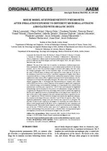
Results Histological analysis of motor cortex, spinal cord, and muscle in KIF5A–/– mice Mice lacking KIF5A have a severe phenotype with death shortly after birth [29]. We analyzed cortex, spinal cord, and muscle paraffin sections of KIF5A–/– embryos (E 18.5) and confirmed normal morphology of cortex sections. Additionally, muscle tissue appeared normal in all three genotypes (exemplary images are shown in Online Resource 2). Previous qualitative histological analysis of brain and spinal cord in KIF5A–/– embryos suggested swelling of lower motor neuron cell bodies and nuclei, but quantitative data were missing [29]. In contrast to these data we found that the nuclear area was significantly smaller in KIF5A–/– lower motor neurons of the spinal cord in comparison to KIF5A+/+ controls (115±22 vs. 141±27 μm2). Quantitative analysis of cell body area did not reveal significant differences between the genotypes, although there was a trend towards smaller cell bodies (241±73 vs. 276±82 μm2) in KIF5A–/– motor neurons. Reduced survival of KIF5A–/– motor neurons Motor and sensory neuron cell cultures were obtained from the lumbar spinal cord and the adjacent DRGs, dissected from day E 12.5 mouse embryos. The cells were grown on coverslips over 4 days in presence of neurotrophic factors. Cells were identified as motor and sensory neurons by morphology and positive anti-islet, anti-AChT, anti-MAP2ab, and antiphospho-tau staining. Typical examples are shown in Online Resource 3. KIF5A–/– motor neurons showed reduced survival rates in comparison to heterozygous and wildtype littermates. After 4 days in vitro (DIV), mean survival of KIF5A–/– motor neurons had declined to 43 %, whereas in KIF5A+/– 77 %, and in KIF5A+/+ mice 84 % of the cells survived (Fig. 1, continuous lines). In sensory neurons, there was no significant difference between the three genotypes (Fig. 1, broken lines). Reduced outgrowth of KIF5A–/– motor and sensory neurons Fluorescence microscopy of KIF5A –/– motor neurons stained with antibodies against phospho-tau and MAP2ab
Neurogenetics (2012) 13:169–179
Fig. 1 Survival of primary motor and sensory neurons from KIF5A+/+, KIF5A+/–, and KIF5A–/– mice. The number of initially counted cells was set 100 % (day 0). The percentage of surviving cells was calculated for every day in culture. Motor neurons are indicated in continuous lines, sensory neurons in broken lines. Squares show KIF5A+/+, triangles KIF5A+/–, and circles KIF5A–/– mice. After 4 days in culture mean survival of KIF5A–/– motor neurons had declined to 43±6 %, whereas in KIF5A+/– 76±5 %, and in KIF5A+/+ mice 84±6 % of the cells survived. In sensory neurons, there was no significant difference between the three genotypes at any time point. In total, at least 11 mice were analyzed per genotype. Mean and SEM are given. * denotes significant difference with p