International Journal of Clinical Trials Klassen PD et al. Int J Clin Trials. 2016 Aug;3(3):120-131 http://www.ijclinicaltrials.com
pISSN 2349-3240 | eISSN 2349-3259
DOI: http://dx.doi.org/10.18203/2349-3259.ijct20162794
Research Article
A multicenter, prospective, randomized study protocol to demonstrate the superiority of a bone-anchored prosthesis for anular closure used in conjunction with limited discectomy to limited discectomy alone for primary lumbar disc herniation Peter Douglas Klassen1*, Robert Hes2, Gerrit Joan Bouma3, Sandro Eustacchio4, Martin Barth5, Adisa Kursumovic6, Senol Jadik7, Volkmar Heidecke8, Richard Bostelmann9, Claudius Thomé10, Peter Vajkoczy11, Hans-Peter Köhler12, Javier Fandino13,Richard Assaker14, Erik Van de Kelft15, Susanne Fröhlich16, Wimar van den Brink17, Jason Perrin18, Jasper Wolfs19, Mark Arts20, Frederic Martens21 Department of Neurosurgery, 1St. Bonifatius Hospital, Lingen, Germany, 2AZ Klina, Brasschaat, Belgium, 3Sint Lucas-Andreas Ziekenhuis and Academic Medical Center, Amsterdam, The Netherlands, 4Medical University Graz, Graz, Austria, 5University Hospital, Bochum, Germany, 6Deggendorf, Germany, 7University Hospital, Kiel, Germany, 8 Klinikum Augsburg, Augsburg, Germany, 9University Hospital, Düsseldorf, Germany, 10Innsbruck Medical University, Innsbruck, Austria, 11Charité Universitätsmedizin, Berlin, Germany, 12Asklepios Westklinikum Hamburg, Hamburg, Germany, 13Kantonsspital Aarau, Aarau, Switzerland, 14Centre Hospitalier Régional Universitaire de Lille, Lille, France, 15AZ Nikolaas, Sint-Niklaas, Belgium, 16 Department of Orthopedics, Orthopädische Klinik und Poliklinik der Universität Rostock, Rostock, Germany, 17Neurochirurgisch Centrum Zwolle, Zwolle, The Netherlands,18Department of Neurosurgery, Universitätsmedizin Mannheim, Mannheim, Germany, 19MCH Westeinde, Den Hague, The Netherlands, 20 MCH Antoniushove, Leidschendam, The Netherlands, 21OLV Ziekenhuis, Aalst, Belgium Received: 11 July 2016 Accepted: 29 July 2016 *Correspondence: Dr. Peter Douglas Klassen, E-mail:
[email protected] Copyright: © the author(s), publisher and licensee Medip Academy. This is an open-access article distributed under the terms of the Creative Commons Attribution Non-Commercial License, which permits unrestricted non-commercial use, distribution, and reproduction in any medium, provided the original work is properly cited. ABSTRACT Background: Same-level reherniation and progressive degeneration with disc height loss are main causes of poor outcome after discectomy and may necessitate reoperation. A novel prosthesis for anular closure was developed to address these causes. Methods: The design of a multicenter, prospective, randomized, post-market superiority trial comparing limited lumbar discectomy augmented with this device (intervention group) with limited lumbar discectomy alone (control group) is presented. Results: Patients with single-level (L1-S1) posterior or posterolateral disc herniation and radiologic confirmation of neural compression for whom at least six weeks of conservative treatment has failed are eligible. Patients must have posterior disc height ≥5 mm at index level and baseline Oswestry and VAS leg pain scores of ≥40/100. Intraoperatively, subjects meeting anular defect size criteria post-discectomy (4-6 mm tall and 6-10 mm wide) will be randomized to study groups in a 1:1 ratio using centralized, web-based software. A Bayesian statistical approach will be used to enroll 400 to 800 subjects who will be followed for at least 24 months. Two co-primary endpoints will be assessed at 24 months: 1) a composite of leg pain, clinical function, disc height maintenance, and absence of reherniation, reoperation, and device failure; and 2) absence of reherniation based upon independent radiologic analysis. Conclusions: This type of analysis is becoming increasingly important as governments and health insurers continue to be pressured to spend limited healthcare funding wisely. Keywords: Disc herniation, Reherniation, Anular closure, Discectomy International Journal of Clinical Trials | July-September 2016 | Vol 3 | Issue 3
Page 120
Klassen PD et al. Int J Clin Trials. 2016 Aug;3(3):120-131
INTRODUCTION Despite high rates of safety and success in relieving pain and improving function, up to 30% of discectomy patients experience unsatisfactory results.1-8 Same-level recurrent disc herniation and progressive degeneration with loss of disc height are the most common causes of poor outcome and may require reoperation.5,7,9-15 The incidence of recurrent disc herniation reported in literature ranges between 0% and 27%.1,4,7,9,10,12,13,15-22 A major risk factor for recurrence is anular defect size observed at time of surgery, with defect widths greater than 6 mm identified as being at particularly high risk.13,18 The majority of patients lose more than 25% of preoperative disc height after surgery; this loss has been associated with poor clinical outcomes, particularly low back pain.5,9,12,13 A novel bone-anchored prosthesis for anular closure was developed in an effort to address these complications. It is hypothesized that the device will minimize the risk of recurrent sciatica and disc herniation by blocking the defect in the disc anulus. The device has been CE-marked since 2009, and initial clinical results have been reported at various conferences. The current post-market study, whose design is the subject of this report, compares limited posterior lumbar discectomy with and without this device. Study goals and objectives The goal of this study is to demonstrate the superiority of limited discectomy augmented with a bone-anchored prosthesis for anular closure compared with limited discectomy alone in preventing recurrent pain, dysfunction, and herniation. Success of each subject and overall study success will be evaluated at 24 months according to the co-primary endpoints defined below: 1. A composite of safety and effectiveness. To be considered a success, a subject must achieve success as follows: 15/100-point improvement in Oswestry Disability Index (ODI) score compared with baseline; 20/100-point improvement in visual analog scale (VAS) leg pain score compared with baseline; Maintenance of at least 75% of baseline disc height; No deterioration of neurological status (femoral stretch or straight leg raise (SLR); motor; reflex; and sensory) at the index level. Mixed neurological outcomes will be adjudicated by the data safety monitoring board (DSMB). Radiologic confirmation of device integrity (not fractured or disassembled) and lack of migration (intervention group only). Migration is defined as the presence of anteroposterior (AP) or lateral motion of the device > 2 mm relative to its initial position, and/or motion of the radiopaque marker(s) beyond the margin of the disc space,
associated with extrusion of the occlusion component through the anulus. No spontaneous fusion; No reherniation at the index level (see definition below); and No secondary surgical interventions at the index level.
2. Reherniation. To be considered a success, a subject must not exhibit evidence of recurrent herniation at the index level (on either side) at any time previous to, and including, the 24 month follow up evaluation. Recurrent herniation may be determined surgically by the investigator or radiologically through independent analysis (unless surgical confirmation that the suspected herniation was not an actual herniation, e.g., scar tissue).The intervention group will be determined to be superior to the concurrently randomized control group regarding safety and effectiveness if the rates of overall success are statistically superior for the intervention group compared with the control group for both endpoints. In addition, safety will be evaluated through a comparison of the type(s) and rate(s) of occurrence of adverse events (AEs) between the two groups. Study design This study is a multicenter, prospective, randomized, post-market superiority trial designed to demonstrate superiority of limited posterior discectomy augmented with the bone-anchored prosthesis for anular closure (intervention group) compared with limited posterior discectomy alone (control group) in preventing recurrent pain, dysfunction, and herniation. Randomization is 1:1 and occurs intraoperatively, following completion of the limited discectomy, to ensure homogeneity between study groups. The trial is registered at the United States National Institutes of Health Clinical Trials Registry (Identifier: NCT01283438), which may be accessed online at http://www.clinicaltrials.gov. Patients between 21 to 75 years with posterior or posterolateral disc herniations at one level between L1 and S1; radiculopathy with a positive SLR [23] (L4/5, L5/S1) or femoral stretch test (L1/2, L2/3, L3/4); radiologic confirmation of neural compression; baseline ODI and VAS leg pain scores of at least 40/100; and six weeks of failed conservative treatment are eligible for enrollment. Comprehensive listings of the inclusion and exclusion criteria are provided in Tables 1 and 2. All investigators must complete the device manufacturer’s training program prior to study participation to gain familiarity with the surgical technique for implantation of the device. To minimize learning curve bias, each investigator must have performed or participated in at least three limited posterior lumbar discectomies augmented with the prosthesis for anular closure prior to study participation. Study recruitment was initiated in December 2010, with a completed enrollment goal of approximately three years and planned follow-up of at least two years on all subjects.
International Journal of Clinical Trials | July-September 2016 | Vol 3 | Issue 3
Page 121
Klassen PD et al. Int J Clin Trials. 2016 Aug;3(3):120-131
Table 1: Inclusion criteria. Inclusion Criteria 1. Age 21 to 75 years and skeletally mature (male or female); 2. Posterior or posterolateral disc herniations at one level between L1 and S1 with confirmation of neural compression using MRI. [Note: Intraoperatively, only post-discectomy anular defects between 4 and 6 mm tall and 6 and 10 mm wide shall qualify]; 3. At least six weeks of failed, conservative treatment prior to surgery, including physical therapy, use of antiinflammatory medications at maximum-specified dosage, and/or administration of epidural/facet injections; 4. Minimum posterior disc height of 5 mm at the index level; 5. Radiculopathy (with or without back pain) with positive straight leg raise (0 – 60 degrees) (L4/5, L5/S1) or femoral stretch test (L1/2, L2/3, L3/4 only); 6. Oswestry Disability Index score of at least 40/100 at baseline; 7. Visual analog scale leg pain (one or both legs) score of at least 40/100 at baseline; and 8. Psychosocially, mentally, and physically able to fully comply with the clinical protocol and willing to adhere to follow-up schedule and requirements. Table 2: Exclusion criteria. Exclusion Criteria 1. 2. 3. 4. 5. 6. 7. 8. 9. 10. 11. 12.
13. 14. 15. 16. 17. 18. 19. 20. 21. 22. 23. 24. 25. 26. 27. 28. 29. 30.
Spondylolisthesis grade II or higher (25% slip or greater); Requires spinal surgery other than a discectomy (with or without laminotomy) to treat leg/back pain (scar tissue and osteophyte removal is allowed); Back or non-radicular leg pain of unknown etiology; Prior surgery at the index lumbar vertebral level; Patients with a SCORE of 6 or greater and a subsequent spine DXA T-score less than -2.0 at the index level. For herniations at L5/S1, the average T-score of L1-L4 shall be used; Clinically compromised vertebral bodies in the lumbosacral region due to any traumatic, neoplastic, metabolic, or infectious pathology; Pathologic fractures of the vertebra or multiple fractures of the vertebra or hip; Scoliosis of greater than 10 degrees (both angular and rotational); Any metabolic bone disease; Active infection, either systemic or local; Cauda equine syndrome or neurogenic bowel/bladder dysfunction; Severe arterial insufficiency of the legs or other peripheral vascular disease (Screening on physical examination for subjects with diminution or absence of dorsalis pedis or posterior tibialis pulses. If diminished or absent by palpation, then an arterial ultrasound is required with vascular plethysmography. Absolute arterial pressure below 50 mm Hg at the calf or ankle level results in exclusion.); Significant peripheral neuropathy, defined as Type I or II diabetes or similar systemic metabolic condition causing decreased sensation in a stocking-like or non-radicular and non-dermatomal distribution in the lower extremities; Insulin-dependent diabetes mellitus; Morbidly obese, defined as a body mass index > 40 or weighing more than 100 lbs over ideal body weight; Active hepatitis, AIDS, or HIV; Rheumatoid arthritis or other autoimmune disease; Known allergy to titanium, polyethylene, or polyester materials; Baseline MRI cannot be obtained; Pregnant or interested in becoming pregnant in the next three years; Active tuberculosis or history of tuberculosis in the past three years; History of active malignancy, defined as any invasive malignancy, except non-melanoma skin cancer, unless treated with curative intent with no signs or symptoms of malignancy for at least two years; Immunologically suppressed, defined as receiving steroids for more than one month over the past year; Current anticoagulation therapy other than aspirin, unless anticoagulation therapy may be suspended for surgery; Current chemical/alcohol dependency or significant psychosocial disturbance; Life expectancy of less than three years; Current involvement in active spinal litigation; Current involvement in another investigational study; Incarceration; and Any contraindication for MRI or CT scan (e.g., claustrophobia, contrast allergy).
International Journal of Clinical Trials | July-September 2016 | Vol 3 | Issue 3
Page 122
Klassen PD et al. Int J Clin Trials. 2016 Aug;3(3):120-131
METHODS
Neurological evaluation
Screening and preoperative assessment
Within 30 days prior to surgery, femoral stretch (L1/2, L2/3, L3/4) or SLR (L4/5, L5/S1), reflex, motor muscle strength, and sensory neurological evaluations will be performed. These results later will be compared with those obtained postoperatively.
Screening All subjects who meet the entry criteria will be considered for inclusion. Any subject meeting one or more of the exclusion criteria will not be permitted to participate in the trial. After informed consent is obtained, subjects will be assigned a study patient identification number. Subjects may become screen failures at any step until randomization occurs, at which point the subject will be considered enrolled. No further study-related follow-up evaluations will be required for non-randomized subjects. Radiologic assessment Within three months prior to surgery, magnetic resonance imaging (MRI) with T1 and T2 weighted axial and sagittal images and low-dose, multiplanar computed tomography (CT) at the index level only must be performed. Disc degeneration and extent of disc herniation are assessed by MRI; the pre-existing state of the vertebral bodies is documented by CT. Within 60 days prior to surgery, neutral AP, lateral, and flexionextension radiographs must be obtained to determine baselines for disc height and range of motion at the involved level. Medical history Within 30 days prior to surgery, demographic information will be collected; a detailed medical history, including documentation of prior treatments for back and leg pain, will be obtained; a physical examination will be conducted; and work status, current pain medications, and other drug therapies will be recorded. All patients will complete the simple calculated osteoporosis risk (SCORE) validated risk assessment tool for osteoporosis.24,25 Patients with a SCORE of 6 or greater will undergo a subsequent dual energy x-ray absorptiometry (DXA) scan to evaluate bone mineral density. A pregnancy test will be performed for all female subjects of childbearing potential. Clinical assessment Within 30 days prior to surgery, subjects will undergo the following pain and function assessments: 1) Oswestry Disability Index.26 2) Visual Analog Scales for back and right and left leg pain using a 100 mm scale; and 3) SF-36v2™ Health Survey.27,28 The baseline scores of the above assessments will be compared with postoperative scores.
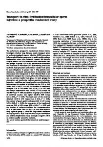
Surgery and randomization All subjects will be prepared for surgery according to hospital and investigator protocol and a single-level, limited discectomy as described by Spengler will be performed.29 This technique will remove any nucleus that has migrated within the anular defect or beyond the anular wall, including sequestered fragments. Loose fragments of nucleus from within the disc will be removed in subjects with extrusions or protrusions. Any nuclear material removed, either from within or outside the disc, will be placed dry in a syringe and the volume will be measured and recorded. After completion of the discectomy, the size of the anular defect will be measured and recorded. If the defect is between 4- and 6-mm tall and 6- and 10-mm wide, the subject will qualify for randomization in the study. Any subject whose defect size does not meet this requirement will not be considered enrolled, but will have data collected and reported through the day of surgery. Subjects who meet the intraoperative criteria will be randomized to study groups in a 1:1 ratio using a computer-generated randomization scheme maintained by a centralized randomization center, at which point no further removal of nucleus is allowed. Subjects randomized into the intervention group then will undergo implantation of the bone-anchored anular closure prosthesis under fluoroscopic control and per the manufacturer’s surgical technique and instructions for use. Subjects for whom the device is not successfully implanted will be considered treatment failures. Parameters, such as duration of surgery, blood loss, length of hospitalization, and complications, will be recorded. Anteroposterior and neutral lateral radiographs will be obtained perioperatively for all randomized subjects. Immediate postoperative care, discharge, ambulation, and any physical therapy will be per hospital and investigator protocol. Device description The Barricaid® (Intrinsic Therapeutics, Inc., Woburn, MA, USA) is an adjunct to lumbar discectomy designed to maintain the relative position of nucleus within the disc space. The device is comprised of a flexible polymer (polyethylene terephthalate (PET) or “Dacron”) mesh that prevents migration of the nucleus from within the disc and a titanium (Ti6Al4V ELI) anchor that secures the mesh to one of the adjacent vertebral bodies
International Journal of Clinical Trials | July-September 2016 | Vol 3 | Issue 3
Page 123
Klassen PD et al. Int J Clin Trials. 2016 Aug;3(3):120-131
as given in Figure 1. The mesh has a platinum iridium (radiopaque) marker for radiologic confirmation of mesh position and is attached to the anchor using suture. The implant is provided pre-assembled onto the delivery tool and has been CE-marked since 2009.
Figure 1: Graphic representation of the anular closure prosthesis, with a titanium bone anchor holding the polyester mesh in place.
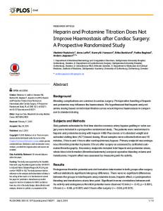
Figure 2: Neutral lateral radiograph obtained six weeks post-operatively showing the implanted device.
Table 3: Summary of radiologic evaluation parameters evaluated pre and postoperatively. Radiologic Evaluation Pre Post Parameter operative operative Quantitative measures Disc angle X X Angular motion (index and X X adjacent) Translational motion X X (index and adjacent) Disc height (index and X X adjacent) Change in disc height X (index and adjacent) Spondylolisthesis X X Change in X spondylolisthesis Qualitative measurements Heterotopic ossification X X Osteophyte formation X X (index and adjacent) Anular tears/fissures X X Disc signal intensity X X Endplate changes/reactions X X (MRI-based) Endplate sclerosis (index X X and adjacent) Device condition X Device migration X Device subsidence X Reherniation X Quantitative and qualitative measurements Modic change X X Bone resorption (each X X vertebral body, CT-based): number of lesions and lesion type X Spontaneous fusion
RESULTS Outcome assessment
Trial subgroups
Current work status and pain medication intake, as well as clinical, neurologic, and radiologic assessments, will be obtained at all follow-up intervals per preoperative protocols. Postoperative CT and MRI per preoperative protocols will be obtained at all annual follow up intervals until the final patient enrolled reaches their 24 month follow-up.
Single blinding subgroup
Independent radiologic analysis All radiologic imaging will be analyzed by independent certified radiologists. The specific parameters that will be assessed are summarized in Table 3.
Investigational sites in the Netherlands will blind subjects (120 minimum) to their treatment arm as part of a singleblind cohort to assess any possible placebo effect. Such blinding is not possible in most other locations due to patient ownership of radiographic images. The investigators agree not to disclose randomization determination to the subject until completion of the study or subject withdrawal unless an emergency un-blinding is necessary. Accidental un-blinding will be documented, with continued monitoring per protocol.
International Journal of Clinical Trials | July-September 2016 | Vol 3 | Issue 3
Page 124
Klassen PD et al. Int J Clin Trials. 2016 Aug;3(3):120-131
Economic data subgroup Selected sites will collect economic data from subjects to augment healthcare utilization data from the study and to support a cost-effectiveness analysis. Subjects shall be consented to the additional data collection. Using cost diaries, subjects will report admissions to hospital, visits (specialists, general practitioner, physical therapy, and alternative health care), home care, paid domestic help, informal care, drugs and aids, and out-of-pocket expenses as a result of sciatica and/or back pain, as well as hours of absenteeism from work. Utilities represent the valuation of patient quality of life on a scale from 0 (as bad as death) to 1 (perfect health). The EuroQol classification system (EQ-5D), collected at the follow-up intervals established in the study protocol, and SF-6D utilities, calculated from the SF-36 data, will be used for societal valuation.30-32
Patient valuation will be determined by transforming VAS and ODI scores to a utility scale.33 The total utility during each follow-up period will be calculated from the area under the utility curve as quality-adjusted life years (QALYs). Strict cost minimization analyses, comparing the costs of the two treatments, as well as cost utility analyses, comparing the cost per QALY gained/lost between the two groups, will be performed. Data will be analyzed by country, between countries, and across the entire data-set. A sample size of 150 subjects is anticipated for the cost analysis, but an interim analysis will be performed to determine if significance may be achieved earlier. Summary of study assessments and procedures The study assessments and procedures performed for each evaluation interval are outlined in Table 4.
Table 4: Outline of study assessments and procedures performed for each evaluation interval. Assessment/Procedure
Signed informed consent/enrollment Demographic information Medical history Current pain medication intake Work status Clinical assessment Neurological evaluation Neutral Lateral and AP Radiographs Flexion-extension radiographs CT evaluation MRI evaluation Patient randomization Perioperative details Economic data (subgroup only)
Preoperative†
Follow-up examinations* 6 3 6 weeks months months
12 months
24 months
Additional annual
X X
X
X
X
X
X
X
X X X
X X X
X X X
X X X
X X X
X X X
X X X
X
X
X
X
X
X
X
X
X
X
X X
X X
X X
X X
X
X
X
Surgery
X X
X
X
X X X
X
X
*The allowed window for completing follow-up examination is ± 2 weeks, 1 month, and 2 months for weekly, 6-month, and annual examinations, respectively. †The baseline radiographic evaluation must be conducted within 60 days prior to surgery, CT and MRI within 90 days, and all others within 30 days.
Trial status Participants currently are being recruited for this trial. Safety considerations Adverse and serious adverse events: All adverse clinical events that occur during the study, having been absent at
baseline, or were present at baseline and appear to worsen during the study, will be documented as AEs using definitions established by Good Clinical Practice (GCP) and the Food and Drug Administration (FDA) Guidelines. In addition to Institutional Review Board (IRB)/medical ethics committee (EC) reporting requirements, AEs will be reported to the sponsor within the following timeline requirements once the investigator
International Journal of Clinical Trials | July-September 2016 | Vol 3 | Issue 3
Page 125
Klassen PD et al. Int J Clin Trials. 2016 Aug;3(3):120-131
learns of the event: all serious AEs, including subject deaths, within 24 hours; unanticipated adverse device effects within 24 hours; and all other AEs in a timely manner. The investigator must notify the respective IRB/EC of all unanticipated adverse device effects as soon as possible, but no later than 10 days after learning of the event. Subsequent surgical intervention Any surgical intervention performed at the treated level after the index surgery will be categorized as a revision (intervention group only), removal (intervention group only), supplemental fixation, or other reoperation. Reoperated subjects will remain in the study.
Sample size calculation A Bayesian approach to sample size selection will be used. A minimum total sample size of 400 and a maximum of 800 will be considered. An interim analysis will be performed when 400 subjects have been accrued. If trial success is determined to be highly likely, per the statistical analysis plan, then accrual will be stopped. If accrual continues, another interim analysis will be performed after 50 additional subjects have been accrued. These 100-subject incremental analyses will continue until accrual is stopped or 800 subjects have been accrued. Statistical analysis
Follow-up
Interim analyses
Subjects will be followed for at least 24 months, with routine follow-up examinations at six weeks; three, six, 12, and 24 months; and annually thereafter until the last subject enrolled reaches the 24-month evaluation interval or the study is concluded. Besides the routine follow-up examinations conducted as part of the clinical trial, any adverse event that is related to the study and is continuing at the end of the study will be followed until the event has resolved or is determined to be irreversible.
The interim analyses performed before patient enrollment is stopped will be conducted as sample size determination analyses. The joint predictive probability of superiority for both co-primary endpoints will be calculated. All interim results available will be used to calculate the predictive probability of trial success for the currently accrued subjects. A decision then will be made to stop the trial at the current sample size or to continue enrollment.
Data management and statistical analysis
Early claims interim analyses
Data management
After the sample size is determined, early claim interim analyses will be performed. These analyses will occur when accrual is stopped and at 6, 12, and 18 months after accrual is stopped, yielding a possible total of four early claim analyses. No early claim analysis will be performed until at least 200 intervention group subjects have reached the 24 month follow-up interval. If the predictive probability of success (joint probability for each endpoint) in an early claim analysis is at least 0.99, then an immediate claim of superiority will be made.
The study sponsor is responsible for data management, which involves an electronic data capture (EDC) system. All data itemized in the trial protocol will be documented in the subjects’ records and on provided source worksheets that follow standardized electronic case report forms (CRFs). Only authorized investigational site personnel will complete the source worksheets and electronic CRFs.
Primary statistical analysis Electronic CRFs must be reviewed and approved by an investigator. Since there is a potential for errors, inaccuracies, and misinterpretation in transcribing data from source documents into the EDC system, originals or photocopies of all relevant worksheets, records and reports, and copies of test results must be available at all times for inspection and comparison to the electronic data capture data by the study monitor. The study sponsor is responsible for database development and data acquisition, storage, and validation. Data validation involves controls of completeness, consistency, and plausibility of data documented on CRFs using a query system between data management and investigators. After resolution of queries and upon trial closure, the database will be closed and forwarded to a biostatistician for analysis. Following completion of this trial, final reports will be issued as required.
For each of the two co-primary endpoints, the claim of superiority will be accepted if the posterior probability of superiority is larger than 0.95. The design of the mesh was modified slightly toward the beginning of the study (after 45 intervention group subjects had been enrolled). This design change was implemented to strengthen the attachment of the mesh to the anchor in response to a small number of failures (