the sputum samples stained with auramine 0 or auramine rhodamine, by a fluorescence microscope. [1,2]. An inexpensive version involves a visual examination ...
Proceedings of 2010 International Conference on Systems in Medicine and Biology 16-18 December 2010, liT Kharagpur, India
A New Algorithm for Automatic Assessment of the Degree ofTB-infection Using Images of ZN-stained Sputum Smear Rohit Nayak
Ramesh R. Galigekere
Vishnu Prasad Shenoy Dept. of Microbiology
Dept. of Biomedical Engg.
M.LT., Manipal University
K.M.C., Manipal University
M.LT., Manipal University
Manipa1576104, India
Manipa1576104, India
Manipa1576104, India
Dept. of Elec. & Electronics Engg.
Abstract-This paper describes a new algorithm for automatic
In the most common form of TB, i.e., pulmonary TB, active
assessment of the degree of TB-infection, by counting the number
infection is diagnosed by examining the stained sputum smears
of Mycobacteria i.e., acid fast bacilli (AFB) in the color images of
of subjects, by well-trained technicians. The technician looks
ZN-stained sputum smear. This algorithm consists of two stages. The
first
("pre-processing")
stage
involves
exploiting
color
information to segment the candidate AFB in the image from the
for the presence of Mycobacterium tuberculosis or acid-fast bacillus (AFB) in highly magnified microscopic images of the
background, based on classification of pixels in the HSI color
sputum smear, in which the bacillus-count is an indicator of
space using Mahalanobis distance. In this context, we introduce a
the
novel "divide and conquer" strategy to improve the robustness of
staining/microscopy in practice. The faster and more sensitive
color-classification.
The
pre-processing
stage
is
followed
by
connected component labeling, size-thresholding to remove noisy objects,
proximity-grouping
by
using
a
novel
proximity-test
algorithm, and area-based classification. Our algorithm identifies
degree
of
infection.
There
are
two
methods
of
(although expensive) approach involves manual screening of the sputum samples stained with auramine 0 or auramine rhodamine,
by
a
fluorescence
microscope
[1,2].
An
and counts the number of AFB irrespective of their shapes, can
inexpensive version involves a visual examination of highly
handle bacilli with beaded structure (which are important and
magnified microscopic fields of sputum smears stained by the
are specific to TB) and has shown reasonable success in isolating
Ziehl-Neelsen (ZN) procedure, for the presence of AFB.
clumps. A total of 205 images of ZN-stained sputum smears taken from more than 12 patients were considered in our experiments. Results on 169 images, based on HSI clusters built from 36 other images, are encouraging. Some of the images used in building the
Although the method is commonly practiced, particularly in India, it may not give accurate results because of its being tedious - only a few sub-regions can be examined as there is a
data-base and also in validation, include those sent by the RNTCP
practical limit to the number of images that can be checked by
(Govt. ofIndia) for training-purposes.
a human operator.
Keywords- Mycobacterium tuberculosis, microscopy, Ziehl Neelsen (ZN), sputum smear, beadedness, clump, color model, segmentation, Multivariate analysis, Mahalanobis distance,
recognized by researchers around the world, who have started
The
connected component labeling. L
INTRODUCTION
to vital organs with time. It is the main cause of death from an infectious disease, and currently, it kills about 3 million an [1]. Not surprisingly, the world health organization
(WHO) has declared TB as a global health emergency. The disease is said to be on the increase due to various factors including multi-drug-resistance of the microorganisms causing it and co-infection with HIV. The WHO report also mentions that 26% of the preventable adult deaths in the (so called) developing countries are due to TB [2]. Early diagnosis is, therefore, critical in controlling this disease. Further, as TB affects those with lower immunity and/or malnutrition and hence the poorer sections of the society, it is important to develop cheaper methods of diagnosis. Early and accurate diagnosis and subsequent monitoring would reduce the trauma and cost to the patient (by saving the number of visits, tests
10:115
the
manual
procedure
has
been
applying image processing techniques towards automating the detection of AFB in sputum-smear [1-6]. In the case of fluorescence microscopy, such a task is challenging because of
morbidity and mortality if neglected, as the disease can spread
and treatment).
of
a complex background - due to the presence organisms other
Tuberculosis (TB) is a serious illness that can result in
year
tediousness
than those specific to TB, acid-fast artifacts and debris, as reported in Ref. [1,2]. Consequently, the potential for false positives is high. Forero et al. [3] proposed the use of color based
segmentation
on
fluorescence-microscopic
images
captured through an autofocus algorithm, and extensive post processing involving shape-based feature-extraction, in an effort
to
improve
discrimination.
However,
fluorescence
microscopy is expensive, and the work in Refs. [1-3] involve extensive processing. Note that, in practice, the results of fluorescence-screening may have to be confirmed through ZN stained screening [7]. Sadaphal et al. [4], in their 'first report', proposed
the
use
of
image
processing
techniques
for
identifying Mycobacterium tuberculosis in the images captured from inexpensive ZN-stained sputum smears. Specifically, they use color information to label "TB objects", followed by morphological operations and the extraction of shape-related features
(axis-ratio
&
eccentricity)
size
(to
refine
identification). Classification involves a "Bayesian method",
978-1-61284-038-3/10/$26.00 ©2010 IEEE
294
Proceedings of 2010 International Conference on Systems in Medicine and Biology 16-18 December 2010, liT Kharagpur, India
clear details regarding which are not available. No quantitative results were provided, and the method does not address "V", "Y" and "T" shaped bacilli. Subsequently, Khutlang et al. [5], whose work is on lines similar to those of [1,2] involving shape-based classification, also presented the results of pixel classification based on a product of classifiers, with the best combination yielding correct and incorrect pixel-classification rates of 89.38% and 39.52%, respectively. However, they consider "T", "V" and "Y" shaped objects as "non-bacilli", although bacilli with such shapes are not uncommon - as reported in the literature, and also as per our observations. Further, their method does not address bacilli with beaded appearance (which are not only common but also specific to TB), and clumps of bacilli (whose shape is arbitrary). Note that the presence of large clumps or many small clumps generally indicates a high degree of infection. Recently, Makkipati et al [6] have presented an algorithm for segmenting AFB from images of ZN-stained sputum smears. They report the use of a hue-range for segmenting the red shaped AFB, and a saturation-range to discriminate beaded structures. However, we find that the ranges of both hue and saturation associated with the AFB overlap with those of the background-objects (as may be seen in Fig. 2 in Section V). Further, [6] asserts that all valid AFB are beaded - contrary to our observations. Also, there is no information regarding the presence of "V", "Y" and "T" shapes, and about precisely how the shape-parameters in [6] would tackle them. Finally, the paper provides only visual segmentation-results associated with 4 images, but neither quantitative analysis nor a comparison with the manual count. In this paper, we propose a new method of identifying and counting Mycobacterium tuberculosis from inexpensive ZN stained sputum smears, using color image processing, irrespective of Mycobacterial morphotypes or beaded appearance. We do not use shape descriptors, and hence our method is not restrictive. Our procedure involves segmentation of candidate Mycobacterial objects using color information, followed by connected component labeling, proximity grouping and application of size constraints. Color-based segmentation involves classification in the HSI color-space, using Mahalanobis distance [8,9]. The motivation for this method is our recent work on wound-image processing [10]. Note that our method of classification utilizes all the color space parameters (H, S & 1) together, unlike in [6] (wherein only 2 of the 3 parameters are used, separately). Further, we propose a novel "divide and conquer" strategy to improve the performance of classification in the HSI space, and a novel "proximity-test algorithm" to handle AFB with beaded appearance and to identify clumps. Our results have been presented recently at the 41 St Union World Conference on Lung Health [11]. The rest of the paper is organized as follows: Section II describes the extraction of candidate AFB by color-
10:115
2
segmentation. Section III describes post-processing steps towards identifying the true AFB. Section IV summarizes the overall procedure for counting the number of AFB. Section V presents the results of implementing our procedure on the images of ZN-stained sputum smears. Section VI concludes the paper. PRE-PROCESSING
II.
The first step in attempting to identifying the AFB in images of ZN-stained sputum-smear involves exploiting color information to identify candidate regions of AFB i.e., color based segmentation. Such an approach is natural, and follows that of a human operator i.e., color is the most important aspect that is utilized in identifying the stained AFB. We propose color-segmentation based on classification in the HSI (hue, saturation and intensity) space, as explained in the following. A.
The Color Model
The method proposed in this paper was motivated by our recent work [10], and utilizes the fact that the distribution of pixels in the bacterial region and the rest of the background are fairly distinct in the HSI space. We selected the HSI model to describe colors, because this model is close to the way humans (and hence the operators who work with images of the stained sputum smear) perceive color. Hue describes pure color, saturation refers to the "degree of dilution from purity" by white light, and intensity is similar to that associated with gray level pictures (and is decoupled from the color-information embedded in hue & saturation). Since color-images from the microscope are available in the RGB-format, the HSI parameters would have to be extracted by the following transformations: H-
B:G
I
B
=
COS-I
S=l-
-[(R-G)+(R-B)
{
2
J(R-G)2 +(R-B)(G-B) 3
(R+G+ B)
}
(2)
Min{R,G,B}
1 1 =-(R+G+ B) 3
B.
Color-based Classification: The Basic Approach
The procedure for segmenting the candidate bacterial regions based on color consists of two phases: (i) Building up a knowledge/data-base (clusters) corresponding to the colors associated with the bacilli and the background, and (ii) using the database to classify the pixels into the class of (candidate) bacteria or the background. This approach is similar, in principle, to the method used by us in our recent work for assessing wound-composition [10]. Specifically, one maps a large number of pixels belonging to bacteria and the background from several images of ZN-stained sputum smears, into the HSI space. These pixels form clusters in the HSI 295
Proceedings of 2010 International Conference on Systems in Medicine and Biology 16-18 December 2010, liT Kharagpur, India
space, representing the knowledge regarding the color-content associated with the bacteria and the background, respectively.
III. A.
Once the color-knowledge-base is built, segmentation may be achieved using the minimum distance rule. Specifically, a pixel is considered to belong to the bacterial region if it is closer - in terms of its color-content - to the cluster associated with the bacterial region in the HSI space. The measure we employ to assess proximity to a cluster is the Mahalanobis distance [8,9]. Specifically, if x is the vector whose elements are the H, Sand 1 components of a given pixel, its distance in the HSI space from the cluster indexed by i, is given by: (3) where C is the covariance matrix and P, is the mean, respectively, of the cluster under consideration. The index (subscript) i is used to indicate the cluster representing the color associated with the stained bacteria and those present in 1 and 2, respectively. The rationale the background i.e., i behind choosing the aforementioned measure lies in the fact that the clusters are not spherical, and the covariance matrix takes care of the cluster-shapes. Indeed, the contours of constant density (3D histogram) are hyper-ellipsoids of =
constant Mahalanobis distance from Pi [8].
C.
3 POST-PROCESSING
Connected Component Analysis
The pre-processing stage described in the preceding reduces the color images of sputum-smear to a binary version. The binary image generally consists of regions (connected components or "blobs") representing the bacilli in individual or beaded appearance or in clumps, and noise. The first step in post-processing involves connected component labeling [8,12], which involves segmenting and labeling the set of all connected-components in the binary image. This is achieved by scanning the binary image and recording the set of all 8adjacent pixels, which results in another binary image consisting of a set of labeled objects/blobs. B.
Noise-elimination
Errors due to color-segmentation, debris, or bacteria which may have got splintered in the pre-processing stage are likely to leave blobs which are generally tiny in size. They can be eliminated from consideration for further processing by retaining only blobs of size (area, in number of pixels) greater than a predefined value, Amin• The value of Anlln will have to be determined empirically. At this juncture, we wish to note that the use of divide-and-conquer strategy-assisted color-based classification reduced the size of noisy blobs significantly, in comparison with that associated with the background un-split.
Divide and Conquer Strategy
The clusters, associated with the bacteria and the background, happen to be fairly distinct. Although they suffer a minor overlap due to similarity in color-content, regions with such a similarity may not occur often - as evident by the smaller number of pixels in the overlapping region (Fig. 2). Nevertheless, we propose a strategy to address this issue. The strategy consists of dividing/splitting the large cluster associated with the background to several smaller sub-clusters so that segmentation of pixels associated with the AFB (i.e., of correct classification) becomes more robust. The logic behind this operation is based on the fact that the expression for Mahalanobis distance involves distance from the mean, apart from the inverse of the covariance. If the cluster associated with, for example, the bluish objects (sometimes with some pink-content) e.g., epithelial cells, possibly interfering with the bacterial cluster - is considered a separate cluster, any point that is truly from the epithelial region lies very close to the mean associated with the that sub-cluster but far from the mean associated with the bacterial cluster, and would be classified as a non-bacterial object. Indeed, as seen from the results presented subsequently, this approach improves the robustness of segmentation. As the technique involves dividing the background-information into several sub-clusters towards improving the performance by aiding background rejection, it has been termed as a "divide and conquer" strategy.
C.
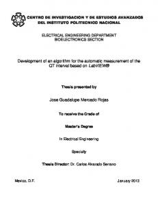
Proximity-test Algorithm
Acid fast bacillus with beaded appearance is common in, and also specific to, Mycobacterium tuberculosis. Detecting beadedness is therefore important as it is specific to AFB, and also because, a bacillus with beaded-appearance should not be counted more than once. To handle AFB with beaded appearance (and hence with a beaded structure i.e., a chain of closely spaced blobs - in the color-segmented binary image), we propose a new "proximity-test algorithm", towards counting as one, blobs/objects that are very close to each other. The steps in the proximity-test algorithm are given in the following: 1. (a) Get the coordinates of the extreme-points associated with the first object:
Pll ==(xminl'Yll),PI2 ==(xmaxl'YI2)' where, Yll and YI2 are the y-coordinates of PI I and PI2 respectively. Similarly,
�3 ==(xI 3,Yminl)'PI 4 ==(xI 4,Ymaxl) (b) Get the coordinates of the extreme points associated with the second object:
P21 ==(xmin2'Y21),P22 ==(Xmax2'Y22)' P23 ==(x23,Ymin2),P24 ==(x24,Ymax2)
10:115
296
Proceedings of 2010 International Conference on Systems in Medicine and Biology 16-18 December 2010, liT Kharagpur, India
4
2. Find the distance between each of the extreme points
the operation in Step-I. This results in a set of labeled
associated with object-I, and those associated with object-2.
objects/lobs.
3. Find dmm i.e., the minimum of all the distances calculated in
3. Size-thresholding: Eliminate noisy objects of size (area) less
step-2.
than the specified minimum, to remove noisy objects.
4. If the value of dmin is less than a pre-defmed value, the two
4. Proximity Grouping: Run the proximity-test algorithm, pair
objects are grouped together.
wise, over the set of all blobs in the image, to detect groups of closely-spaced objects/blobs. Store the indexed array of blobs
An
illustration
for
the
proximity-test
algorithm
(for
i.e., an indexed set of groups/chains. 5. Apply size-constraints: Chains/groups are classified into
"proximity-pairing" of blobs) is provided in Fig. l.
different forms of appearance, based on the specific size-range D.
to which they belong.
Proximity-grouping
6. The number of members in each of the area-ranges yields
A run of the proximity-test algorithm performs proximity
the
count
of
the
respective
types
of
appearance
of
pairing of two closely spaced blobs/objects. Proximity-pairing
Mycobacterium tuberculosis i.e., the number of (i) unbeaded
can be used over the set of all blobs in the image, to perform
AFBs, (ii) beaded AFBs, or (iii) AFB in clumps, respectively,
"proximity-grouping" of closely spaced set of blobs i.e.,
in the image.
generation of indexed array of chains/groups of proximal V.
blobs/objects, in which each blob is close to at least another in
RESULTS
the group. Such a grouping paves the way for identifying
Thirty six images of ZN-stained sputum smears, captured by
beaded structures, and also clumps; clumps are groups of
the Leica DFC 320 Camera at a magnification of 1000x at the
bacteria - beaded
or
Dept. of Microbiology, Kasturba Hospital, Manipal, India,
overlapping, that clear counting is not possible. The presence
or
unbeaded - that are so close
were used for building the data-base. Some of them included
of large clumps or even many small clumps in a smear
smears sent by the Revised National TB Control Program
generally indicates a higher degree of infection. It is important
(RNTCP), Govt. of India, for training technologists as a part of
to note that an image of sputum-smear can contain AFB in the
a quality-control program. The images of size
form of: (i) an unbeaded bacillus, (ii) beaded-bacillus, and (iii)
were captured under a standardized set of imaging parameters.
a
clump
of
bacilli
(since
noisy
objects
are
1550x2088
removed
The procedure for classification explained in Section III, has
immediately after connected-component labeling). Note that,
been implemented on MATLAB, by first building the data
the number of beads per individual AFB has been found
base in the HSI space as described in
empirically, to be less than 5.
background was split into two clusters: one corresponding to
Section II.
The
the bluish objects (epithelial cells) and the other pertaining to E.
the rest of the background. Note that experiments without such
Size-constraints and Counting
As described in the previous subsection, proximity-grouping generates an indexed array of chain/groups of blobs, which will have to be classified further into one of the following forms of presence of AFB: (i) individual, (ii) beaded, or (iii) clumps. Such a classification is possible by noting that size range associated with unbeaded individual bacillus, beaded
a splitting of the background can be performed by simply combining the two clusters and considering it as one. The clusters in the HSI space, with the background split, are shown in Fig. 2. Subsequently,
169 images of sputum-smears, ZN
stained according to the RNTCP guidelines (in conformity with the WHO standard), were considered for testing (the set
individual bacillus, and clump, are different. We label the
of 205 images used in our experiments, 36 of which were used
ranges as Rj,R2 and R3,respectively.
for building the data-base, involved more than 12 patients). The results of color-segmented images were then subject to
After imposing size-constraints, the number of members
post-processing as described in Section III. Two samples of
in each of the size-range yields the count of the respective
images of ZN-stained sputum smear, and the result of color
forms in which the AFB appear: unbeaded, beaded or in
segmentation are given in Fig. 3 - showing the efficacy of our
clumps.
method. The range of areas, in terms of number of pixels, for classifying the blobs into noisy objects, IV.
THE OVERALL PROCEDURE
individual AFB,
beaded AFB and clumps, were found empirically (based on a
detecting and counting the
careful examination of a large number of images). Specifically,
instances of AFB in a digital image j(m,n) of ZN-stained
objects less than 30 pixels in size were discarded, and the rest
sputum smear, is summarized in the following steps.
of the area-ranges were found to be: R), = [ 150, 900], R2
The
overall
procedure
for
[400,900], and R3 1.
Color-based segmentation: Extract the candidate AFB
=
[90I,Amax]. Currently, Amax has been set to
be the size of the image itself, as we did observe some large clumps. The program "bwlabel" in MATLAB was utilized for
regions by color-based segmentation. 2.
=
connected
connected component labeling [8,12]. The computer-estimated
component labeling, on the binary image which results from
counts, along with the respective visual-counts, associated with
Connected
Component
Labeling:
Perform
169 images, are displayed as plots in Fig. 4.
10:115
297
Proceedings of 2010 International Conference on Systems in Medicine and Biology 16-18 December 2010, liT Kharagpur, India
5
The efficacy of our algorithm in detecting unbeaded & beaded
AFB, and clumps of AFB, may be quantified by observing the percent correctness ("%correct") of detection of the respective forms of appearance, as listed in Table-I. The quantity "%Correct" is defined as follows:
%Correct
where,
=
r
N
1
LIE", E , I 1
-
i�l
.
N
c
-
LE", f=!
.
xl 00
Ev; is the visual (manual) count of the AFB present in
x
the image with index ''1"', Ec; is the respective computer
Figure I. Illustration for the proximity-test algorithm
assessment (count extracted by our algorithm) and N is the total number of images (169 in the present case). Note that the two cases of color-segmentation i.e., those with the background split and un-split, have been considered. The improvement afforded by the divide & conquer algorithm may be observed in this table. The overall success of detecting AFB was 93.5%. The quantitative results do indicate that the overall procedure has worked fairly well.
VI.
CONCLUSION
We have developed a new algorithm towards identifying and counting the number of Mycobacteria (AFB) in the images of inexpensive ZN-stained sputum smears. The algorithm aims to work irrespective of the shape of the AFB, and also to detect AFB with beaded appearance and/or in clumps. The basic pre-processing involved color-segmentation of candidate bacterial regions, by classification in the HSI space using
HUE
Mahalanobis distance. A novel "divide and conquer" method has been found improve the robustness of color-based classification. subject
to
The
color-segmented
connected
component
binary
images
labeling
and
were size
thresholding, and grouped by using the new proximity-test algorithm. Finally, size-constraints were imposed to extract the
Figure
2.
Scatter-plot of the data-base in the HSI space, with the background
cluster split into two sub-clusters. Legend: Red - bacteria
(16,70,542 pixels) (12,57,309 pixels).
Blue - bluish epithelial cells rest of the background
(51,839
pixels),
in the background, Green -
counts of AFB in unbeaded and beaded appearance, and in clumped presence, respectively. Results on 169 images of ZN stained sputum smears have been found to be good. A major factor contributing to the success of our algorithm is the excellent color-segmentation. Future work involves identifying additional factors that characterize clumps, towards improved detection of clumps.
298
Proceedings of 2010 International Conference on Systems in Medicine and Biology 16-18 December 2010, liT Kharagpur, India [2]
6
K. Veropouloset
al .,
"Image Processing and Neural Computing Used in
the Diagnosis of Tuberculosis",
Proc. Colloqium on Intelligent Methods in Healthcare and Medical Applications, Digest No. 98/514, Smith &
y
Nephew Research Center, York, Control Division, LEE, Oct. 1998. [3]
Shape and Color",
Yonaxl
[4]
[5]
P"1
[6]
10:115
Yonin] Yonax2
et al., "Identification of Tuberculosis Bacteria Based on Real Time Imaging, Vol. 10, 2004, pp. 251-262 P. Sadaphal et al., "Image Processing Techniques for IdentifYing Mycobacterium Tuberculosis in Ziehl-Neelsen Stains", Int. J. Tuber. Lung Dis., Vol. 12, No. 5, pp. 579-582. R. Khutlang, et al., "Classification of Mycobacterium Tuberculosis in Images of ZN-Stained Sputum Smears", IEEE Trans. Inform. Tech. Biomedicine, Vol. 14, No. 4, July 2010, pp. 949-957. V. Makkapati et al., "Segmentation and Classification of Tuberculosis Bacilli from ZN-stained Sputum Smear Images", Proc. 5Th Annual Conf Automation and Engg., Bangalore, India, Aug. 2009. M.G. Forero
[7]
I I I I I I ---,-------------
Revised National Tuberculosis Control Program (RNTCP), Manual for Sputum
Smear
Fluorescence
Microscopy,
Central
TB
Division,
Directorate General of Health Services, Ministry of Health and Family Welfare, Govt. of India, New Delhi. [8]
R.C. Gonzalez, R.E. Woods, and S.L. Eddins,
Using MATLAB,
Digital Image Processing
Pearson Education Inc. and Dorling Kindersley (India)
Pvt. Ltd., Delhi, 2004. [9]
T.
Yakov
and
G.A.
Multivariate Analysis,
Marcoulides,
An Introduction to Applied
Routledge, Taylor and Francis Group, New York,
2008. [10] Rohit Nayak, Pramod Kumar and Ramesh R. Galigekere, 'Towards a Comprehensive Assessment of Wound Composition Using Color Image Processing", Proc. Figure 3. (a) & (b) are two color images of ZN-stained sputum-smears. The small, reddish objects are the bacteria. (c) & (d) show the color-segmented
iEEE Int. Conf Image Process.
(ICIP'09), Nov. 7-
11, Cairo, Egypt, 2009, pp. 4185-4188. [11] Rohit Nayak, V.P. Shenoy and Ramesh R. Galigekere, "Automatic Assessment of the Degree of TB- infection Using Images of ZN-stained
binary versions of (a) and (b), respectively.
Sputum Smear" presented at the 41st
Health,
[12]
Union World Conference on Lung
ICC, Berlin, Germany, 11-15 November 20 I o.
A.K. Jain,
Fundamentals of Digital Image Processing,
Prentice Hall,
Englewood Cliffs, NJ, 1989.
Figure 4. A plot showing the assessment of the degree of TB-infection, in terms of number of AFB patients) with index
i.
in each of 169 images (from more than 12
(N)
Solid green line shows the true count, and the dotted
line shows the computer-estimated count.
TABLE I.
Percent-correctness
of the detection of AFB with unbeaded &
beaded appearance, and in clumps. The results for the two cases
i.e.,
of
classification with the background split as well as un-split, are shown.
Background
Individual AFB (beaded & unbeaded)
Beaded AFB
Split
93.488
83.5979
Unsplit
87.57
53.549
Clumps 82.4427 51.1450
REFERENCES [1]
K. Veropoulos
et al.,
"The Automated Identification of Tubercle Bacilli Th
Using Image Processing and Neural Comuting Techniques", Proc. 8
Int. Conf Artificial Neural Networks,
10:115
Sep. 1998, pp. 797-802.
299