ORIGINAL RESEARCH
A New Approach That Improves Range of Motion Without Stretching or Pain Comparison With PNF Vinicius Monteiro Diederichs
Abstract Objective: the purpose of this study is to verify the efficiency of a new approach to improve range of motion of joints without stretching. In this comparison proprioceptive neuromuscular facilitation (PNF) is used as standard approach. Methods: A total of 22 subjects suffering from lumbo-pelvic stiffness, 9 females and 13 males (mean age 40 years, range 20-63 years) are selected. Subjects are randomly allocated in two groups, A and B. The bending test is used to verify the range of motion before and after the intervention. Both groups are submitted to the new approach, and to the standard PNF for comparison, performed in two different sequences; data is then collected for analysis.
Vinicius Monteiro Diederichs; Ronin Institute for Independent Scholarship. Keywords: joint stiffness, thixotropy, muscle spindles, stretch reflex, recalibration, joint impedance, motor control, effort. Correspondence: Vinicius Monteiro Diederichs; Ronin Institute for Independent Scholarship; Via Giuseppe Zanardelli, 36; Cap: 00186 Roma; Italy. E-mail address:
[email protected] Introduction This paper introduces a new approach able to improve ROM without stretching. Earlier approaches, such as proprioceptive neuromuscular facilitation (PNF) and static stretching, appear effective in improving ROM.1 Theoretically however, forced stretching of skeletal muscles causes muscle spindles to become insensitive to muscle tension, thus preventing normal proprioceptive functioning. Moreover, in the case of a multi-joint segment, use of standard stretching is questionable. This is an important issue raising questions as to whether 48
Functional Neurology, Rehabilitation, and Ergonomics • Vol. 7, No. 4
Results: in the first sequence, both PNF and the new approach are significantly effective P < .001 while in the second sequence only the new approach demonstrates to be effective. Conclusions: while both PNF and the new approach are effective in improving range of motion (ROM), the new approach is more efficient and avoids pain in execution. The new approach is also effective in resolving the pain related to postural maladaptation, such as pelvic misalignment. Strikingly, after the performance of the new approach, all subjects reported a pleasant sensation of lightness, often described as little bubbles touching the skin of the exercised limb, a benefit awaiting further investigation.
stretching should be used to improve ROM. In fact, excessive muscle stiffness is not caused only by the biomechanical properties of muscle, such as elasticity and viscosity, but is also the result of a neural strategy to maintain an equilibrium position during functional tasks. The above ideal ROM depends on the proper functioning of mechanoreceptors, especially the muscle spindles, which must continuously send feedback information toward the central nervous system (CNS), that is, to the spinal cord. By sending this continuous flow of feedback information, muscle spindles are able to close the loop of the neural circuitry,2 thus turning on the normal muscle reflex. In subjects suffering excessive stiffness of lumbopelvic muscles, however, integration of the neural circuitry seems to be partially disrupted, especially as a result of diminished abdominal oblique muscle (AOM) ability to contract.3 The following paragraphs address two specific phenomena that cause malfunction of proprioception. These are followed for further clarification by an analogy between the normal pattern of a muscle’s activation during gait and the new approach. To increase the precision of movement of a multi-joint segment, the neural controller must stiffen certain joints reducing degrees of freedom (DOF), a condition termed joint impedance.4 It would appear that Diederichs—Improve ROM Without Stretching
subjects suffering postural discomfort and pain often present significant lumbopelvic stiffness which reduces the DOF. In some way, this exaggerated impedance in the pelvic joints seems to be necessary. It can be a normal neural strategy to overcome a malfunction in a population of stretch receptors, such as muscle spindles, suggesting that the neural controller does not have all the proprioceptive feedback required. Taking this case, improvement in lumbopelvic ROM is unlikely so long as proprioceptive drift is present. Proprioceptive drift is a range of joint angle that is unresponsive to the subject’s perception5 and also to the stretch reflex. To overcome proprioceptive drift, the neural controller must adapt the body posture by shortening the extrafusal muscles6 where spindles signal a background. In order to identify this proprioceptive drift, it is necessary to understand the normal pattern of muscle activation during a functional task such as gait. Indeed electromyographic (EMG) study performed on healthy subjects during a normal gait has found high levels of activity on the contralateral AOM relative to the limb, then in the swing phase.7 This is in contrast to subjects suffering back pain, who manifest significant asymmetric stretch reflexes comparing right and left side AOM. By manually pressing simultaneously both right and left sides, it is possible for the clinician to verify if one side’s AOM shows an asymmetrical latency response or even an absence of response to the stretch reflex.8 In consequence, during gait one side’s AOM is insensitive to stretching (i.e. to the stretch reflex), falling slack and causing proprioceptive drift on only one side of the trunk. Here proprioceptive drift must require motor adaptation, such as stiffness in the lumbopelvic muscles. Proprioceptive drift is caused by the insensitivity of muscle spindles as a result of a biophysical property of the muscle fibres themselves; this is termed thixotropy. Thixotropy is a biophysical property of all muscles. It is a dry, friction-like, passive stiffness which is distinct from both a muscle’s elastic (length-dependent) and viscous (velocity-dependent) properties, being contraction history dependent. It affects the status of cross-bridges between myosin filaments, affecting both extrafusal fibres of skeletal muscles and the intrafusal fibres of muscle spindles. Thus thixotropy applies to materials that change their physical properties as a result of movement. A further consequence of thixotropic behaviour is the vulnerability of muscle fibres to slacking, as a result of their previous history of contraction and length changes. This has implications for reflex action and roles in proprioception.9 The term “slack” refers to the condition in which a muscle fibre’s actual length is greater than the distance between its points of attachment. Slackness can be demonstrated by the presence of a delay in the onset of a response to fusimotor stimulation, with significant consequences for the neural control of muscles. If the muscle is contracted and its elastic limit is reached Diederichs—Improve ROM Without Stretching
however, the latency shift of stretch reflex is reduced and disappears rapidly.10 This thixotropic behaviour is both muscle-length and time dependent, thus only a slow, wide amplitude, movement resets the normal functionality of muscle spindles. A further phenomenon is the behavioural dynamic dominance of motor control, causing motor adaptation which appears to exacerbate the thixotropic effect. Having the possibility of various motor solutions, human motor behavior simplifies motor actions seeking efficiency in terms of energy consumption. This, in turn, leads to a tendency to prefer one side of the body rather than the other when performing specific motor tasks, such as fine movements or stabilization. This body side preference was previously termed brain lateralization,11 but now the term used is dynamic dominance. The theoretical framework for dynamic dominance originates in the work of Sainburg et al. This has the dominant arm performing dynamic movement tasks based on neural controller predictions and optimizations, while the non-dominant arm is more specialized, performing movement to achieve the desired goal position by specifying impedance in that position. Dynamic dominance in motor control is a body side preference to perform fine movements, while the other, non-dominant, side is used as a beam or stabilizer.12 This arm dynamic dominance can affect the conditioning state of AOM, with impact on gait. In fact, a characteristic of gait is the prevalent coronal plane rotation, which requires side-to-side measurement to ascertain whether there is an asymmetry in the mediolateral rotation. Significant asymmetry was found in the frontal plane trunk rotation in back pain subjects.13 This rotational asymmetry seems to relate to thixotropic effects. Empirically, my study finds significant asymmetry in the coronal plane rotation among those people suffering postural discomfort and pain. When a subject passively rotates the trunk in a sitting position, the rotation on one side of the trunk is significantly wider than on the other. This is in agreement with the impact of dynamic dominance on motor control, causing motor adaptation and exacerbating the thixotropic effect, thus causing significant slackness in one side’s AOM. At this point, the dynamic dominance of one arm causes an unusual top to bottom asymmetry of AOM that can provoke problems in gait. Turning to empirical analysis of a normal gait pattern, gait is essentially a sequence of mediolateral movements of the body. Moving the foot in a mediolateral direction also creates a mediolateral moment of the COM. Indeed, when the subject moves the foot forward, the COM gains moment in a mediolateral direction. At the end of the swing phase, the foot is ready for the strike, which in turn generates an anterior-posterior reaction force that counteracts the mediolateral moment of COM, thus resulting in a zero-moment force. It is this counteracting force that allows equilibrium control during gait. In a normal speed gait, equilibrium control is achieved by maintaining the COM on the base of support,14 oscillating Functional Neurology, Rehabilitation, and Ergonomics • Vol. 7, No. 4
49
between feet. This oscillation begins when one foot accumulates energy as a spring to thrust the COM forward towards the opposite foot, which is in a terminal swing phase. In the stance phase, the ground energy is accumulated by plantar flexion and then transferred by coupling rotational forces. This coupling starts with the leg rotating externally, the thigh internally and the iliac externally.15 The iliac external rotation seems to be controlled by an eccentric contraction of AOM. The hypothesis of this study is that slackness of one side’s AOM causes a proprioceptive drift which prevents the normal functional pattern of iliac rotation during stance phase of gait, thus causing motor maladaptation due to an excessive stiffness of lumbopelvic muscles. To resolve the motor maladaptation, the new approach expounded here uses major aspects of functional motor control, such as the mediolateral rotation of the body, and an ideal base of support. But this base of support uses a top to bottom, rather than bottom to top process, to control a fine point-to-point movement using the foot (see Figure 1). Indeed, the designated position to be assumed in this technique is a quasi-crawl position. As a result the base of support is kept firm by holding a fixed bar which allows the subject to maintain the forearm stable. The forearm’s position diminishes the DOF by impeding elbow joint rotation, thus reducing the possibility of movement of the shoulder girdle. The position therefore turns the normal impedance of the body upside down, releasing lower limb impedance, thus facilitating performance of more dynamic movement tasks, as predicted by one arm dynamic dominance. More specifically, in a normal standing position impedance of joints progresses from bottom to top; when assuming the crawl position and ensuring stability of the shoulder girdle however, impedance progresses inversely from top to bottom. This progressive top to bottom impedance is possible because of a volitional point-to-point movement of the foot. While the foot moves in a mediolateral direction, the pelvis shifts in a contralateral direction in an innate strategy to maintain the COM on the base of support (Figure 2). This is analogous to the movement observed in the pelvis when a contralateral foot is in the swing phase during gait. From the quasi-crawl position above, the mediolateral movement of the foot and the contralateral shift of the pelvis cause eccentric contraction of oblique muscles. This seems effective in recalibrating muscle spindles, which activate the neuronal mechanism, having the effect of immediately improving ROM by releasing lumbopelvic muscles. The position assumed in the new approach stabilizes the body from top to bottom, allowing one foot to move for the performance of a volitional movement point-to-point. To stimulate efference copy,16 elastic tubing is used on the foot. The action must be slow, allowing the subject to track the foot during all its range of motion, and visual feedback is also helpful. Here, the subject rotates the head to visually track the foot displacement. To verify efficacy of the new approach I 50
Functional Neurology, Rehabilitation, and Ergonomics • Vol. 7, No. 4
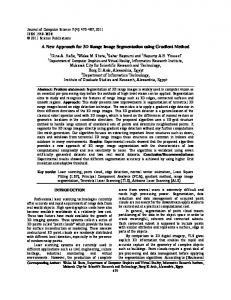
Figure 1. The equipment is specifically designed to be portable, allowing the experiment to be performed at the subject’s home or office. The equipment can, however, be anything (see notes) that allows firm grip of hands and elbows in order to stabilize the shoulder girdle. The diagram at the rear indicates the body’s position with the foot at point A; the diagram at the forefront indicates the body’s position with the foot at point B. In both figures the upper and lower limbs are indicated by thick continuous lines, the empty spheres indicate joints; the thoracic spine and pelvis are indicated by the rectangular box; feet are indicated -in red; the elastic tubing is indicated by the spring; thick arrows indicate movement direction and rotation; the narrow zigzag line indicates concentric contraction of oblique muscles (CCOM), while the broad zigzag line indicates eccentric contraction of oblique muscles (ECOM). The exercise is performed by slowly moving the foot from point A to point B, the other leg remaining in the kneeling position, the knee under the hip in order to bear the weight of the trunk. Note the difference in the contraction state of oblique muscles when comparing point A to point B.
Figure 2. This shows the shift of the pelvis as the foot moves to point B. The rectangular box indicates the pelvis from a transverse view; the line on the left indicates the thigh, the knee bearing the weight of the trunk; the line on the right indicates the thigh and the shank at maximal knee extension; empty spheres indicate joints; the foot is indicated in red; the arrow indicates the direction of movement of the pelvis when the foot is at point B; the arrow close to the foot indicates external rotation of the limb, which requires the toes to point upwards.
Diederichs—Improve ROM Without Stretching
Figure 3. Indicates performance of the bending test.
use PNF, this being frequently cited in the literature to be effective in improving ROM of the lumbopelvic complex. Data collection was obtained using the bending test. Procedure for performing the new approach From a quasi-crawl position, the subject with supine hands holds a fixed bar to maintain the elbows in contact with the equipment, while ensuring that both shoulders are perpendicular to the elbows and that the bearing hip is perpendicular to the knee. Light resistance tubing should be used on the foot free to move. Before trying the action, the subject must understand the procedure, by watching a video or by analysing a sketch (see notes). Clarification must be given informing the subject that the exercise will challenge the body’s equilibrium resulting in perception of high-level effort. Thus the clinician must instruct the subject to hold the bar firmly, keeping the elbows down to stabilize the spine and then perform the action by moving the foot. It is necessary that the subject understands that he or she needs to support the high level of effort during the action of the foot from point A to B, varying from 3 to 5 repetitions. It must also be emphasized that it is extremely important to perform the movement slowly in order to be able to track visually the foot displacement. After providing the instructions, the clinician assists the subject in performance of the action by holding the foot and positioning it at point A. To ensure confidence without assistance, the clinician asks the subject to hold the position in point A steady in place for few seconds, asking: Can you hold the position alone? With an affirmative response, the clinician assists the subject to move the foot toward point B, asking the subject to rotate the head to watch the foot. Further assistance can be given by holding and guiding the subject’s foot displacement (see notes). Methods and Methodology Subjects Twenty-two subjects suffering lumbopelvic extensors stiffness, 9 females and 13 males (mean age 40 years, range 20-63 years), participated in the study. Many of these subjects were sedentary; a few subjects practice some recreational sport Diederichs—Improve ROM Without Stretching
at the weekend, while the majority work as office workers or factory workers; only two are students. The inclusion criterion was stiffness in the lumbopelvic extensors, defined as hands remaining far from the floor when performing the bending test. The experiment was performed at their home or office. An ethics committee formed by the local community of sports and exercise scientists approved the study. All subjects signed an informed consent form agreeing to participate in the study prior to testing, in accordance with the principles expressed in the Declaration of Helsinki. Procedures The New Approach. Ideally a simple, portable, but carefully designed, piece of equipment is used to limit the DOF of certain joints, but this can be anything that allows firm grip of hands and elbows in order to stabilize the shoulder girdle (see notes). The quasi-crawl position allows sufficient stability when holding the bar while keeping both elbows as well as one knee firmly on the equipment, allowing the other limb to move (figure 1). Elastic tubing on the foot is used to increase efference copy. Thus the subject is required to perform a volitional movement between two points, A and B, maintaining a slow motion to be able to track the foot displacement. The new approach uses major aspects of functional motor control (as previously cited), and slow, wide-amplitude, movements for recalibration of muscle spindles. The PNF Approach. From a supine position the subject uses a belt to hold the limb and then to draw it toward the chest. Using the belt under the foot, the subject raises the limb until it reaches the stiffness barrier and pulls it toward the chest, counteracting the movement with the limb extensors. The counteracting forces are held for 6 seconds, followed by relaxing the limb extensors and then pulling the limb toward the next barrier for 5 to 10 seconds. Thus, in the first phase, the counteracting forces produce an isometric contraction of the limb extensors, while in the second phase the subject applies a forced stretching of the relaxed limb extensors until the next stiffness barrier is reached. This is performed in three sets, increasing the duration of the second phase up to 30 seconds. To collect data, the bending test is used in both approaches to measure the distance of hands from the floor. From the standing position the subject keeps the knees locked and bends the trunk down with arms relaxed, perpendicular to the floor, see figure 3. Both the new and the standard approach are self-performing, with some assistance where necessary. The stiffness of the lumbopelvic extensors are measured before and after the performance of both approaches. The bending test is used, measuring the distance of the hands from the floor. For greater accuracy, all subjects must receive precise instructions with a demonstration of how to perform the bending test, the new approach and the standard approach. Subjects are crossover allocated to two groups, A and B. Both groups perform both approaches but in a different Functional Neurology, Rehabilitation, and Ergonomics • Vol. 7, No. 4
51
order: Group A starts with the new approach, followed by a resting time of 5 minutes, then moving to the standard approach; Group B does the opposite. The ROM is checked before and after the performance of each approach. Instructions In performing the bending test, it is essential to keep the knees locked in maximal extension and then, as the subject starts to bend, not to force the bend beyond the stiffness barrier, but to release the body down, keeping the arm relaxed, until data is collected, see Figure 3. To perform the new approach, subjects are first shown an illustration better to understand how to position themselves on the equipment. The researcher then demonstrates the exercise, and a further explanation is delivered, using the illustration, while the subject positions him or herself on the equipment. An additional important explanation to be given is the high level of effort required to perform the new approach, since the exercise position is challenging for the body equilibrium when moving the foot between points A and B. A source of motivation is the number of repetitions. This varies between 3 to 5, depending on the movement’s quality. To perform the standard approach, an illustration was again used as explanation and then the approach was demonstrated. From the supine position, the leg is raised until the stiffness barrier is reached; a belt is placed on the plantar foot and then tightened. The action starts by performing a maximal isometric contraction for 6 seconds, and then relaxing the limb so as to apply a forced stretching by pulling it toward the next barrier for 5 to 10 seconds, increasing the duration of the forced stretching up to 30 seconds from the second set. Means and standard deviations of pre-intervention and post-intervention data are then calculated for each group. In addition, the mean difference between before-intervention and after-intervention is calculated; before-intervention and after-intervention is calculated also for the second approach. The paired t-test is used to verify statistical significance pre-intervention and post-intervention. The level of significance is P < .05. Statistical analyses are performed using SPSS version 21. Results In this comparative study, subjects were tested three times, at pre-intervention, after the new approach and after the standard approach, the level of significance being 0.05. In Group A, the paired t test indicates strong correlation, score .682 for improvement in ROM after the performance of the new approach; the value of the test statistic is 5.562, P-value associated to the t test, P < 0.001, which indicates significant improvement in the ROM post-new approach. On completion of the new approach, the same subjects performed the standard approach; the value of the test statistic is 2.250, p-value associated to the t test .051 > .05. This finding indicates non-significant improvement after the performance of the standard approach. 52
Functional Neurology, Rehabilitation, and Ergonomics • Vol. 7, No. 4
In Group B, the paired t-test indicates a strong correlation, score .801 for improvement in ROM after performance of the standard approach; the value of the test statistic: 6.520, P value associated to the t test P < .001, which indicates significant improvement in ROM poststandard approach. Again, on completion of the standard approach, the same subjects performed the new approach; the value of the test statistic is 9.402, P value associated to the t test is P < .001. This indicates greater significant improvement in ROM after the new approach. Discussion This study demonstrates that the new approach is as effective as the PNF approach in improving ROM in the first sequence of intervention. In the second sequence of intervention however, only Group B improves ROM significantly. In the first intervention, Group B performed PNF and then the new approach, whereas Group A first performed the new approach and then PNF. However this result established that the new approach is more effective than PNF. Moreover the PNF approach uses direct stretches of stiff muscles by alternating isometric maximal voluntary contractions, followed by limb extension for a passive stretch of muscles. Theoretically, when a muscle is held extended for several seconds its spindles lose their sensitivity as a result of slackness on the spindles themselves. This is detrimental to the body’s proprioception. Hence, recalibration of muscle spindles is possible only by performing a volitional wide amplitude movement, and not by stretching. Analytically the new approach takes into account major aspects of functional motor control such as the stereotype mechanism of gait. An example is the action of moving the foot toward point B, causing both the AOM and the hip abductor muscles to contract eccentrically, which allows the pelvis to shift in the opposite direction with regard to point B, see figures 1 and 2. This shifting of the pelvis maintains the COM on the base of support, as during the stance phase of gait. First cited here, this normal stereotype mechanism is not easily visible when one-side hip abductor muscles are stiff. Indeed, if the foot is moving toward point B the abductor muscles of the bearing hip should contract eccentrically, allowing the pelvis to shift towards the contralateral side. The first symptom of one-side hip abductor muscle stiffness is a diminished capacity to perform the pelvic shift, compared to the analogous shifting on the contralateral side while attempting the new approach. This hip stiffness is said to deteriorate equilibrium control during gait, especially in older adults.17 In normal conditions, during the stance phase hip abductor muscles are extremely important in performing several tasks: by contracting eccentrically they control the medial forces acting on the hip itself, and by contracting concentrically they support the contralateral hip that is in the swing phase.18 Additionally hip abductors generate a substantial Diederichs—Improve ROM Without Stretching
contribution to knee extension by powerfully accelerating the pelvis, which in turn accelerates the contralateral limb in the swing phase toward knee extension.19 This agrees with the hypothesis of this study that slackness of one side AOM causes maladaptation, resulting in compensational overwork of the ipsilateral hip abductor muscles causing these to become short and stiff. In the case of exaggerated hip abductor muscle stiffness, the clinician must assist the subject performing the new approach first with the ipsilateral foot (stiff hip and its ipsilateral foot) using greater resistance, and then with the contralateral. The new approach - which I term here, the paradoxical swaying equilibrium (PSE) approach - is a straightforward, self-controlled, point-to-point, wide amplitude movement of the foot. The efficacy of the new approach is due particularly to the quasi-stable position of the shoulder girdle which allows top to bottom impedance, permitting fine control of movement actions to be performed with the foot. The sensation of effort required for the volitional movement is important because it increases the sensitivity of the muscle spindles.20,21 Thus the clinician must be trained to describe the sensation of effort to the subject, prior to the protocol’s application. Reinforcing the rationale of the present study is the fact that a joint’s kinematic configuration depends on the amount of muscle torque, the variability of which is regulated solely by the level of body dynamics and peripheral feedback.22 This principle is in line with the fact that recalibration of muscle spindles in the AOM is sufficient to regulate joint impedance in the lumbopelvic complex for an ideal ROM. The population of muscle spindles in AOM seems to excite a spinal neural pathway of Ia inhibitory interneurons which can modulate the excitatory state of α-motoneuron pools, the axons of which target the extensor muscles of the lumbopelvic complex. Additionally EMG study is needed to understand the pattern of lumbopelvic muscle activation during gait before and after the new approach in subjects suffering stiffness. The principal objective of this study has been to highlight an easier, safer, and pain-free functional approach for clinical practice. A phenomenon worthy of further investigation is the sensation of lightness in the limb immediately after performance of the new approach. This sensation is always described by subjects while walking during the interval between performance of one foot and the other. Based on my acquired clinical experience this new PSE approach can be recommended to anyone, independently of kinanthropometric aspects, even when scoliosis is present. It is however not to be encouraged for use in subjects who have undergone hip replacement or who have a rigid spine as a result of scoliosis corrective surgery. Notes • A video is available, (https://www.youtube.com/ watch?v=oUsVoIYDSqY) • By using a gymnastic wall bar and an electrical Diederichs—Improve ROM Without Stretching
physiotherapy plinth. First, adjust the plinth position as low as possible in a manner to guarantee the subject forearm and elbow contact with the plinth while holding the bar. The elastic tube can be hooked in the bar. Acknowledgments
I want to first thank Arkadij Bucci, Marco Iorio and Paolo Varvara for their participation and technical support during elaboration of the study. I should also thank Prof James Ruscoe for commenting and proofreading the final text and Prof Franco Pavoncello for his kind suggestions.
Author Disclosure Statement
The author declared no conflict of interest.
References
1. Feland JB, Myrer JW, Merrill RM. Acute changes in hamstring flexibility: PNF versus static stretch in senior athletes. Phys Ther Sport. 2001;2(4):186–193. 2. Kandel E., Schwartz JH, Jessel TJ, Siegelbaum SA, Hudspeth AJ. Principles of neural science. Fifth edition. USA: McGraw-Hill; 2012:743-766. 3. Hanada EY, Johnson M, Hubley-Kozey C. A comparison of trunk muscle activation amplitudes during gait in older adults with and without chronic low back pain. Phys Med Rehab. 2011;3(10):920–928. 4. Mitrovic D, Klanke S, Osu R, Kawato M, Vijayakumar S. A computational model of limb impedance control based on principles of internal model uncertainty. PLoS One. 2010;5(10):e13601. 5. Tsay A, Savage G, Allen TJ, Proske U. Limb position sense, proprioceptive drift and muscle thixotropy at the human elbow joint. J Physiol. 2014;592(12):2679–2694. 6. Liebetrau A, Puta C, Anders C, de Lussanet MHE, Wagner H. Influence of delayed muscle reflexes on spinal stability: model-based predictions allow alternative interpretations of experimental data. Hum Mov Sci. 2013;32(5):954–970. 7. Anders C, Wagner H, Puta C, Grassme R, Petrovitch A, Scholle H-C. Trunk muscle activation patterns during walking at different speeds. J Electromyogr Kinesiol. 2007;17(2):245–252. 8. Walker HK, Hall WD, Hurst JW. Clinical methods: the history, physical, and laboratory examinations. 3rd ed. Chapter 72 in: deep tendon reflexes. Boston: Butterworths; 1990 9. Gregory JE, Wise AK, Wood SA, Prochazka A, Proske U. Muscle history, fusimotor activity and the human stretch reflex. J Physiol. 1998;513(Pt 3):927–934. 10. Hagbarth KE, Hägglund JV, Nordin M, Wallin EU. Thixotropic behaviour of human finger flexor muscles with accompanying changes in spindle and reflex responses to stretch. J Physiol. 1985;368:323–342. 11. Mutha PK, Haaland KY, Sainburg RL. The effects of brain lateralization on motor control and adaptation. J Mot Behav. 2012;44(6):455–469. 12. Mutha PK, Haaland KY, Sainburg RL. Rethinking motor lateralization: specialized but complementary mechanisms for motor control of each arm. PLoS One. 2013;8(3):e58582. 13. Gombatto SP, Norton BJ, Scholtes SA, Van Dillen LR. Differences in symmetry of lumbar region passive tissue characteristics between people with and people without low back pain. Clin Biomech. 2008;23(8):986–995. 14. Herr H, Popovic M. Angular momentum in human walking. J Exp Biol. 2008;211(4):467–481. 15. Brunner R, Rutz E. Biomechanics and muscle function during gait. J Child Orthop. 2013;7(5):367–371. 16. Pierrot-Deseilligny E, Burke D. The circuitry of the human spinal cord - spinal and corticospinal mechanisms of movement. Cambridge University Press. 2012:481-486. 17. Kerrigan DC, Lee LW, Collins JJ, Riley PO, Lipsitz LA. Reduced hip extension during walking: healthy elderly and fallers versus young adults. Arch Phys Med Rehabil. 2001;82(1):26–30. 18. Kirkwood RN, Gomes H de A, Sampaio RF, Culham E, Costigan P. Biomechanical analysis of hip and knee joints during gait in elderly subjects. Acta Ortopédica Bras. 2007;15(5):267–271. 19. Arnold AS, Thelen DG, Schwartz MH, Anderson FC, Delp SL. Muscular coordination of knee motion during the terminal swing phase of normal gait. J Biomech. 2007;40(15):3314–3324. 20. Ribot-Ciscar E, Rossi-Durand C, Roll J-P. Increased muscle spindle sensitivity to movement during reinforcement manoeuvres in relaxed human subjects. J Physiol. 2000;523(Pt. 1):271–282. 21. Gandevia SC. Mind, muscles and motoneurones. J Sci Med Sport. 1999;2(3):167–180. 22. Buhrmann T, Di Paolo EA. Spinal circuits can accommodate interaction torques during multijoint limb movements. Front Comput Neurosci. 2014;8:144.
Functional Neurology, Rehabilitation, and Ergonomics • Vol. 7, No. 4
53