NOTE Pharmacology
A New Method for Rapid Detection of the Mutant Allele for Chediak-Higashi Syndrome in Japanese Black Cattle Ahmed ABDEEN1, 2)**, Hiroko SONODA1)**, Ikuo KOBAYASHI3), Go KITAHARA4) and Masahiro IKEDA1)* 1)Department
of Veterinary Pharmacology, Faculty of Agriculture, University of Miyazaki, Miyazaki 889–2192, Japan of Forensic Medicine and Toxicology, Faculty of Veterinary Medicine, Benha University, Toukh 13736, Egypt 3)Sumiyoshi Livestock Science Station, Field Science Center, Faculty of Agriculture, University of Miyazaki, Miyazaki 889–2192, Japan 4)Laboratory of Theriogenology, Faculty of Agriculture, University of Miyazaki, Miyazaki 889–2192, Japan 2)Department
(Received 5 February 2013/Accepted 11 April 2013/Published online in J-STAGE 25 April 2013) Chediak-Higashi syndrome (CHS) is an autosomal recessive hereditary disorder in Japanese Black cattle, caused by a mutation of the Lyst gene. So far, the mutation has been detected by PCR-restriction fragment length polymorphism (PCR-RFLP) analysis. However, this method is disadvantaged by its low-throughput performance. Here, we report an alternative method involving real-time PCR with TaqMan minor groove binder probes, which shortens the total assay time by more than 120 min, analyzing 10 samples in a duplicated manner. Using this method, we examined 102 Japanese Black cattle and found that 8.8% of the cattle were CHS-carriers. These data indicate that our technique is useful for routine diagnostic testing for CHS in Japanese Black cattle. KEY WORDS: Chediak-Higashi syndrome, Japanese Black cattle, PCR-RFLP, real-time PCR with TaqMan minor groove binder probes ABSTRACT.
doi: 10.1292/jvms.13-0063; J. Vet. Med. Sci. 75(9): 1237–1239, 2013
Chediak-Higashi syndrome (CHS) is an autosomal recessive disease caused by mutation of the Lyst gene [2, 4, 5]. CHS has been reported in many mammalian species including humans, cattle, mice and rats [6, 7, 9]. In Japan, Japanese Black cattle affected by CHS were frequently seen in the South Kyushu area more than a decade ago [8, 14]. Japanese Black cattle with CHS exhibit variable degrees of oculocutaneous albinism, easy bruisability and a bleeding tendency, causing an economic loss to cattle farms [8, 12, 14]. It has been thought that the clinical manifestations are related to malfunction of the protein encoded by the Lyst gene, a lysosomal trafficking regulator protein, leading to alterations in the size, structure and function of lysosomes [3, 12, 13]. The Lyst gene is located in the proximal region of bovine chromosome 28. In 1999, 2 research groups independently found the Lyst mutation in Japanese Black cattle with CHS as a G to A nucleotide substitution at position 6044, resulting in substitution of the amino acid histidine to arginine [4, 15]. Based on these observations, the PCR restriction fragment length polymorphism (PCR-RFLP) method has been established for detection of heterozygous and homozygous mutant alleles [4, 15] in Japanese Black cattle, and it is now widely used as a gold standard method for diagnosis in Japan. Although PCR-RFLP analysis is specific, sensitive and reproducible, it is disadvantaged by being a slow process, involving several steps, such as restriction enzyme digestion, electrophoresis and DNA staining, taking more than 180 *Correspondence to: Ikeda, M., Department of Veterinary Pharmacology, Faculty of Agriculture, University of Miyazaki, Miyazaki 889–2192, Japan. e-mail:
[email protected] **Equally contributed to this work ©2013 The Japanese Society of Veterinary Science
min to analyze 10 samples in a single sample test. This slow processing limits the method’s routine diagnostic use [1, 11]. Here, we report a new method with a high throughput performance for detection of CHS in Japanese Black cattle. Genomic DNA was isolated using the commercially available QIAamp DNA Blood Mini Kit (Qiagen®, Tokyo, Japan) in accordance with the manufacturer’s instructions. For PCR-RFLP, the DNA was PCR-amplified using the primers: forward (5′-GAAAATTACAGCAGAAGTCCTTGG-3′) and reverse (5′-TGACAAACATAAGTATTAGTAGGAGG-3′). Conditions for cycling were at 94°C for 5 min, followed by 40 cycles of 94°C for 45 sec, 56°C for 45 sec and 68°C for 45 sec. After amplification, amplicons were subjected to digestion with the Fok-I restriction enzyme (Takara, Tokyo, Japan) at 37°C for 1 hr. Finally, fragment length was analyzed after separation by agarose gel electrophoresis (2%) and ethidium bromide staining. For real-time PCR, TaqMan minor groove binder probes and primers were designed using software provided by Life Technologies (Tokyo, Japan). The mutant probe was 5′-AGCAGTTCGTCCTC-3′, the wild type probe was 5′-AGCAGTTCATCCTC-3′ and the 5′ end of each probe was labeled with a different fluorogenic dye, FAM and VIC, respectively. Also, the minor groove binder moiety, which stabilizes the hybridization of the probes with singlestranded DNA targets, and a non-fluorescent quencher were attached to the 3′ end of each probe. The primers used in this study were 5′-CGGATTTGGAATTATTGACGATTA-3′ for forward and 5′-CTATGTGCAAAGAAAAATAGAAGTTTGTG-3′ for reverse, respectively. Cycling conditions were at 95°C for 10 min followed by 40 cycles of 92°C for 15 sec and 60°C for 1 min. All reactions were performed in duplicate with a 7900HT fast real-time PCR instrument, and the data were analyzed using software (Life Technologies). Figure 1A shows the result of PCR-RFLP analysis with
1238
A. ABDEEN ET AL.
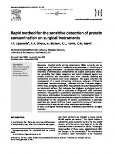
Fig. 1. Genotyping of DNA samples with two different methods. A) The Fok-I restriction patterns of a genomic fragment produced by PCR with primers flanking the CHS mutation in CHS mutant, heterozygous and wild type cattle. (+) lanes are the Fok-I restriction fragments. (−) lanes are the corresponding undigested fragments. B) Allelic discrimination cluster analysis of the wild type and mutant alleles using wild type (VIC-labeled) and mutant (FAM-labeled) TaqMan probes.
samples from 3 Japanese Black cattle that had been diagnosed as a wild-type, heterozygous carrier and homozygous mutant (CHS), by PCR-RFLP analysis, blood coagulation test and blood smear test, at the University of Miyazaki. The animals had no clinical symptoms at the time of the diagnosis. Fok-I-digestion of DNA from wild-type clearly produced the 2 digested fragments. On the other hand, when DNA derived from homozygous mutant was digested with Fok-I, no digested fragments were detectable. In a sample from heterozygous carrier, Fok-I-digestion produced three bands. Next, using these samples in a sextuplicated manner, we verified the method for real-time PCR with TaqMan minor groove binder probes. In a preliminary experiment, the amplification curve obtained with each sample did not show any significant non-specific allelic amplification under the cycling conditions we employed. Figure 1B shows the result of allelic discrimination cluster analysis at the end point of the PCR reaction, indicating 4 separate distinct clusters that represent the no-template controls and three genotypes. The VIC fluorescence for DNA from wild-type was markedly high, whereas that for FAM was rather low. On the other hand, analysis of DNA from the homozygous mutant revealed a mirror-image fluorescence pattern. For the
DNA from heterozygous carrier, the intensities of both types of fluorescence were moderately increased by the PCRreaction. These fluorescence patterns clearly discriminated samples from wild-type, heterozygous carrier and CHS animals. Assessment of the time required for analysis showed that our new technique could be carried out in only 60 min, thus shortening the total assay time by more than 120 min, in comparison with PCR-RFLP analysis. Next, exploiting the high-throughput performance of our real-time PCR with TaqMan minor groove binder probes, we used it to examine the genotypes of 102 Japanese Black cattle from a cattle farm whose manager had approved this study. We found that 93 (91.2%) of these cattle were wild-type, but unexpectedly, 9 (8.8%) of the cattle were heterozygous carriers. These diagnoses were confirmed by PCR-RFLP analysis. No cattle with the homozygous mutant allele were detectable. Based on these data, we investigated the pedigree records of the carriers and found that 8 of the 9 heterozygous animals were offspring of a single known carrier sire (Sire A). Further investigation revealed that Sire A had produced at least three heterozygous carrier dams that had not been diagnosed. For the remaining heterozygous animal, we were unable to find a potentially causative individual harboring the mutant allele in the first two generations.
RAPID DETECTION OF CHS ALLELE
In this study, we have developed a new high-throughput method for accurate detection of heterozygous and homozygous mutant alleles for CHS. In addition to the rapid processing offered by this method, it has other advantages over PCR-RFLP. It has been reported that partial digestion is one of the main problems associated with SNP genotyping by PCR-RFLP, because undigested PCR products can be confusing and hinder accurate judgement [1]. Also, as mentioned earlier, the PCR-RFLP requires several steps, which increase the risk of contamination [1, 10]. Our method simply produces allele-specific fluorescence, and detection can be performed in a single reaction tube or well, thus minimizing any false results or contamination. After the introduction of PCR-RFLP analysis more than a decade ago [4, 15], many animals with heterozygous and homozygous mutant alleles have been excluded from breeding, and therefore, we had initially assumed that heterozygous carriers would have been largely eradicated from the cattle population. However, our findings clearly indicated that some heterozygous dams remained, and that unfortunately they had been used for breeding. This might have been due to the lack of a rapid and convenient molecular method for screening large numbers of cattle. Our present results clearly indicate that this new method for real-time PCR with TaqMan minor groove binder probes is rapid, specific, sensitive and reproducible. We anticipate that this method will be used to screen large numbers of cattle, thus helping to eradicate CHS and its carriers. In conclusion, to our knowledge, this is the first reported study to have used the method for real-time PCR with TaqMan minor groove binder probes for genotyping of heterozygous and homozygous CHS alleles in Japanese Black cattle. The proposed technique would be similarly applicable for rapid genotypic screening of other genetic defects in different breeds and species. ACKNOWLEDGMENT. The authors are grateful to the Egyptian government for providing partial fund support for this work. REFERENCES 1. Abdolmohammadi, A., Atashi, H., Zamani, P. and Bottema, C. 2011. High resolution melting as an alternative method to genotype diacylglycerol O-acyltransferase 1 (DGAT1) K232A polymorphism in cattle. Czech J. Anim. Sci. 56: 370–376. 2. Barbosa, M. D. F. S., Nguyen, Q. A., Tchernev, V. T., Ashley, J. A., Detter, J. C., Blaydes, S. M., Brandt, S. J., Chotai, D., Hodgman, C., Solari, R. C. E., Lovett, M. and Kingsmore, S. F. 1996. Identification of the homologous beige and Chediak-Higashi syndrome genes. Nature 382: 262–265. [Medline] [CrossRef] 3. Huizing, M., Anikster, Y. and Gahl, W. A. 2001. Hermansky-
1239
Pudlak syndrome and Chediak-Higashi syndrome: disorders of vesicle formation and trafficking. Thromb. Haemost. 86: 233–245. [Medline] 4. Kunieda, T., Nakagiri, M., Takami, M., Ide, H. and Ogawa, H. 1999. Cloning of bovine LYST gene and identification of a missense mutation associated with Chediak-Higashi syndrome of cattle. Mamm. Genome 10: 1146–1149. [Medline] [CrossRef] 5. Nagle, D. L., Karim, M. A., Woolf, E. A., Holmgren, L., Bork, P., Misumi, D. J., McGrail, S. H., Dussault, B. J., Perou, C. M., Boissy, R. E., Duyk, G. M., Spritz, R. A. and Moore, K. J. 1996. Identification and mutation analysis of the complete gene for Chediak-Higashi syndrome. Nature Genet. 14: 307–311. [Medline] [CrossRef] 6. Nieuwenhuis, H. K., Akkerman, J. W. N. and Sixma, J. J. 1987. Patient with a prolonged bleeding time and normal aggregation tests may have storage pool deficiency: studies on one hundred six patients. Blood 70: 620–623. [Medline] 7. Nishimura, M., Inoue, M., Nakao, T., Nishikawa, T., Miyamoto, M., Kobayashi, T. and Kitamura, Y. 1989. Beige rat: a new animal model of Chediak-Higashi syndrome. Blood 74: 270–273. [Medline] 8. Ogawa, H., Tu, C. H., Kagamizono, H., Soki, K., Inoue, Y., Akatsuka, H., Nagata, S., Wada, T., Ikeda, M., Makimura, S., Uchida, K., Yamaguchi, R. and Otsuka, H. 1997. Clinical, morphologic, and biochemical characteristics of Chediak-Higashi syndrome in fifty-six Japanese Black cattle. Am. J. Vet. Res. 58: 1221–1226. [Medline] 9. Padgett, G. A., Leader, R. W., Gorham, J. R. and O’Mary, C. C. 1964. The familial occurrence of the Chediak-Higashi syndrome in mink and cattle. Genetics 49: 505–512. [Medline] 10. Riikka, J., Teija, K. and Auli, H. 2008. Group-specific PCRRFLP and real-time PCR methods for detection and tentative discrimination of strictly anaerobic beer-spoilage bacteria of the class Clostridia. Int. J. Food Microbiol. 125: 162–169. [Medline] [CrossRef] 11. Shabat, M. B., Mikula, I., Gerchman, I. and Lysnyansky, I. 2010. Development and evaluation of a novel single-nucleotidepolymorphism real-time PCR assay for rapid detection of fluoroquinolone-resistant Mycoplasma Bovis. J. Clin. Microbiol. 48: 2909–2915. [Medline] [CrossRef] 12. Shiraishi, M., Ogawa, H., Ikeda, M., Kawashima, S. and Ito, K. 2002. Platelet dysfunction in Chediak-Higashi syndrome-affected cattle. J. Vet. Med. Sci. 64: 751–760. [Medline] [CrossRef] 13. Spicer, S. S., Sato, A., Vincent, R., Eguchi, M. and Poon, K. C. 1981. Lysosome enlargement in the Chediak-Higashi syndrome. Fed. Proc. 40: 1451–1455. [Medline] 14. Tu, C. H., Takahashi, Y., Kaseda, Y., Uchida, K., Yamaguchi, R., Suzuki, K., Ogawa, H. and Otsuka, H. 1996. Inheritance of Chediak-Higashi syndrome in Japanese Black cattle. J. Vet. Med. Sci. 58: 501–504. [Medline] [CrossRef] 15. Yamakuchi, H., Agaba, M., Hirano, T., Hara, K., Todoroki, J., Mizoshita, K., Kubota, C., Tabara, N. and Sugimoto, Y. 2000. Chediak-Higashi syndrome mutation and genetic testing in Japanese Black cattle (Wagyu). Anim. Genet. 31: 13–19. [Medline] [CrossRef]