THE JOURNAL OF BIOLOGICAL CHEMISTRY © 1999 by The American Society for Biochemistry and Molecular Biology, Inc.
Vol. 274, No. 18, Issue of April 30, pp. 12383–12390, 1999 Printed in U.S.A.
A New b Subtype-specific Interaction in a1A Subunit Controls P/Q-type Ca21 Channel Activation* (Received for publication, December 21, 1998)
Denise Walker‡, Delphine Bichet‡, Sandrine Geib‡, Emiko Mori§, Ve´ronique Cornet‡, Terry P. Snutch¶, Yasuo Mori§, and Michel De Waard‡i From ‡INSERM Unite´ 464, Institut Fe´de´ratif Jean Roche, Faculte´ de Me´decine Nord, Boulevard Pierre Dramard, 13916 Marseille cedex 20, France, the §Department of Information on Physiology, National Institute of Physiological Science, Okazaki 444, Aichi, Japan, and the ¶Biotechnology Laboratory, Department of Zoology, University of British Columbia, Vancouver, British Columbia, Canada V6T 1Z3
The cytoplasmic b subunit of voltage-dependent calcium channels modulates channel properties in a subtype-specific manner and is important in channel targeting. A high affinity interaction site between the a1 interaction domain (AID) in the I-II cytoplasmic loop of a1 and the b interaction domain (BID) of the b subunit is highly conserved among subunit subtypes. We describe a new subtype-specific interaction (Ss1) between the amino-terminal cytoplasmic domain of a1A (BI-2) and the carboxyl terminus of b4. Like the interaction identified previously (21) between the carboxyl termini of a1A and b4 (Ss2), the affinity of this interaction is lower than AID-BID, suggesting that these are secondary interactions. Ss1 and Ss2 involve overlapping sites on b4 and are competitive, but neither inhibits the interaction with AID. The interaction with the amino terminus of a1 is isoform-dependent, suggesting a role in the specificity of a1-b pairing. Coexpression of b4 in Xenopus oocytes produces a reduced hyperpolarizing shift in the I-V curve of the a1A channel compared with b3 (not exhibiting this interaction). Replacing the amino terminus of a1A with that of a1C abolishes this difference. Our data contribute to our understanding of the molecular organization of calcium channels, providing a functional basis for variation in subunit composition of native P/Qtype channels.
Despite their functional diversity, high voltage-gated Ca21 channels have three subunit types in common (1, 2). The a1, pore-forming component of the channel is associated with a cytoplasmic b subunit of 52–78 kDa and a largely extracellular a2d component, anchored by a single transmembrane domain. These subunits are encoded by at least 7 a1, 4 b, and 1 a2d genes, respectively, of which numerous splice variants exist (3). The b subunit, when coexpressed with the a1 subunit, results in an increase in current density, alteration of the voltage dependence and kinetics of both inactivation and activation, and an increase in the number of recognition sites for channelspecific ligands (for review, see Refs. 4 and 5). These effects reflect not only conformational modulation but also an increase in the number of channels properly addressed to the cell sur-
* This work was supported by an INSERM postdoctoral fellowship (Poste Vert) (to D. W.). The costs of publication of this article were defrayed in part by the payment of page charges. This article must therefore be hereby marked “advertisement” in accordance with 18 U.S.C. Section 1734 solely to indicate this fact. i To whom correspondence should be addressed: INSERM U464, Institut Fe´de´ratif Jean Roche, Faculte´ de Me´decine Nord, Bd Pierre Dramard, 13916 Marseille cedex 20, France. Tel.: 33-4-9169-8860; Fax: 33-4-9109-0506; E-mail:
[email protected]. This paper is available on line at http://www.jbc.org
face, suggesting multiple roles for the b subunit. Although the effects of b are highly conserved, significant differences are seen depending on the combination of a1 and b subunits studied. For example, the kinetics of inactivation shows a general trend of variation with b subtype (6 –9), whereas a shift in the voltage dependence of inactivation has been reported only for non-L-type, A, B, and E (10 –12), and not L-type channels (13). b subunits also seem to differ in the mechanism by which they become localized to the plasma membrane (14, 15), perhaps suggesting that they are differentially targeted. Finally, a1 and b subtypes differ in their potential (based on sequence predictions) to be phosphorylated by various protein kinases. These factors together point to a functional explanation for the growing evidence that the in vitro promiscuity of a1-b interactions is reflected by a heterogeneity of combinations in native channels (N (16), P/Q (17), and L type (18)). Preliminary studies (10, 19) have identified a high affinity interaction between a highly conserved region in the cytoplasmic loop linking transmembrane regions I and II of a1 (AID,1 or a1 interaction domain) and a 30-residue region in the second conserved domain of b subunits (BID, or b interaction domain). This interaction occurs with a stoichiometry of 1:1 (20) and (at least in vitro and in expression systems) occurs between all combinations of a1 and b subtypes tested so far. We have since reported (21) the existence of a subunit-specific interaction between the carboxyl-terminal domain of a1A and the most carboxyl-terminal 109 residues of b4, and a similar interaction has been reported (22, 23) between a1E and b2a. The comparative high affinity of the AID-BID interaction (20, 21), coupled with the abolition of all b modulatory effects by mutation of residues critical to the interaction between AID and BID (10, 19), suggests that this interaction represents a primary, anchoring interaction upon which further, secondary, interactions might depend. The specificity of such secondary interactions, or at least differences in affinity, represents a potential source for the variation seen for different a1 and b combinations, in terms of both the electrophysiological properties of the channel and potential differences in control by other cellular factors, such as protein kinases and G proteins. We therefore set out to determine whether further secondary interaction sites exist. The present report describes the identification of an interaction between the amino-terminal cytoplasmic region of a1A and the b4 subunit of P/Q channels providing a refreshed 1 The abbreviations used are: AID, a1 interaction domain; BID, b interaction domain; GST, glutathione S-transferase; PCR, polymerase chain reaction; MBP, maltose-binding protein; 35S-b4, [35S]methioninelabeled b4 subunit; PAGE, polyacrylamide gel electrophoresis. Fusion proteins are referred to as, for example, GST-NTA for that containing the entire amino-terminal region of a1A, and GST-NTA,2–52 for the truncated form of this which contains only residues 2–52.
12383
12384
Role of a New a1A-b4 Interaction Site in Activation
understanding of the molecular organization of voltagedependent calcium channels. The interaction plays a critical role in the precise positioning of the channel activation process on the voltage axis. It constitutes yet another molecular determinant underlying functional differences among various b subunits and, by extension, probably among various native P/Q channel subtypes. EXPERIMENTAL PROCEDURES
GST Fusion Proteins—Regions of the rabbit brain a1A cDNA (BI-2 (24)) corresponding to residues 2–98 (i.e. the entire amino-terminal region), 2–52, 43–77, 66 –98, and 76 –98 were amplified by PCR and, with the aid of BamHI and EcoRI restriction sites included in the primers, were subcloned into pGEX2TK (Amersham Pharmacia Biotech). The resulting recombinant plasmids were expressed in Escherichia coli BL21, and the GST fusion proteins were purified as described previously (20). Fusion proteins expressing the entire amino-terminal regions (minus start codon) of a1B (amino acids 2–95, GenBank M92905 (25)), a1C (amino acids 2–151, M57974 (26)), and a1S (amino acids 2– 49, M23919 (27)) were constructed and purified similarly. The resulting fusion proteins are referred to as, for example, GST-NTA for that containing the entire amino-terminal region of a1A and GST-NTA,2–52 for the truncated form of this which contains only residues 2–52. In Vitro Translation of b Subunits—b1b, b2a, b3, and b4 cDNA clones were as described previously (21). Truncated derivatives of b4 were constructed by PCR amplification of the corresponding regions of cDNA and subcloning into pcDNA3 (Invitrogen), using HindIII and BamHI sites (added to the PCR primers) with the addition of a Kozak sequence (28) and initiation codon (ACCATGG) or termination codon (TGA) as necessary. The b3/4 chimera construct (b3 1–360/b4 402–519 in pcDNA3) is as described previously (21). 35S-Labeled b subunits were synthesized in vitro using the TNTTM-coupled Transcription/Translation System (Promega). Non-incorporated [35S]methionine was removed by purification on a PD10 column (Amersham Pharmacia Biotech). Binding Assays—These were carried out using fusion proteins coupled to glutathione-agarose in Tris-buffered saline as described previously (21). Binding reactions were incubated for 5 h unless otherwise stated. Peptides—A 21-amino acid peptide containing the AIDA sequence QQQIERELNGYMEWISKAEEV and a 21-amino acid peptide containing residues 76 –96 of the a1A amino-terminus (RSLFLFSEDNVVRKYAKKITE) were synthesized by Genosys (United Kingdom). Competition Experiments—For competition experiments, a maltosebinding protein (MBP) in fusion with the carboxyl-terminal binding site of a1A (amino acids 2120 –2275) was constructed using the BamHI/SalI sites of pMAL-c2 (MBP-CTA,2120 –2275). The effects of b-AIDA and b-CTA associations on b-AIDA, b-CTA, and b-NTA interaction were analyzed by saturating each b site by preincubating the [35S]methionine-labeled b4 subunit (35S-b4) with 10 mM AIDA peptide (1 h) or 2 mM MBP-CTA,2120 – 35 2275 (4 h). Binding of S-b4 to various a1A binding sites was then tested by a 4-h incubation with 250 nM GST (control), 250 nM GST-AIDA (AID site), 2 mM GST-NTA, and 2 mM GST-CTA,2090 –2424, precipitation of glutathione-agarose beads, gel electrophoresis, and autoradiography. Chimera a1A Subunit—Pairs of primers CSl-N1(1) 59-GGGTCGACTAAAACGTAAAGTATTACTAAAACCTCAATTTGCAG-39 and BIC-N1(2) 59-GTACTCAAAGGGTTTCCACTCGACGATGCT-39, and primers BIC-N1(1) 59-GTCGAGTGGAAACCCTTTGAGTACATGATT-39 and BINt-N1(2) 59-GAGCGGCCGCAGCACCCGCACTGC-39 were combined with the templates pCARD3 (29) and pSPBI-2 (24), respectively, in PCR amplification using the Advantage PCR kit (CLONTECH). The resulting PCR products and the primers CSl-N1(1) and BINt-N1(2) were subjected to subsequent PCR amplification to yield a chimeric sequence that contains nucleotides 2191 to 462 from the a1C sequence (29) and 336 – 650 from the a1A sequence (24). The chimeric fragment was digested with SalI and NotI and ligated with the 11.8 kilobase NotI (partially digested)/SalI fragment from pSPBI-2 to yield pSP72C(N)BI-2 (a1A(NT)C subunit). Electrophysiology—Xenopus oocytes were prepared as described previously (6). Stage V and VI oocytes were injected with a1A (BI-2 (24)) or a1A(NT)C-specific mRNA (0.3 mg/ml) either alone or in combination with b3- or b4-specific mRNA (0.15 mg/ml) and maintained for 3– 4 days before recording in defined nutrient oocyte medium (6). Two-electrode voltage clamp recording was performed at room temperature (18 –20 °C) using a GeneClamp amplifier (Axon Instruments, Foster City, CA). The extracellular recording solution was of the following composition (in mM): Ba(OH)2, 40; NaOH, 50; KCl, 3; HEPES, 5; niflumic acid, 0.5; pH 7.4 with methanesulfonic acid. Electrodes filled with 3 M KCl had a resist-
FIG. 1. In vitro binding of 35S-labeled b4 to the amino-terminal region of a1A. Panel A. Left, Coomassie Blue-stained SDS-PAGE showing the GST fusion proteins used (5 mg). Right, autoradiogram of the binding assay. In vitro translated b4 was assayed for binding to the fusion proteins indicated (5 mM). GST, glutathione S-transferase alone; GST-AIDA, GST fused to AID region (residues 369 – 418) of a1A; GSTNTA, GST fused to entire amino-terminal cytoplasmic domain (residues 2–98) of a1A. After binding interactions as described under “Experimental Procedures,” washed beads were analyzed by SDS-PAGE and autoradiography. Panel B, various concentrations of GST-NTA fusion protein were assayed for binding to 35S-b4, and binding was quantified by counting. Specific binding was calculated by subtraction of binding to GST (at the same concentration) and normalized by expression as a proportion of maximal binding. Error bars indicate normalized S.D. Data are described by a logistic function f 5 [(a 2 d)/(1 1 (x/c)b] 1 d where a 5 1. 019 and d 5 0. 01022 are the asymptotic maximum and minimum, respectively; x is the fusion protein concentration; c 5 336 nM is the Kd; and b 5 22,816 is the slope of the curve. For comparison purposes, the saturation curve for AIDA-GST (Kd 5 3 nM, dashed line) is also shown (20). ance of 0.1 megohm. Current records were filtered at 1 kHz, leaksubtracted on-line by a P/6 protocol, and sampled at 5 kHz. Residual capacitative currents were blanked. Data were analyzed using pCLAMP version 6.03 (Axon Instruments). All values are mean 6 S.D. RESULTS
A GST fusion protein, GST-NTA, expressing the entire amino-terminal cytoplasmic region of a1A (splice variant BI-2) was assayed for in vitro binding to 35S-b4. As Fig. 1A shows, the NTA region exhibits a significant and specific interaction with b4 which is comparable to the binding observed to a GST fusion protein carrying the AIDA sequence. The binding of GST-NTA to 35S-b4 appears slightly stronger than the binding of GSTAIDA, but the relative efficiency of binding of these fusion proteins varied slightly depending on the b-translation reactions used. The affinity of this interaction was determined by carrying out similar binding assays using a range of concentrations of GST-NTA fusion protein. Fig. 1B shows the resulting saturation curve, which is compared with that observed previously for the interaction of 35S-b4 with GST-AIDA. The affinity of interaction of GST-NTA is 100-fold lower (kD 5 336 nM) than that for the AID interaction (close to 3 nM). These data are in favor of the idea that the AID-BID interaction represents a primary anchoring site of interaction between the two subunits which allows secondary interactions of lower affinity to occur. As already mentioned, it is interesting that in Fig. 1A, GST-NTA
Role of a New a1A-b4 Interaction Site in Activation
FIG. 2. Localization of the interaction site in a1A. Panel A. Top, schematic diagram of the a1A subunit. Amino acid positions are shown above, transmembrane domains (each composed of six membrane-spanning segments) are shown as dark boxes and numbered (I–IV) above. Bottom, enlargement of the amino-terminal domain, showing GST fusion proteins constructed with amino acid positions in a1A marked at the extremities. Panel B. Top, Coomassie Blue-stained SDS-PAGE showing fusion proteins used (5 mg). Bottom, capacity of 5 mM purified fusion proteins to interact with 35S-b4. After the binding reactions, washed beads were analyzed by SDS-PAGE and autoradiography. Panel C, amino-terminal binding site of a1A (BI-2, amino acids 1– 66) and its alignment with sequences of other calcium channels (GenBank accession codes M92905, a1B; X67855, a1E; X15539, a1C; M57682, a1D; and M23919, a1S).
demonstrates greater binding than GST-AIDA to 35S-b4. Given that both fusion proteins are at concentrations giving maximal binding (Fig. 1B), this demonstrates a difference in maximal binding which appears to reflect a difference in conformational requirements, coupled with conformational heterogeneity in the 35S-b4 preparation (permissive and nonpermissive binding states; data not shown). To characterize more precisely the region of a1A responsible for interaction with b4, a series of GST fusion proteins carrying truncations of the region concerned (depicted in Fig. 2A) was constructed, and the proteins were assayed for their capacity to interact with 35S-b4. As Fig. 2B shows, removal of the most
12385
carboxyl-terminal amino acids, or of the 42 most amino-terminal, does not abolish the capacity to interact with b4. Concomitantly, fusion proteins corresponding to the most carboxylterminal region, which is most highly conserved among a1 subtypes, are incapable of binding. In addition, the interaction between GST-NTA and 35S-b4 was not inhibited by addition to the binding reaction of a peptide (500 mM) corresponding to amino acids 76 –98. These data suggest that the b4 binding site concerns a region between residues 1 and 66 of a1A, maybe comprising, but not necessarily limited to, residues 42–52. The reduced binding to NTA,2–52 and NTA,42–77 compared with full-length NTA probably reflects instability and/or sequence reduction of the interaction site. The reduction in binding of smaller deleted derivatives meant that we were unable to pursue this approach further. A sequence alignment of this a1A binding domain with equivalent domains of other a1 subunits (a1B, a1E, a1C, a1D, and a1S), some used in this investigation, suggests a relatively low level of sequence conservation, although a1B and a1A show some similarity (Fig. 2C). This observation implies that the interaction may not be conserved, a prediction that we went on to test (see Fig. 5). To identify the region of b4 which interacts with the aminoterminal region of a1A, we initially analyzed the binding capacity of several deleted derivatives of b4, translated in vitro (Fig. 3, A and B). These derivatives lacked either the amino-terminal, carboxyl-terminal, or both regions, which shows a low level of conservation among b subunit subtypes. As Fig. 3B shows, removal of the amino-terminal region had no effect, whereas removal of the carboxyl-terminal abolished binding completely, illustrating the importance of this region in the interaction. We also found that although b3 does not interact with GST-NTA (see Fig. 5), the opposite is true for a b3-b4 chimera, in which the nonconserved carboxyl terminus of b3 is replaced by the equivalent domain of b4 (Fig. 3B). We have shown previously (21) that the carboxyl-terminal region of b4 also interacts with the carboxyl-terminal cytoplasmic domain of a1A (BI-2). We therefore wanted to map the two interaction sites more precisely, for which we constructed two additional derivatives of b4, lacking a third (residues 483–519) and two-thirds (residues 447–519) of the carboxyl terminus (Fig. 3, A and B). As Fig. 3C shows, deletion of residues 483– 519 of b4 had no effect on its capacity to bind to GST-NTA, whereas truncation of the carboxyl terminus of b4 up to residue 446 resulted in a total loss of binding capacity. This indicates that the NTA binding region is located between residues 446 and 482 of b4. Analysis of the capacity of these truncates to bind to a GST fusion protein of the carboxyl-terminal region (residues 2090 –2424) of a1A (GST-CTA) resulted in binding capacity being gradually lost with each further deletion. This suggests that the binding site of CTA spans a wider region than the NTA binding site, is dependent on secondary or tertiary structures that are disrupted by the deletions, or consists of a series of dispersed sites. It is noteworthy that the previously characterized a1A carboxyl-terminal binding site was also difficult to define, in that deleted derivatives over a long region retained binding capacity, giving support to the hypothesis that there are microdomains of interaction between these two sites (21). In contrast, the NTA site and corresponding domain on b4 are shorter and seem more easily delineated. In any case, the different patterns of interaction capacities seen for GSTNTA and GST-CTA suggest that these two regions of a1A occupy different but overlapping sites on b4. The involvement of overlapping regions of b4 in interactions with the amino- and carboxyl-terminal domains of a1A also raised the question as to whether these interactions could occur simultaneously or whether they were mutually exclusive. To
12386
Role of a New a1A-b4 Interaction Site in Activation
FIG. 3. Localization of the interaction site in the b4 subunit. Panel A. Top, schematic map of b4 subunit, dividing the protein into five domains. Darker domains (II and IV) represent sequences of highest conservation among b subtypes (10). Bottom, autoradiography of SDSPAGE showing b4 and deleted derivatives translated in vitro in the presence of [35S]methionine. Panel B, in vitro translated full-length, truncated b4 derivatives and a b3-b4 chimera (T) were assayed for their capacity to interact with GST and GST-NTA fusion proteins (2.5 mM) as described under “Experimental Procedures,” and washed beads were analyzed by SDS-PAGE and autoradiography. Panel C, 35S-b4 and carboxyl-terminal deleted derivatives were assayed for their capacity to interact with 2.5 mM GST-NTA and a GST fusion protein containing residues 2090 –2424 of a1A (GST-CTA (21)). Specific binding was calculated by subtraction of binding to GST (at the same concentration) and normalized by expression as a percentage of maximal binding to GST-AIDA. Error bars represent normalized S.D.
investigate this as well as their relationship with the AID-BID interaction, we tested whether the binding of AIDA (21-amino acid peptide) or GST-CTA to 35S-b4 could prevent its interaction with GST-NTA. The results, illustrated in Fig. 4, show that although the AID peptide was effective in preventing the interaction of b4 to GST-AIDA, it did not prevent the concomitant interaction with either GST-NTA or GST-CTA,2070 –2275 (Fig. 4A). On the other hand, the association of MBP-CTA,2120 –2275 with b4 blocked the ability of b4 to interact with GST-CTA,2070 – 2275 and also significantly reduced the binding of b4 to GSTNTA (Fig. 4B), suggesting that b4 is able to interact with AID and only one of the secondary interaction sites at a time. We have shown previously (21) that b subtypes differ in their capacity to interact with the carboxyl-terminal region of a1A, with b4 interacting with greatest affinity, b2A with a lesser affinity, and b1b and b3 showing no significant interaction. We therefore wished to determine whether the same was true for interaction with the amino-terminal domain. In addition, because the b interaction site in the amino-terminal region of a1A shows a variable level of conservation among a1 subtypes, we wished to investigate whether b interaction capacities were conserved among them. Both of these questions were addressed by constructing a series of GST fusion proteins carrying the amino-terminal cytoplasmic region of a1B, a1C, and a1S (Fig. 5A). These fusion proteins, along with GST alone, GST-AIDA (for comparison purposes), and GST-NTA, were assayed for their ability to interact with four different b subtypes, translated in vitro in presence of [35S]methionine (Fig. 5B). Interestingly, interaction with GST-NTA showed a pattern similar to that observed for the carboxyl-terminal region of a1A (21) in that b4 exhibited the most significant interaction, b2a interacted to a lesser degree, and b1b and b3 showed no significant interaction. The amino-terminal domains of a1B showed no significant interaction despite its closer sequence relatedness to a1A. GST-NTS, on the other hand, showed significant interaction with all four b subunits, whereas GST-NTC, another L-type channel member, showed no interaction with any of the b subunits. Because the amino-terminal sequences of a1A and a1S are very different, we checked whether binding of b4 to NTS involved the same interaction domain of b4. Fig. 6 demonstrates
FIG. 4. Multiple site occupancies on b4 subunit. Panel A, effect of 1-h AIDA peptide (10 mM) preincubation with 35S-b4 on control GST (250 nM), GST-AIDA (250 nM), GST-NTA (2 mM), and GST-CTA (2 mM) binding to 35S-b4. The 4-h binding reaction was conducted in the continued presence of AIDA peptide. Panel B, effect of 4-h MBP-CTA (2 mM) preincubation with 35S-b4 on control GST (250 nM), GST-NTA (2 mM), and GST-CTA (2 mM) binding to 35S-b4. The 4-h binding reaction was conducted in the continued presence of MBP-CTA. Complete inhibition of GST-NTA binding to 35S-b4 by MBP-CTA was difficult to achieve because MBP-CTA bound only a subset of the available 35S-b4, which was presumably in a more favorable conformation.
that, as for NTA, the carboxyl terminus of b4 was required for binding to NTS, and the use of deleted derivatives of the carboxyl terminus of b4 also indicates an important role for residues 446 – 482 of b4 in this interaction. These results suggest that the interaction site is defined more by the tertiary structure of the a1 amino-terminal region than by its primary sequence, also explaining why the NTA site could not be localized more precisely than to residues 1– 66 (Fig. 2). Finally, we questioned the relevance of the interaction between the amino terminus of a1A and the carboxyl terminus of b4 in terms of channel functioning. First, because b3, in contrast to b4, does not interact with the amino terminus of a1A, we investigated whether there were significant differences in terms of channel regulation by these two subunits. We found that in addition to triggering different inactivation kinetic behaviors (6), the two subunits differed in terms of their ability to shift the activation curve toward hyperpolarized potentials (Fig. 7A). Although both b subunits shifted the activation curve
Role of a New a1A-b4 Interaction Site in Activation
12387
FIG. 5. a1 amino-terminal specificity of interaction with b subunits. Panel A, Coomassie Blue-stained SDSPAGE showing various GST fusion proteins used (5 mg). Panel B, in vitro translated b subunits were assayed for their capacity to interact with 5 mM GST fusion proteins, and the remaining radioactivity associated with washed beads was quantified by counting. GST, control; AIDA, GST-AIDA; NTA, NTB, NTC, NTS, GST fusion proteins containing amino-terminal cytoplasmic domains of a1A, a1B, a1C, and a1S, respectively. Error bars represent S.D.
FIG. 6. The carboxyl terminus of b4 is also involved in NTS binding. 35S-b4 and deleted derivatives were assayed for their capacity to interact with GST-NTS (5 mM). Specific binding was calculated by subtraction of binding to GST (at the same concentration) and normalized by expression as a percentage of maximal binding to GST-AIDA (500 nM). Error bars represent normalized S.D.
along the voltage axis, the shift induced by b3 was significantly more pronounced than the one produced by b4. The estimated half-activation potential shifted from 17 mV (a1A-expressing
oocytes) toward 213 mV (a1Ab3 oocytes) and 1.5 mV (a1Ab4 oocytes). There is thus an approximately 14 –15 mV difference in the shift induced by the b3 and b4 subunits. In addition, we found that depending on the b subunit being expressed, the channels differed in their voltage dependence of inactivation with half-inactivation at 250 and 237 mV for a1Ab3 and a1Ab4 channels, respectively (data not shown). Because these differences in functional regulation by the various b subunits may be the result of differences in interaction levels between a1A and the two b subunits, we determined the role of the NTA site in b-induced channel regulation. We took advantage of the observation that essential differences were found in b subunit association with amino-terminal sequences of various a1 subtypes. We constructed a chimera a1A subunit (a1A(NT)C), in which we replaced the amino terminus of a1A (interacts with b4 but not b3) with the amino terminus of a1C (does not interact with either b4 or b3). Coexpression of this chimeric channel with b3 or b4 triggers high voltage-activated currents in Xenopus oocytes (Fig. 7B). The amplitude of the currents elicited by membrane depolarization are reduced slightly compared with those obtained for the wild-type a1A channel. Cells expressing a1Ab3, for instance, have a peak current amplitude of 1,001 6 651 nA (n 5 7, S.D.), whereas cells expressing a1A(NT)Cb3 peak at 423 6 655 nA (n 5 12), which corresponds to a 2.37-fold
12388
Role of a New a1A-b4 Interaction Site in Activation
FIG. 7. Role of the NTA-b4 interaction in the control of voltage dependence of activation. A, b3 and b4 differ in their ability to shift the voltage dependence of activation of a1A. Left and center, currents elicited by various membrane depolarizations (230, 220, 210, and 0 mV) illustrating differences in threshold, intermediate, and peak activation of a1Ab3 and a1Ab4 channels expressed in Xenopus oocytes. Right, corresponding average current-voltage (I-V) relationship for a1Ab3 (n 5 7) and a1Ab4 (n 5 6) expressing cells. The I-V curve for cells expressing a1A channel alone is shown for comparison purpose (n 5 8). The experimental data were fitted with a modified Boltzmann equation IBa5 (gz(V 2 E))/(1 1 exp(2(V 2 V1/2)/k)), where g is the normalized conductance (g 5 0.032, no b; 0.018, 1b3; and 0.026, 1b4); V1/2 is the half-activation potential (V1/2 5 17 mV, no b; 213 mV, 1b3; and 1. 5 mV, 1b4); E is the reversal potential (E 5 67 mV, no b; 63 mV, 1b3; and 58 mV, 1b4); and k is the range of potential for an e-fold change around V1/2 (k 5 7. 9 mV, no b; 4. 2 mV, 1b3; and 5. 8 mV, 1b4). B, change in difference in the b-induced I-V shift by a1A amino-terminal sequence substitution. Left and center, currents elicited by various membrane depolarizations (230, 220, 210, and 0 mV) showing the absence of a difference in channel activation for a1A(NT)Cb3 and a1A(NT)Cb4 channels. Right, corresponding average I-V curves for a1A(NT)Cb3 (n 5 13) and a1A(NT)Cb4 channels (n 5 12). The fit of the experimental data yields V1/2 5 213 (1b3) and 29 mV (1b4); k 5 4. 2 (1b3) and 4. 4 mV (1b4); g 5 0. 017 (1b3) and 0. 018 (1b4); and E 5 60 (1b3) and 63 mV (1b4).
reduction. A similar 2.06-fold reduction in current amplitude is seen when b4 is coexpressed with a1A(NT)C (peak current 542 6 240 nA, n 5 12) rather than a1A (peak current 1,118 6 871 nA, n 5 6). These results suggest that the amino terminus plays a role in channel expression levels at the plasma membrane but that b subunits and the NTA interaction site have little influence on this process. Also, the amino-terminal substitution induced an important shift in the voltage dependence of inactivation with half-inactivation occurring at 252 mV for a1A(NT)Cb4 channels compared with 237 mV for a1Ab4 channels (data not shown). Because a similar shift is seen with b3 (not shown), this supports the idea that b subunit interaction with the amino terminus plays a minor role in this modification. In contrast, we found that the difference in the shift of voltage dependence of activation of a1A(NT)Cb3 and a1A(NT)Cb4 channels was reduced significantly (Fig. 7B). The average halfactivation potential of a1A(NT)Cb3 channels was 213 mV and thus remained identical to that of the a1Ab3 channels, whereas the V1/2 of a1A(NT)Cb4 channels was 29 mV, a significant hyperpolarizing shift compared with the a1Ab4 channels. These data suggest that in the absence of an NTA/b4 interaction, the
I-V shift induced by the b4 subunit resembles the shift induced by the b3 subunit. Finally, the substitution of the a1A NTA sequence by NTC produced a slowing of channel inactivation with b4 but not with b3. The decay of a1A(NT)Cb4 currents occurred along two components with time constants of t1 5 80 6 4 ms and t2 5 368 6 39 ms (at 10 mV, n 5 7) compared with t1 5 51 6 9 ms and t2 5 246 6 27 ms (n 5 6) for a1Ab4 currents. In contrast, no significant differences were seen in inactivation kinetics of a1Ab3 or a1A(NT)Cb3 channels with time constants at 10 mV of t1 5 65 6 15 ms and t2 5 243 6 47 ms (n 5 10) for a1A(NT)Cb3 currents and t1 5 62 6 14 ms and t2 5 222 6 13 ms (n 5 7) for a1Ab3 currents. These data further confirm a functional role in inactivation kinetics of the carboxyl terminus of b4 by its interaction with the carboxyl terminus (21) and amino terminus of the a1A subunit. DISCUSSION
We describe the identification of a specific interaction site between the amino-terminal cytoplasmic region of the calcium channel a1A subunit and the b4 subunit. The b4 subunit is widely expressed in the brain, especially in the cerebellum (30).
Role of a New a1A-b4 Interaction Site in Activation
12389
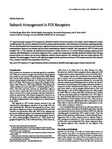
FIG. 8. Schematic a1A-b4 interactions. Shown are the AID-BID anchoring site (As) implicating the I-II loop of a1A and the first 30 residues of domain IV of b4; and the two secondary interactions sites, the first comprising NTA (Ss1) and the second CTA (Ss2), involving overlapping regions of domain V of b4.
On the basis of their colocalization in many tissue types, b4 appears largely to be associated with the a1A subunit in native channels (17, 31). However, coimmunoprecipitation studies demonstrate that a1A is also found associated with b1b, b2, and b3 (17) and that b4 is also found associated with a1B (16). The importance of the b4 subunit is illustrated by the recent demonstration that a lethargic phenotype in mice results from a deletion of approximately 60% of the b4 coding sequence (32). This truncated b subunit would lack all three of the interactions described with a1A (AIDA (19) and NTA and CTA (21)), although such a deletion is also likely to result in severe conformational perturbation and probably degradation of the protein. This mutation is not entirely lethal, however, which is reminiscent of a growing number of experiments in which knockout of proteins of central importance does not turn out to be lethal. This is probably explained by a partial compensation by a related protein, in this case suggesting that other b subunits are expressed in parallel or that their expression is switched on to compensate for this deficiency (33). In fact, b3 is known to be a normal constituent of about one-third of P/Q-type channels (17). Because b3 expression is high in brain and parallels that of b4 (34), it would be the most likely candidate for b4 substitution in the lethargic mice. Since b3 lacks both secondary interaction sites described so far in b4, such a substitution would not be functionally equivalent, perhaps explaining some of the neurological defects encountered in these mice. The NTA interaction identified is of relatively low affinity, supporting the idea that this is one of several secondary interactions between the two subunits that rely on the initial, high affinity interaction between the AID and BID sites identified previously. This idea is supported by the observation that mutagenesis of AID or BID to disrupt interaction between the two sites also disrupts the ability of the b subunit to modify channel properties (10). It also stems from the fact that this is the third interaction site mapped between a1A and b4 and that binding of multiple b subunits to a1 does not seem very plausible. The new interaction site that we describe involves the amino terminus of a1A (residues 1– 66) and carboxyl terminus (residues 446 – 482) of b4. This is particularly interesting given the rather low level of sequence conservation in the two regions identified. With regard to a1A splice variants, the sequence of the amino-terminal cytoplasmic region is identical in BI-1 and BI-2 subtypes, indicating conservation of this interaction (24). This is in contrast to the b4 interaction site that we have identified previously in the carboxyl-terminal region of the BI-2 splice variant. The low degree of sequence conservation observed for the respective interaction sites identified in the amino-terminal region of a1A and the carboxyl-terminal of b4 is reflected by the high degree of subtype specificity exhibited by this interaction
with respect to both a1 and b isoforms. Our results indicate that the equivalent amino-terminal regions of a1B and a1C did not interact with any of the b subunits tested. Because other b subtypes exist, we cannot rule out that this may reflect the use of an inappropriate a1-b combination. Interestingly, we found that a fusion protein expressing the entire amino terminus of a1S could interact with all four different b subunits tested. This is in contrast to the NTA binding, which occurs only on b4 and to a lesser extent on b2a. These results are indicative of a potential interaction of a1S with b subunits other than b1a, the major b subunit of skeletal muscle, and parallel recent findings that b3 (7, 32) and a b1 splice variant other than b1a (2) are also expressed, albeit at low levels, in skeletal muscle. Overall, our results are indicative of evolution to provide for a1-b interaction specificity both within the a1 amino-terminal and the b carboxyl-terminal sequences. Fig. 8 summarizes what is now known about a1A-b4 interactions in terms of structure. One interesting aspect is that the b4 subunit can interact simultaneously with AID and, via its carboxyl-terminal region, with either the amino- or carboxylterminal regions of a1A, thereby defining two patterns of interactions. These interactions probably impose conformational constraints on the molecule which appear to affect channel function. It is also tempting to speculate that the conformational constraints are different depending on the patterns of interaction in use by the channel. The importance of the amino and carboxyl termini of a1A are underlined by the observation that truncations of equivalent domains in a1C result in enhanced current levels of the channel (35, 36). These enhanced current levels occur either by a greater membrane incorporation (amino terminus) or enhanced open probability (carboxyl terminus). Because b subunits also increase channel expression, and this effect varies in amplitude depending on the a1 and b subtype studied, it is tempting to speculate that the secondary interaction sites described so far also intervene in a1A channel expression by one of the two mechanisms described for a1C. We did indeed find that substitution of the amino terminus of a1A by the equivalent sequence of a1C resulted in an important reduction in current density. This effect was, however, b subtype-independent, and it is therefore unlikely that the NTA interaction site described here plays a role in b-induced enhancement of current amplitude. Despite this, secondary interactions appear to play other roles in several aspects of control of channel activity. We have shown previously (21) the importance of the carboxyl-terminal region of a1A in the control of channel inactivation kinetics. Here, we demonstrate that the amino-terminal interaction site of a1A is required for fine tuning the voltage dependence of activation. The NTA interaction with b4 appears to limit the amplitude of the hyperpolarizing b-induced shift of channel activation. By
12390
Role of a New a1A-b4 Interaction Site in Activation
this unique mechanism, it can be predicted that the b3-containing P/Q channel subtype is activated more easily than the b4-containing P/Q channel subtype. In addition, secondary interaction sites may serve to protect or uncover phosphorylation sites in the a1A subunit, thereby altering the regulatory input of these. Another obvious possibility is that they play a role in the antagonistic relationship between the b subunit and Gbg complex. In this respect, it is interesting that Qin et al. (23) have recently shown that, in addition to interacting with a region overlapping with the AID site (37, 38), Gbg also interacts with the carboxyl-terminal domain of a1A, a1B, and a1E and that the amino terminus has recently been recognized as another determinant for Gbg regulation in a1E subunits (39). Finally, the existence of secondary interactions in addition to the AID-BID interaction could serve to favor certain combinations of subunits in cells where several subtypes are expressed. Given that b subunits also play a role in the surface targeting of a1 and a2d (14), an interesting possibility is that specific b subunits serve to target a1 subunits to specific regions of the cell surface. REFERENCES 1. Flockerzi, V., Oeken, H.-J., Hofmann, F., Pelzer, D., Cavalie, A., and Trautwein, W. (1986) Nature 323, 66 – 68 2. Witcher, D. R., De Waard, M., Sakamoto, J., Franzini-Armstrong, C., Pragnell, M., Kahl, S. D., and Campbell, K. P. (1993) Science 261, 486 – 489 3. Birnbaumer, L., Campbell, K. P., Catterall, W. A., Harpold, M. M., Hofmann, F., Horne, W. A., Mori, Y., Schwartz, A., Snutch, T. P., Tanabe, T., and Tsien, R. W. (1994) Neuron 13, 505–506 4. Catterall, W. A. (1995) Annu. Rev. Biochem. 64, 493–531 5. Walker, D., and De Waard, M. (1998) Trends Neurosci. 21, 148 –154 6. De Waard, M., and Campbell, K. P. (1995) J. Physiol. (Lond.) 485, 619 – 634 7. Hullin, R., Singer-Lahat, D., Freichel, M., Biel, M., Dascal, N., Hofmann, F., and Flockerzi, V. (1992) EMBO J. 11, 885– 890 8. Perez-Reyes, E., Castellano, A., Kim, H. S., Bertrand, P., Baggstrom, E., Lacerda, A. E., Wei, X. Y., and Birnbaumer, L. (1992) J. Biol. Chem. 267, 1792–1797 9. Castellano, A., Wei, X. Y., Birnbaumer, L., and Perez-Reyes, E. (1993) J. Biol. Chem. 268, 12359 –12366 10. De Waard, M., Pragnell, M., and Campbell, K. P. (1994) Neuron 13, 495–503 11. Soong, T. W., Stea, A., Hodson, C. D., Dubel, S. J., Vincent, S. R., and Snutch, T. P. (1993) Science 260, 1133–1136 12. Singer, D., Biel, M., Lotan, I., Flockerzi, V., Hofmann, F., and Dascal, N. (1991) Science 253, 1553–1557 13. Tomlinson, W. J., Stea, A., Bourinet, E., Charnet, P., Nargeot, J., and Snutch,
T. P. (1993) Neuropharmacology 32, 1117–1126 14. Brice, N. L., Berrow, N. S., Campbell, V., Page, K. M., Brickley, K., Tedder, I., and Dolphin, A. C. (1997) Eur. J. Neurosci. 9, 749 –759 15. Chien, A. J., Zhao, X., Shirokov, R. E., Puri, T. S., Chang, C. F., Sun, D., Rios, E., and Hosey, M. M. (1995) J. Biol. Chem. 270, 30036 –30044 16. Scott, V. E. S., De Waard, M., Liu, H., Gurnett, C. A., Venzke, D. P., Lennon, V. A., and Campbell, K. P. (1996) J. Biol. Chem. 271, 3207–3212 17. Liu, H., De Waard, M., Scott, V. E. S., Gurnett, C. A., Lennon, V. A., and Campbell, K. P. (1996) J. Biol. Chem. 271, 13804 –13810 18. Pichler, M., Cassidy, T. N., Reimer, D., Haase, H., Kraus, R., Ostler, D., and Striessnig, J. (1997) J. Biol. Chem. 272, 13877–13882 19. Pragnell, M., De Waard, M., Mori, Y., Tanabe, T., Snutch, T. P., and Campbell, K. P. (1994) Nature 368, 67–70 20. De Waard, M., Witcher, D. R., Pragnell, M., Liu, H., and Campbell, K. P. (1995) J. Biol. Chem. 270, 12056 –12064 21. Walker, D., Bichet, D., Campbell, K. P., and De Waard, M. (1998) J. Biol. Chem. 273, 2361–2367 22. Tareilus, E., Roux, M., Qin, N., Olcese, R., Zhou, J., Stefani, E., and Birnbaumer, L. (1997) Proc. Natl. Acad. Sci. U. S. A. 94, 1703–1708 23. Qin, N., Platano, D., Olcese, R., Stefani, E., and Birnbaumer, L. (1997) Proc. Natl. Acad. Sci. U. S. A. 94, 8866 – 8871 24. Mori, Y., Friedrich, T., Kim, M.-S., Mikami, A., Nakai, J., Ruth, P., Bosse, E., Hofmann, F., Flockerzi, V., Furuichi, T., Mikoshiba, K., Imoto, K., Tanabe, T., and Numa, S. (1991) Nature 350, 398 – 402 25. Dubel, S. J., Starr, T. V., Hell, J., Ahlijanian, M. K., Enyeart, J. J., Catterall, W. A., and Snutch, T. P. (1992) Proc. Natl. Acad. Sci. U. S. A. 89, 5058 –5062 26. Perez-Reyes, E., Wei, X. Y., Castellano, A., and Birnbaumer, L. (1990) J. Biol. Chem. 265, 20430 –20436 27. Tanabe, T., Takeshima, H., Mikami, A., Flockerzi, V., Takahashi, H., Kangawa, K., Kohima, M., Matsuo, H., Hirose, T., and Numa, S. (1987) Nature 328, 313–318 28. Kozak, M. (1986) Cell 44, 283–292 29. Mikami, A., Imoto, K., Tanabe, T., Niidome, T., Mori, Y., Takeshima, H., Narumiya, S., and Numa, S. (1989) Nature 340, 230 –233 30. Tanaka, O., Sakagami, H., and Kondo, H. (1995) Mol. Brain Res. 30, 1–16 31. Ludwig, A., Flockerzi, V., and Hofmann, F. (1997) J. Neurosci. 17, 1339 –1349 32. Burgess, D. L., Jones, J. M., Meisler, M. H., and Noebels, J. L. (1997) Cell 88, 385–392 33. McEnery, M. W., Copeland, T. D., and Vance, C. L. (1998) Soc. Neurosci. Abstr. 24, 81 34. Witcher, D. R., De Waard, M., Liu, H., Pragnell, M., and Campbell, K. P. (1995) J. Biol. Chem. 270, 18088 –18093 35. Wei, X., Neely, A., Lacerda, A. E., Olcese, R., Stefani, E., Perez-Reyes, E., and Birnbaumer, L. (1994) J. Biol. Chem. 269, 1635–1640 36. Wei, X., Neely, A., Olcese, R., Lang, W., Stefani, E., and Birnbaumer, L. (1996) Recept. Channels 4, 205–215 37. De Waard, M., Liu, H., Walker, D., Scott, V. E. S., Gurnett, C. A., and Campbell, K. P. (1997) Nature 385, 446 – 450 38. Zamponi, G. W., Bourinet, E., Nelson, D., Nargeot, J., and Snutch, T. P. (1997) Nature 385, 442– 446 39. Page, K. M., Canti, C., Stephens, G. J., Berrow, N. S., and Dolphin, A. C. (1998) J. Neurosci. 18, 4815– 4824