Methods Article
published: 02 November 2010 doi: 10.3389/fneur.2010.00127
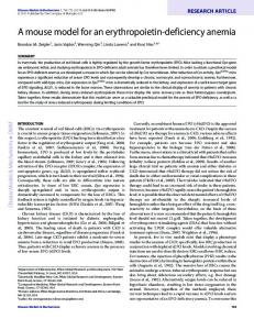
A non-anesthetized mouse model for recording sensory urinary bladder activity Peter Zvara1*, Andrew J. Wright1, Kristopher Roach1, Michal Ursiny1, Bennett Shapiro1, Lawrence M. Dagrosa1, Mark T. Nelson 2 and Thomas J. Heppner 2 Department of Surgery, University of Vermont College of Medicine, Burlington, VT, USA Department of Pharmacology, University of Vermont College of Medicine, Burlington, VT, USA
1 2
Edited by: Stuart Mazzone, University of Queensland, Australia Reviewed by: Lu-Yuan Lee, University of Kentucky, USA Michael Ruggieri, Temple University, USA *Correspondence: Peter Zvara, Department of Surgery, College of Medicine, University of Vermont, D319 Given Building, 89 Beaumont Avenue, Burlington, VT 05405, USA. e-mail:
[email protected]
The goal of this study was to develop an in vivo awake mouse model for extracellular bladder sensory nerve recording. A bipolar 125-μm silver electrode was positioned under a single postganglionic bladder nerve. Efferent nerve signals were eliminated by tying off the postganglionic bladder nerve between the major pelvic ganglion and the recording electrode. Sensory nerve activity was measured in the conscious animals 48 h after surgery during continuous intravesical infusion of 0.9% saline/0.5% acetic acid followed by 0.5% acetic acid with capsazepine (10 μM) at a rate of 0.75 ml/h. Continuous infusion of 0.9% NaCl led to a gradual increase in the frequency of sensory nerve firing that peaked upon reaching threshold pressure. Non-micturition contractions were observed in some animals during filling and other animals exhibited only minimal pressure fluctuations; both types of events were associated with a rise in sensory nerve activity. Intravesical infusion of 0.5% acetic acid reduced the intermicturition interval. This was associated with a 2.1-fold increase in bladder pressure during filling and a two-fold increase at both threshold and micturition pressures. Concurrent with these changes, sensory activity increased 2.8-fold during filling and 2.4-fold at threshold pressure. Subsequent intravesical infusion of capsazepine in 0.5% acetic acid reduced filling and threshold pressures by 21 and 31.2%, respectively, and produced corresponding decreases of 36 and 23.4% in sensory nerve activity. The current study shows that multifiber sensory nerve recordings can be reproducibly obtained from conscious mice. Keywords: urinary bladder, sensory nerves, conscious mouse
Introduction The afferent limb of the micturition reflex presents an attractive target for pharmacological intervention in the treatment of bladder dysfunction, but the animal models currently available for testing potential therapeutic agents have limitations (Andersson, 2002). Two types of in vitro preparations have been used to study bladder sensory activity in response to mechanical and chemical stimulation: an en block bladder/pelvic nerve preparation (Sengupta and Gebhart, 1994; Rong et al., 2002) and flat-sheet bladder preparation (Zagorodnyuk et al., 2006). Studies employing these models have contributed significantly to defining the peripheral neuronal mechanisms involved in bladder sensation. Studies focusing on central neuroregulation of the micturition reflex during bladder filling and micturition require an intact micturition reflex pathway and can therefore only be conducted in vivo. Animal models used to date require the use of anesthesia, which exerts its effects by enhancing inhibitory and/or suppressing excitatory neural output, likely compromising nerve activity recordings (Habler et al., 1990; Hara and Harris, 2002; Malley and Vizzard, 2002). To address these limitations, we developed a new method for recording sensory nerve activity in vivo in the conscious animal. We have adapted the ex vivo technique of multifiber sensory nerve recording to in situ recordings from the postganglionic bladder nerve in a conscious mouse. Bladder-specific afferent neural discharge and corresponding changes in intravesical pressure during
www.frontiersin.org
bladder filling were recorded. The validity of this model was tested by recording extracellular bladder sensory nerve activity in healthy animals and after inducing bladder overactivity with 0.5% acetic acid. The functional contribution of C-fibers to the bladder sensory discharge during noxious/chemically induced bladder irritation was assessed using the TRPV1 receptor antagonist, capsazepine (Daly et al., 2007).
Materials and Methods Male 6- to 8-week-old BalbC mice (Charles River Laboratories, St Constant, QC, Canada) were maintained under standard laboratory conditions with free access to food and water. All animal use procedures were approved by the University of Vermont Institutional Animal Care and Use Committee, and appropriate measures were taken to minimize pain and discomfort of the animals. After anesthetizing mice with isoflurane, a lower midline abdominal incision was made and PE-10 tubing (BD®) with a heat-flared end was implanted into the dome of the urinary bladder. Postganglionic bladder nerves running on the lateral aspect of the prostate were identified (Figure 1A), and a single branch was bluntly dissected approximately midway between the major pelvic ganglion (MPG) and the bladder neck. A 0.125-mm diameter, bipolar Teflon-coated silver electrode (WPI, Sarasota, FL, USA, catalog number AGT0510) with hook-shaped poles was positioned under the single postganglionic nerve. Isolation from surrounding
November 2010 | Volume 1 | Article 127 | 1
Zvara et al.
Bladder sensory signaling – awake mouse
exposed hook-shaped poles lying beneath the postganglionic bladder nerve (marked by white arrow), embedded in the clear transparent silicone adhesive compound. Black arrow marks the 10-0 nylon ligature tied between the MPG and the recording electrode (total magnification ×30).
Figure 1 | (A) Postganglionic bladder nerves (two branches are marked by black arrows) running between the MPG and the bladder neck alongside the bladder vein (marked by white arrow) on the lateral aspect of the prostate (total magnification ×40). (B) Depiction of the teflon-coated silver electrode with
The animal was allowed to acclimate 15–30 min before initiating continuous intravesical infusion of isotonic 0.9% NaCl at a rate of 0.75 ml/h. After three reproducible micturition cycles, the NaCl solution was replaced by 0.5% acetic acid (Sigma Chemical Co., St. Louis, MO, USA) diluted in 0.9% saline. In a subsequent set of experiments, 10 μM capsazepine (Sigma) was added to the infusate after initial exposure to acetic acid. Intravesical pressure and sensory nerve activity were recorded simultaneously. At the conclusion of the sensory nerve activity recording, the viability of the postganglionic bladder nerve was assessed. Neurostimulation, as square-wave pulses (1.0 ms, 12 Hz, 4 V), was applied through the bipolar silver electrode using a Grass stimulator (Grass Technologies, MA, USA). A segment of the postganglionic bladder nerve overlying the bipolar electrode was subsequently removed and evaluated histologically.
tissues was achieved by slightly lifting the nerve and the electrode of the prostatic surface using a micromanipulator Narishige MX-1 (Narishige Scientific Instrument Lab., Tokyo, Japan). After achieving a stable recording, the nerve was ligated between MPG and the electrode to eliminate efferent nerve signaling. The nerve-electrode complex was then embedded in a biocompatible silicone adhesive compound (KWIK-SIL; WPI, Sarasota, FL, USA; Figure 1B). This non-toxic compound secures the contact between the electrode and the nerve, anchors the nerve-electrode complex to the surrounding tissues, thereby preventing electrode movement and damage to the nerve. It also guarantees electrical isolation. The recording electrode and the ground reference electrode wire which was positioned within the abdominal cavity, were then externalized at the animal’s back and secured to paravertebral muscles with 6-0 nylon sutures. The bladder tubing and the electrode were coiled into two separate subcutaneous pouches, and the animal was allowed to recover for 48 h. Postoperative analgesia was maintained with buprenorphine (0.05 mg/kg s.c.). Analgesia was stopped 24 h prior to sensory nerve recording to prevent its suppressive effects on sensory neurotransmission. The activity of sensory bladder nerves was recorded in situ in conscious animals. Under brief inhalation anesthesia with isoflurane, PE-10 tubing and the electrode and ground wire were removed from the subcutaneous pouches on the animal’s back. The mouse was placed in a flat bottom mouse restrainer lined with an absorbent pad; the tubing and electrode were then connected to a pressure transducer (Grass Model PT300, Quincy, MA, USA) and a Neurolog head stage (NL100, Digitimer Ltd, UK), respectively. Intravesical pressure and sensory nerve activity were recorded simultaneously during continuous bladder filling. Intravesical pressure was recorded and analyzed using LabVIEW software (Texas Instruments, Dallas, TX, USA). Electrical activity was transmitted through the head stage, amplified (NL104), captured and analyzed via a power 1401 interface and Spike 2 software (version 6.03, CED, UK). Nerve activity was sampled at a rate of 25,000 Hz and stored on a PC. Off-line analysis was performed using the Spike 2 wavemark function; action potentials were discriminated based on their waveform.
Frontiers in Neurology | Autonomic Neuroscience
Data analysis
The animal was continuously monitored and its physical activity was recorded. Artifacts in sensory recordings caused by movement were eliminated by analyzing only that data obtained during intervals when the animal was not moving. Whole-nerve multifiber afferent activity was expressed as impulses per second (Hz) and is referred to as frequency of afferent nerve discharge. Data was quantified using a spike processor, which counts the number of action potential units per second that cross a preset threshold. The threshold value was set to the level of the smallest identifiable spike (approximately twice the baseline noise level). Nerve activity is expressed as a mean frequency of firing obtained over the course of 10 s. Quantitative analyses compare data from three areas of interest within the micturition cycle before and after pharmacological intervention: (1) at the lowest pressure point during the filling phase (filling pressure), (2) at the voiding contraction threshold (threshold pressure), and (3) at peak micturition pressure. Quantitative values are presented as the mean ± SEM, with differences between means determined using a paired Student’s t-test. P-values ≤0.05 were considered statistically significant.
November 2010 | Volume 1 | Article 127 | 2
Zvara et al.
Bladder sensory signaling – awake mouse
Results
capsazepine (Figure 3). In the presence of capsazepine, the bladder response to acetic acid was reduced as evidenced by an increase in intermicturition interval and a reduction in filling and threshold pressures.
Intravesical pressure during bladder filling and micturition
During continuous filling with 0.9% NaCl, cystometrograms showed two distinct patterns: (a) a steady intravesical pressure with pressure fluctuations of less than 2 cm H2O (Figure 2A), and (b) a filling phase with pressure fluctuations ranging from 2 to 10 cm H2O (Figure 2B). Intravesical infusion of 0.5% acetic acid evoked a significant reduction in intermicturition intervals coupled with an increase in average filling and threshold pressures. The same mouse utilized for the basal and acetic acid urodynamic comparison was further challenged with a mixture of 0.5% acetic acid and 10 μM
Multifiber bladder sensory nerve activity during bladder filling
Continuous filling with saline in vivo generated a reproducible pattern of afferent nerve discharge (n = 8). In all animals, baseline sensory activity oscillated between 20 and 60 Hz, and repeated bursts of afferent discharge were observed throughout the entire filling phase (Figure 2). In some animals, low amplitude non-voiding bladder
Figure 2 | Two distinct patterns recorded with cystometry: (A) Steady intravesical pressure during the filling phase with minimal pressure fluctuations; (B) Phasic components (small fluctuations in pressure) during the entire filling
phase. The phasic intravesical pressure components were associated with bursts of afferent discharge. Phasic bursts of afferent activity were observed in all recordings regardless of pressure fluctuations.
Figure 3 | Continuous recording of bladder pressure and afferent nerve activity. Infusion of 0.9% NaCl led to regular micturition cycles with steady pressure during filling. 0.5% acetic acid led to higher micturition frequency, multiple non-voiding contractions along with elevated filling, threshold and micturition
pressures. This change in bladder function was associated with increased nerve activity. Addition of capsazepine to the acetic acid solution reduced the number of non-voiding contractions and nerve activity. The rectangles mark the points in the experiment where the infusate was changed (M, micturition event).
www.frontiersin.org
November 2010 | Volume 1 | Article 127 | 3
Zvara et al.
Bladder sensory signaling – awake mouse
contractions (2–10 cm H2O) seen throughout the filling phase correlated with phasic afferent discharge (Figure 2B). Phasic afferent discharge was also evident in a subgroup of animals that displayed only minimal fluctuations in intravesical pressure (amplitude