Diabetologia (2006) 49:2388–2391 DOI 10.1007/s00125-006-0390-5
SHORT COMMUNICATION
A perfusion protocol for highly efficient transduction of intact pancreatic islets of Langerhans A. R. Barbu & B. Bodin & M. Welsh & L. Jansson & N. Welsh
Received: 9 February 2006 / Accepted: 8 June 2006 / Published online: 30 August 2006 # Springer-Verlag 2006
Abstract Aims/hypothesis Successful gene transfer to pancreatic islets might be a powerful tool for dissecting the biological pathways involved in the functional impairment and destruction of beta cells in type 1 diabetes. In the long run, such an approach may also prove useful for promoting islet graft survival after transplantation in diabetic patients. However, efficient genetic modification of primary insulinproducing cells is limited by the specific compact structure of the pancreatic islet. We present here a whole-pancreas perfusion-based transduction procedure for genetic modification of intact pancreatic islets. Materials and methods We used flow cytometry analysis and confocal microscopy to evaluate the efficiency of in vitro and perfusion-based transduction protocols that use adenoviral and lentiviral vectors expressing green fluorescent protein. Islet cell viability was assessed by fluorescence microscopy and beta cell function was determined via glucose-stimulated insulin secretion. Results In intact rat and human pancreatic islets, adenoviral and lentiviral vectors mediated gene transfer to about 30% of cells, but they did not reach the inner cellular mass within the islet core. Using the whole-pancreas perfusion protocol, we demonstrate that at least in rodent models the Electronic supplementary material Supplementary material is available for this article at http://dx.doi.org/10.1007/s00125-0060390-5 and is accessible for authorized users. A. R. Barbu : B. Bodin : M. Welsh : L. Jansson : N. Welsh Department of Medical Cell Biology, Uppsala University, Uppsala, Sweden A. R. Barbu (*) Department of Medical Cell Biology, Biomedical Centre, P.O. Box 571, S-751 23 Uppsala, Sweden e-mail:
[email protected]
centrally located insulin-producing cells can be transduced with high efficiency, while preserving the structural integrity of the islet. Moreover, islet cell viability and function are not impaired by this procedure. Conclusions/interpretation These results support the view that perfusion-based transduction protocols may significantly improve the yield of successfully engineered primary insulin-producing cells for diabetes research. Keywords Adenoviral vector . Cell death . Islet . Lentiviral vector Abbreviations ESM Electronic supplementary material GFP green fluorescent protein KRBH Krebs–Ringer bicarbonate PFU plaque-forming unit
Introduction Much work is currently directed to finding highly efficient and non-toxic vectors for gene transfer in primary insulinproducing cells. Among these, viral vectors have emerged as first choice for engineering beta cells, mostly due to their capacity to mediate efficient gene transfer in non-dividing cells. Adenoviral vectors in particular offer the advantage of a high gene transfer efficiency as well as relatively longterm gene expression in primary beta cells [1]. However, one important limitation of the adenoviral-mediated gene transfer in beta cell research is the reduced ability to reach and transduce the inner cellular mass of the intact pancreatic islet. This is an important drawback for experimental designs that involve gene therapy approaches
Diabetologia (2006) 49:2388–2391
2389
in which the final target for genetic modification is the insulin-producing cell, since, at least in rodent models, insulin-producing cells are centrally located in the core of the islet, surrounded by a mantle of non-beta cells at the periphery.
Materials and methods Sprague-Dawley rats (Skanbur BK, Solna, Sweden) were anaesthetised with an i.p. injection of thiobutabarbital sodium (120 mg/kg body weight). A catheter (inner diameter 1.40 mm) connected to a peristaltic pump was placed in the abdominal aorta so that the perfusion medium could flow freely into the pancreas. The gland was removed from the animals [2] and placed in a funnel at a constant temperature (30°C). The gland was perfused for 45 to 60 min at a flow rate of 1 ml/min with a continuously gassed (95% O2–5% CO2) Krebs–Ringer bicarbonate (KRBH) buffer, supplemented with 20 mg/ml BSA, 20 mg/ml dextran T70 and 0.3 mg/ml glucose, and containing ∼109 plaque-forming units (PFUs) per pancreas of the green fluorescent protein (GFP)-expressing adenoviral vector, which corresponds to 20 to 50 PFUs/islet cell. In some of the experiments, the capillary endothelium was disrupted before administering the viral vector by preperfusion for 40 s with medium containing 0.1% Triton X-100, followed by a 10-min wash with KRBH buffer only. Following transduction, islets were isolated and subsequently cultured for 2 to 7 days. All other experimental procedures have been previously described [3, 4] or are given in the Electronic supplementary material (ESM).
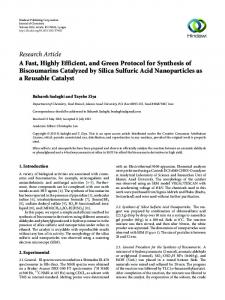
Results Adenoviral-mediated gene transfer to intact rat pancreatic islets High-titre in vitro adenoviral-mediated transduction of intact islets reached efficiencies above 50% (ESM Fig. 1). This approach, however, was paralleled by extensive cellular death [5] (ESM Fig. 1). Low-titre-mediated gene transfer into intact islets, on the other hand, resulted in 31% GFPpositive islet cells and no major increase in cell death (Fig. 1 and ESM Fig. 1). This percentage was increased up to 48% if, prior to the transduction procedure, cell-to-cell contact of the intact islets was transiently disturbed by EGTA treatment. Confocal laser-scanning microscopy sections through adenovirus-transduced isolated rat islets showed that the cells transduced by the in vitro technique were located in the outermost cell layer, while the cells within the core of
Fig. 1 GFP expression in rat islets following adenoviral-mediated transduction. a, b FACS analysis of dispersed islet cells expressing GFP. Rat islets were transduced in vitro (black bars) (a) or by the perfusion protocol (hatched bars) (b) with 50 PFUs/cell and then dispersed into a single-cell suspension. Results are means of six (in vitro) or four (perfusion) experiments±SEM. *** p