Downloaded from ard.bmj.com on April 4, 2013 - Published by group.bmj.com
ARD Online First, published on April 3, 2013 as 10.1136/annrheumdis-2012-202781 Clinical and epidemiological research
EXTENDED REPORT
Association between testosterone levels and risk of future rheumatoid arthritis in men: a population-based case–control study Mitra Pikwer,1 Aleksander Giwercman,2 Ulf Bergström,1 Jan-Åke Nilsson,1 Lennart T H Jacobsson,1,3 Carl Turesson1 Handling editor Tore K Kvien 1
Section of Rheumatology, Department of Clinical Sciences, Lund University, Malmö, Sweden 2 Reproductive Medicine Centre (RMC), Skåne University Hospital, Lund University, Malmö, Sweden 3 Department of Rheumatology and Inflammation Research, The Sahlgrenska Academy, University of Gothenburg, Gothenburg, Sweden Correspondence to Dr Mitra Pikwer, Department of Rheumatology, Lund University, Skåne University Hospital, Malmö SE-205 02, Sweden;
[email protected] Received 4 October 2012 Revised 22 December 2012 Accepted 3 February 2013
To cite: Pikwer M, Giwercman A, Bergström U, et al. Ann Rheum Dis Published Online First: [please include Day Month Year] doi:10.1136/ annrheumdis-2012-202781
ABSTRACT Objectives Rheumatoid arthritis (RA) is less common among men than women, and sex hormones have been suggested to play a part in the pathogenesis. Lower levels of testosterone have been demonstrated in men with RA, but it is not known if these changes precede the disease. Methods In a nested case–control study, using information and blood samples from a population-based health survey, we identified incident cases of RA by linking the cohort to local and national RA registers. Two controls for each validated case, matched for age, sex and year of screening, were selected from the health survey. Using stored blood samples, collected between 08:00 and 10:00 am after an overnight fast, we analysed levels of testosterone and other reproductive hormones. Results Serum was available from 104 cases (median time from screening to RA diagnosis 12.7 years (range 1–28); 73% rheumatoid factor (RF) positive at diagnosis or later) and 174 matched controls. In conditional logistic regression models, adjusted for smoking and body mass index, lower levels of testosterone were associated with subsequent development of RF-negative RA (OR 0.31 per SD, 95% CI 0.12 to 0.85), with a weaker association with RF-positive RA (OR 0.87 per SD; 95% CI 0.53 to 1.43). Levels of follicle-stimulating hormone were significantly increased in pre-RF-negative RA ( p=0.02), but decreased in pre-RF-positive RA ( p=0.02). Conclusions Lower levels of testosterone were predictive of RF-negative RA, suggesting that hormonal changes precede the onset of RA and affect the disease phenotype.
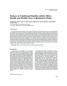
suppress the hypothalamic–pituitary–gonadal (HPG) axis, suggesting that low testosterone levels might be a consequence of the inflammatory disease.12 Alternatively, the measured low testosterone levels may reflect a role for androgens in the pathogenesis of RA, as suggested by the age-related sex differences in the epidemiology of the disease. A large prospective study of women did not find any association between androgen levels measured at a single time point or polymorphism in the sex hormone receptors and risk of RA. The only prospective study on men, based on 32 incident male RA cases from Finland, did not show any significant differences in testosterone levels before RA onset compared with controls.13 However, this study did not adjust for potential confounders and had limited power for stratification by different phenotypes of RA.13 Taken together, there are limited data on the importance of androgen levels for the development of RA, in particular in men. Based on the literature, testosterone is of particular interest, but other hormones such as follicle-stimulating hormone (FSH) and luteinising hormone (LH) are necessary to interpret the origin of differences in testosterone. The interplay of these hormones in the HPG axis is illustrated in figure 1. Data on sex hormone-binding globulin (SHBG) are needed to calculate free testosterone levels. The aim of this study was to measure testosterone and other sex hormones in a larger sample of men who subsequently developed RA, to investigate if differences in hormone concentrations from matched controls could be detected years before diagnosis and if such patterns differed between subtypes of RA.
INTRODUCTION
PATIENTS AND METHODS Source population: the Malmö Preventive Medicine Program
Risk factors for rheumatoid arthritis (RA) include genetic,1 environmental2 and hormonal factors.3 Autoimmunity affects men to a lesser extent than women,4 and, during the fertile years, RA has a female/male incidence ratio of 4–6 : 1.5 With increasing age, the sex difference in incidence narrows.6 In cross-sectional studies, lower levels of serum testosterone have been found in both male and female patients with RA compared with healthy controls,7–10 and low levels of testosterone have been observed in synovial fluids in both female and male patients with active disease.11 Proinflammatory cytokines are known to stimulate the hypothalamic–pituitary–adrenal axis but
Between 1974 and 1992, the Malmö Preventive Medicine Program (MPMP), a preventive casefinding programme, was conducted in Malmö, Sweden ( population 235 000 in 1974).14 The programme included a total of 22 444 males born between 1949 and 1921 and 10 902 females born between 1938 and 1925. The aim of the health survey was to screen large strata of the adult population in order to identify individuals for preventive intervention. The overall attendance rate was 71.2%. The vast majority of participants were Caucasians of Scandinavian origin. The subjects
Pikwer M, Article et al. Ann Rheum Dis 2013;0:1–7. 1 Copyright author (or theirdoi:10.1136/annrheumdis-2012-202781 employer) 2013. Produced by BMJ Publishing Group Ltd (& EULAR) under licence.
Downloaded from ard.bmj.com on April 4, 2013 - Published by group.bmj.com
Clinical and epidemiological research Figure 1 A schematic overview of the hypothalamus–pituitary–gonadal (HPG) axis and the impact of testosterone on immune cells. FSH, follicle-stimulating hormone; GnRH, gonadotropin releasing hormone, LH, luteinising hormone.
underwent physical examination including height and weight measurements and laboratory tests, and completed a selfadministered questionnaire on health and lifestyle factors.15 The subjects were invited to leave blood samples in the morning, between 08:00 and 10:00 am, after an overnight fast. The samples were stored at −20°C.16
Selection of cases and controls In a previous survey,17 we identified individuals who developed RA after inclusion in this cohort and up to 31 December 2004, by linking the MPMP register to a community-based RA register,18 19 the local outpatient clinic administrative register for Malmö University Hospital, the National Hospital Discharge Register and the National Cause of Death Register.17 The community-based RA register has been shown to include more than 90% of patients in 2
the catchment area.19 The Swedish national inpatient register includes more than 99% of all hospital discharges and has a high validity for RA and many other diagnoses.20 In a structured review of all medical records, possible cases were validated and classified according to the 1987 American College of Rheumatology criteria for RA.21 Four controls for each validated case, matched for sex, year of birth and year of screening, who were alive and free from RA when the index person was diagnosed with RA, were selected from the MPMP cohort. Vital status and information on emigration were retrieved from the national census, and controls who were not alive or living in Sweden through the index date were excluded. For the present study, serum samples from two controls per case were collected from the MPMP bio bank. For various reasons, samples were missing from a subset of cases as well as Pikwer M, et al. Ann Rheum Dis 2013;0:1–7. doi:10.1136/annrheumdis-2012-202781
Downloaded from ard.bmj.com on April 4, 2013 - Published by group.bmj.com
Clinical and epidemiological research controls. For cases with available serum, the retrieval of control samples was extended to include the two remaining matched controls, when such were available. This study was approved by the regional research ethics committee for southern Sweden. All participants gave their informed consent to be included in the MPMP and the Malmö RA register, respectively. No informed consent was obtained specifically for the present study.
Socioeconomic background and comorbidities Data on socioeconomic status were derived from self-reported job titles in the Swedish national censuses, as previously described.17 Briefly, occupations were coded and converted into standardised social class categories, and subjects were classified as ‘blue-collar workers’ (manual workers, both skilled and unskilled), ‘white-collar workers’ (non-manual employees and self-employed professionals) and ‘others’. Housewives, students and unemployed without any other self-reported job title during the study period were excluded from this classification.22 Smoking was identified as current smoking versus current nonsmoking (ie, never smoked or past smoking). Data on self-reported overall health and self-reported cancer, diabetes and cardiovascular disease (the latter classified as selfreport of hospitalisation for stroke, physician diagnosis of angina pectoris, or current use of heart medication) at baseline were extracted from the self-administered questionnaire.
Laboratory tests Serum total testosterone, SHBG, LH and FSH concentrations were quantified by ElectroChemiLuminiscence Immunoassay based on a ruthenium derivative according to routine methods used at the Department of Laboratory Medicine, Skåne University Hospital. Free testosterone was calculated from total testosterone and SHBG levels using the Vermeulen formula.23 The detection limits for testosterone, SHBG, LH and FSH were 0.0087 nmol/l, 0.35 nmol/l, 0.10 IU/l and 0.10 IU/l, respectively. Imprecision levels for low and high levels were 2.4% and 4.0% for testosterone, 1.0% and 1.1% for SHBG, 2.0% and 2.2% for LH, and 3.3% and 2.2% for FSH, respectively. The erythrocyte sedimentation rate (ESR) was measured at screening according to the standard Westergren method. Data on rheumatoid factor (RF) tests were collected from the databases of the two clinical immunology laboratories in the area.
Statistical analysis The impact of baseline hormone levels on the risk of RA was examined by bivariate conditional logistic regression analysis, taking into account the matched design of the study. For comparability of the impact of hormones with different concentrations, ORs for RA were calculated per SD of testosterone, FSH, LH and SHBG. Potential confounders were examined in a similar manner. Correlations between body mass index (BMI) and testosterone were examined using Pearson’s test. Multivariate logistic regression analysis was used to adjust for potential confounders. Analyses were stratified by RF status at diagnosis or later (ever positive vs negative) and also by time from screening to RA diagnosis (above vs below the median). Statistical significance was set at p