Sleep Apnea Assessment
A Portable Automated Assessment Tool for Sleep Apnea Using a Combined Holter-Oximeter Conor Heneghan, PhD1,2; Chern-Pin Chua, BE1; John F. Garvey, MB, MRCPI3; Philip de Chazal, PhD2; Redmond Shouldice, PhD2; Patricia Boyle, BS3; Walter T. McNicholas, MD, FRCPI3 School of Electrical, Electronic and Mechanical Engineering, University College Dublin, Dublin, Ireland; 2BiancaMed Ltd, NovaUCD, University College Dublin, Dublin, Ireland; 3The Respiratory Sleep Disorders Unit, St. Vincent’s University Hospital, Dublin, Ireland. 1
Study Objectives: Resource limitations have raised interest in portable monitoring systems that can be used by specialist sleep physicians as part of an overall strategy to improve access to the diagnosis of sleep apnea. This study validates a combined electrocardiogram and oximetry recorder (Holter-oximeter) against simultaneous polysomnography for detection of sleep apnea. Design: Prospective study. Setting: A dedicated sleep disorders unit. Participants: 59 adults presenting for evaluation of suspected sleep apnea. Interventions: NA. Measurements and Results: An automated algorithm previously developed for sleep apnea detection was applied to the electrocardiogram and oximetry measurements. The algorithm provides (a) epochby-epoch estimates of apnea occurrence and (b) estimates of overall per-subject AHI. Using separate thresholds of AHI ≥15 and AHI 20 and 0.04 respectively, with 16.7% of subjects having intermediate test results (AHI 5–14/h). Regardless of AHI, 85.3% of respiratory events were correctly annotated on an epoch-by-epoch basis. AHI underestimation bias was 0.9/h, and the antilogs of log-transformed limits of agreement were 0.3 and 2.7. Correlation between estimated and reference AHI was 0.95 (P < 0.001). Conclusion: Combined Holter-oximeter monitoring compares well with polysomnography for identifying sleep apnea in an attended setting and is potentially suitable for home-based automated assessment of sleep apnea in a population suspected of having sleep apnea. Keywords: Sleep apnea, Holter monitoring, oximetry, apnea screening, polysomnography Citation: Heneghan C; Chua CP; Garvey JF; de Chazal P; Shouldice R; Boyle P; McNicholas WT. A portable automated assessment tool for sleep apnea using a combined holter-oximeter. SLEEP 2008;31(10):1432-1439.
OBSTRUCTIVE SLEEP APNEA SYNDROME (OSAS) IS A MAJOR HEALTHCARE PROBLEM AFFECTING AT LEAST 4% OF MEN AND 2% OF WOMEN.1 FIFTEEN million people in the United States alone are believed to have sleep apnea,2 but currently an estimated 80% to 90% of moderate-tosevere OSAS is undiagnosed.3 OSAS is a recognized independent risk factor for the development of cardiovascular diseases.4 Hypertension is found in up to 50% of patients with OSAS, and there is growing evidence supporting an association with ischemic heart disease, stroke, heart failure, atrial fibrillation, and cardiac sudden death.5,6 Long-term observational studies of effectively treated OSAS patients have also shown a significant benefit in reducing cardiovascular mortality and non-fatal cardiovascular events7-9 when compared to untreated patients. Growing recognition of the significance of OSAS has increased pressure on specialist sleep facilities, and in many countries resource constraints have restricted access to in-laboratory polysomnography.10 The diagnosis of OSAS requires the combined assessment of relevant clinical features and the objective demonstration of abnormal breathing during sleep; current evidence indicates that
basing the diagnosis on either feature alone is unreliable.11-13 Thus, there is considerable interest in limited diagnostic tools that can be used by sleep physicians as part of an overall strategy to improve access to OSAS diagnosis and treatment.14 In this report we present a system that addresses one component of diagnosing OSAS, namely objective measurement of OSA. We have previously developed a combined single-channel electrocardiogram (ECG) and oximetry analysis system for apnea detection.15 In a retrospective study of 125 patients with suspected sleep apnea, the system achieved a sensitivity and specificity of 96.5% and 95.0%, and positive and negative likelihood ratios of 19.30 and 0.04. The present study prospectively validates the performance of the proposed automated ECG-oximetry system by comparing its performance with simultaneous diagnostic polysomnography in subjects with suspected OSAS. The ability of the system to (a) identify apnea events on an epoch-by-epoch basis and (b) identify or exclude subjects with clinically significant sleep apnea was evaluated. ECG and oximetry measurements were obtained using a portable combined Holter-oximeter to establish the practicality of the recorder and associated software as a portable automated assessment tool. The study was conducted and the results reported with reference to the recommended methodology for study design and measurement of agreement between polysomnography and portable monitors set out in the AASM/ACCP/ATS joint review16 and its companion article.17
Submitted for publication May, 2008 Accepted for publication June, 2008 Address correspondence to: Conor Heneghan, PhD, School of Electrical, Electronic and Mechanical Engineering, Engineering and Material Sciences Centre, University College Dublin, Belfield, Dublin 4, Ireland; Tel: (353 1) 716 1909; Fax: (353 1) 283 0921; E-mail:
[email protected] SLEEP, Vol. 31, No. 10, 2008
1432
Portable Automated Assessment —Heneghan et al
METHODS
“SDB” (sleep disordered breathing). The second is an estimated apnea hypopnea index (AHI) derived on the basis of the above annotations. The total number of SDB epochs was first divided by the total time in bed to obtain a respiratory disturbance index (RDI), and this RDI was then transformed by a scaling factor to obtain the estimated AHI. The total time in bed was obtained as the time difference between 2 event markers, one entered just before PSG and one entered just after PSG. The event marker was logged by pressing a button on the Holter-oximeter. In a home setting, total time in bed can be obtained by having subjects press the event button once before they get into bed and once after they wake up. The scaling factor was obtained from a regression constant obtained using known AHI during the development of the ECG-oximetry system.15 The core of the system is a pattern recognition algorithm that identifies occurrences of sleep disordered breathing by analyzing (1) the cyclic variations in heart rate (CVHR) associated with apnea,23 (2) ECG-derived respiration, and (3) SpO2. CVHR can be detected rather reliably using time and frequency-domain heart rate variability analysis of the RR interval time series. In addition, respiratory effort fluctuations during apnea events can be detected from an ECG-derived respiratory signal, as the magnitude of QRS peaks is typically modulated by ribcage movements. Lastly, apnea events are frequently associated with oxygen desaturation, which can be detected by analyzing the SpO2 signal. Briefly, the ECG-based features include • First 5 serial correlation coefficients • Standard deviation of the RR intervals • Standard deviation of the change in RR intervals (delta RR) • Mean epoch RR interval The oximetry-based features include • Mean SpO2 value • Minimum SpO2 value • Number of SpO2 values < 92% saturation • Square root of the 5% to 95% spread in sorted SpO2 values • Mean of the absolute differences between successive SpO2 samples The pattern recognition algorithm considers data in 1-min epochs, overlapping at 30-sec intervals. For each epoch, it combines the features obtained from both ECG and oximetry and decides if that epoch is a SDB epoch or not. In the event that either the ECG or oximetry signal is not available (due to artifact, for example) the system continued working, albeit in a degraded mode, using only features from the available signal. Technically, the algorithm employs a technique called “linear discriminant analysis” to work out a (posterior) probability of the epoch being a SDB epoch, given the observed features and prior probability of SDB epochs. As we were deciding between 2 classes, i.e., SDB or normal, a threshold posterior probability of 0.5 was used.
Subjects Subjects were recruited from patients referred to the Sleep Disorders Units at St. Vincent’s University Hospital and St. Vincent’s Private Hospital, Dublin, Ireland for evaluation of possible OSAS. Clinical manifestations ranged from snoring alone to snoring plus daytime features suggestive of OSAS. All patients had been scheduled to have polysomnography prior to enrolment. All subjects gave written informed consent, and the study was approved by the hospital ethics committee. Exclusion criteria for the study included patients with a known cardiac arrhythmia and patients who had a permanent pacemaker in situ. Patients with a previous diagnosis of hypertension and patients on β-blockers were included in the study. Consecutive patients who met the inclusion criteria were invited to participate. Protocol All subjects underwent full, in-laboratory, attended diagnostic polysomnography (PSG) (Jaeger-Toennies system, Erich Jaeger GmbH, Germany) using standard techniques. Simultaneous electrocardiogram (ECG) and pulse oximetry measurements were recorded using a combined Holter-oximeter (NorthEast Monitoring, Inc., USA). As the Holter-oximeter is intended for unattended use, no intervention was made to repair or correct possible data loss from the portable monitor. Diagnostic Polysomnography All PSG studies were scored according to the criteria of Rechtschaffen and Kales18 by a single experienced sleep technologist who was blinded to the output of the automated ECGoximetry analysis system. Likewise, ECG-oximetry analysis was conducted without knowledge of the results of human PSG scoring. Apneas were defined as complete cessation of airflow ≥ 10 sec and hypopneas as reduction of respiratory signals ≥ 10 sec associated with oxygen desaturation > 4% and/or arousal.19 Obstructive events were distinguished from central events by the presence or absence of paradoxical thoracic and abdominal movements during apneas or hypopneas. Thoracoabdominal motion was measured using uncalibrated inductance plethysmography. Automated ECG-Oximetry Analysis Built-in firmware in the combined Holter-oximeter derived oxygen saturation (SpO2) using 4-beat exponential averaging with a 2-beat processing delay. SpO2 was down-sampled and analyzed at 0.1 Hz. The system was fully automated; no manual review or editing of the outputs was performed. The principles and performance of the automated ECG-oximetry analysis system have been described in detail elsewhere,15 and are summarized here for convenience. The system reads one channel of ECG and one channel of oxygen saturation and produces 2 main outputs. The first is a continuous epoch-byepoch sequence annotating each 30-sec epoch as “normal” or SLEEP, Vol. 31, No. 10, 2008
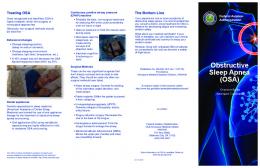
Apnea Hypopnea Index Estimation We performed Bland-Altman analysis as recommended.16 As our system exhibited a “fan-shaped” scatter on the BlandAltman plot, i.e., variability in differences increased with magnitude of averages, we report the antilog of logarithmic 1433
Portable Automated Assessment —Heneghan et al
Table 1—Summary of Subject Characteristics and Polysomnography Results Parameter N (M:F) Age (y) Apnea-hypopnea index (per h) Epworth Sleepiness Scale Body mass index (kg/m2)
No OSAS 11 (8:3) 50 ± 10 2.0 ± 1.4 10.4 ± 6.8 27.7 ± 5.8
Apnea Severity Mild Moderate 17 (13:4) 11 (11:0) 50 ± 8 57 ± 12 9.2 ± 2.9 19.0 ± 3.4 10.2 ± 6.2 11.4 ± 5.1 29.3 ± 3.8 32.1 ± 4.7
Severe 20 (20:0) 48 ± 9 61.8 ± 32.8 12.7 ± 5.6 33.4 ± 6.2
F statistic (P-value) 3.17 (P = 0.03) 32.94 (P < 0.001) 0.62 (P = 0.61) 3.61 (P = 0.02)
Values are mean ± standard deviation. No OSAS: AHI < 5; Mild OSAS: AHÍ 5 to < 15; Moderate OSAS: AHÍ 15 to < 30; Severe OSAS: AHI ≥ 30. F (P value): Results of one-way ANOVA.
transformed bias and limits of agreement between estimated and reference AHI.21 In addition, to help interpretation, we also computed conventional limits of agreement.21 Finally, to facilitate comparison with previous work, we also report the Pearson product moment correlation coefficient.
The oximetry-only system is described in detail in an earlier publication,15 and is summarized here for convenience. The core of the system is a pattern-recognition algorithm that recognizes decreases in oxygen saturation characteristic of apnea events. The system was developed using the same data set as that used to develop the combined ECG-oximetry system. The SpO2 signal was downsampled to 0.1 Hz and analyzed in 1-min epochs. Features used to characterize changes in oxygen saturation and hence identify apnea epochs include (a) mean SpO2 value, (b) minimum SpO2 value, (c) number of SpO2 values < 92% saturation, (d) square root of the 5% to 95% spread in sorted SpO2 values. Based on results obtained from the development of the oximetry-only system, the same AHI thresholds as those chosen for the ECG-oximetry system were preselected to define clinically significant and insignificant sleep apnea.
Per-Subject Automated Assessment for Sleep Disordered Breathing Based on results obtained during the development of the ECG-oximetry system, a PSG AHI ≥ 15 was preselected to define clinically significant sleep apnea, and an AHI < 5 to define no significant sleep apnea. The same thresholds were also preselected to define positive and negative test results from the ECG-oximetry system. Subjects with estimated AHI between 5 and 15 received a nondiagnostic (“Indeterminate”) result. This approach was motivated by the limitations of using a single knife-edge threshold for determining clinically significant SDB,16 as it penalizes small errors due to natural variability. In line with our thresholds, we report the sensitivity, specificity, and likelihood ratios conditional on obtaining a positive or negative result,22 and the proportion of subjects who received an Indeterminate result, even though their PSG-reported AHI values were either fewer than 5 or more than 15. To facilitate comparison with previous studies, we also report the performance if the system operated at 3 commonly used single-threshold points, AHI = 5, 10, and 15.
Holter Analysis Standard Holter reports (Holter 5 software, NorthEast Monitoring, Inc., USA) were obtained on the ECG recordings from the combined Holter-oximeter. These reports were screened by a physician for pathologically significant arrhythmia. RESULTS Sixty-four subjects were eligible for participation; 63 were recruited (one declined); and all 63 successfully completed the protocol. Data from 4 subjects were not analyzed because of technical problems which resulted in premature termination of the recordings. This represents a subject-level data loss rate of 6.3% of all subjects who completed the protocol. Table 1 summarizes the subject characteristics and PSG results. The subjects were predominantly white middle-aged males, and covered the entire spectrum of apnea severity. Eight subjects had > 50% of their respiratory events composed of central apneas and hypopneas.
Epoch-by-Epoch Sleep Disordered Breathing (SDB) Annotation All obstructive, central and mixed respiratory events obtained from PSG (including both apneas and hypopneas) were included, and running 1-min epochs with 30-sec overlaps were used. As respiratory events may straddle epochs, an epoch was marked as an SDB epoch if it contained > 5 sec of a respiratory event. Five sec (half the minimum duration of a respiratory event) was used, so that short respiratory events that straddle epochs are not missed.
Apnea-Hypopnea Index Estimation Figure 1 presents the Bland-Altman plot for AHI estimated from ECG-oximetry analysis and reference AHI obtained from polysomnography. The antilog of logarithmic transformed limits of agreement were 0.29 and 2.72, i.e., 95% of the time, the estimated AHI was between a factor of 0.29 and 2.72 of the PSG AHI. Alternatively, using untransformed data,21 bias was –0.9/h, and conventional limits of agreement were –18.0 to 16.2/h.
Comparison with Oximetry-only System To quantify the benefits of combined ECG-oximetry analysis, we developed, for comparison, a separate system using only the SpO2 signal, using similar principles and procedures employed in developing the combined ECG-oximetry system. SLEEP, Vol. 31, No. 10, 2008
1434
Portable Automated Assessment —Heneghan et al
50
120
Non−OSAS Indeterminate OSAS
30
Non−OSAS Indeterminate OSAS
100
Holter−Oximeter AHI (hr−1)
Holter−Oximeter AHI − PSG AHI (hr−1)
40
20 10 0 −10 −20 −30
80
60
40
20
−40 −50
0
20
40
60
80
0
100
−1
0
20
40
60
80
100
120
−1
Mean AHI (hr )
PSG AHI (hr )
Figure 1—Bland-Altman plot of estimated AHI obtained from ECG-oximetry analysis and reference AHI obtained from PSG. Non-OSAS: AHI < 5, Indeterminate: AHI 5 to < 15; OSAS: AHI ≥ 15. One subject with a PSG AHI of 171/h is not shown to improve the figure’s clarity.
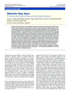
Figure 2—Scatter plot of estimated and reference AHI. PSG AHI: Reference AHI obtained from polysomnography. Holter Oximeter AHI: Estimated AHI obtained from ECG-oximetry analysis. NonOSAS: AHI 2 sec were found in 8 subjects. Despite the significant presence of ectopic beats, inspection of the results for subjects with higher amounts of ectopy or bradycardia indicated that the presence of ectopy or bradycardia did not significantly affect the performance of the ECG-oximetry system in terms of per-subject apnea assessment results. SLEEP, Vol. 31, No. 10, 2008
20
−1
Time (min)
1436
Portable Automated Assessment —Heneghan et al
Table 4—Performance of the ECG-Oximetry System in Epoch-by-Epoch Sleep Disordered Breathing Detection Metric (%) Accuracy Sensitivity Specificity Positive predictive value Negative predictive value
No OSAS 95.6 ± 1.7 27.3 ± 30.7 97.1 ± 1.0 20.9 ± 21.9 97.9 ± 1.7
Apnea Severity Mild Moderate 86.8 ± 3.1 81.0 ± 6.3 35.9 ± 22.0 42.8 ± 21.5 93.4 ± 3.1 91.9 ± 5.2 40.6 ± 23.6 56.9 ± 16.4 92.0 ± 4.0 85.5 ± 7.3
Severe 80.8 ± 6.1 80.3 ± 15.8 74.3 ± 15.0 79.3 ± 10.5 81.3 ± 9.6
Overall 85.3 ± 7.2 51.4 ± 30.3 87.3 ± 13.2 54.2 ± 27.8 88.0 ± 9.1
Values are mean ± SD. No-OSAS: AHI < 5; Mild OSAS: AHI 5 to < 15; Moderate OSAS: AHI 15 to < 30; Severe OSAS: AHI ≥ 30.
and a reasonable nondiagnostic rate, and is comparable with results of previous studies using other modalities. In addition, this approach offers a potential advantage of being more robust in an unattended setting, as sensors measuring respiratory flow and snoring may become detached more easily or be poorly tolerated. The current study adds to the evidence regarding the use of type 4 portable monitors in an attended setting. Since the joint review, 2 recent studies30,31 have compared ECG and oximetry analysis with simultaneous polysomnography as potential tools for apnea assessment. Zamarron et al30 performed spectral analysis on heart rate and oxygen saturation and prospectively obtained a sensitivity and specificity of 94% and 82% using a single PSG AHI threshold of 10. However, the use of this system as an assessment tool may be limited because the system requires manual scoring. In a retrospective study, Raymond et al31 analyzed autonomic arousals based on heart rate and oxygen saturation and obtained a sensitivity and specificity of 52% and 89% using a PSG AHI threshold of 15. Our system compares well with these studies in terms of performance and potential as a tool for portable automated apnea assessment. The AASM has recently announced new guidelines relating to the use of portable monitoring.32 In terms of technology, it has recommended airflow, respiratory effort, and blood oxygenation be recorded using the same sensors as those used in polysomnography. In this context, we acknowledge the proposed system does not meet these recommendations. However, the current study demonstrates the potential clinical utility of limited systems like ours in terms of per-subject apnea assessment and epoch-by-epoch apnea annotation, and it adds to the body of knowledge surrounding limited portable monitors that do not record respiratory effort and airflow directly. The patient population in this study had a high likelihood of OSAS, having already been referred to a specialist center for assessment of possible OSAS. The utility of this device for assessing OSA in a general population (e.g., self-reported snorers) in a primary care setting is presently unproven. However, this paper was not targeted at providing evidence for the use of ECG-oximetry as a primary screening tool in a general population; that is a secondary study which we will perform once we are satisfied as to the technical and clinical robustness of the system in a well-controlled setting with a high pretest OSAS probability. Our automated system provides an estimated AHI value, which is the most commonly used index for quantifying respiratory disturbances. In addition, we also provide detailed epoch-by-epoch annotations of apnea occurrence at 30-sec resoSLEEP, Vol. 31, No. 10, 2008
lution. This feature is of potential clinical utility; most systems do not provide this information. The results also indicated that the combined ECG-oximetry system appears more robust than oximetry alone in terms of overcoming data loss—the combined ECG-oximetry system continued to provide output using ECG information alone when the oximetry channel failed. We expect artifact to be more severe in a home setting, which will further degrade the performance of the oximetry-only system. However, we acknowledge that although the ECG is relatively more robust than the oximetry signal, and in this study was robust enough to ensure all data were analyzed: it is not immune to artifact. Indeed, the system has built-in capabilities to flag artifact epochs that could not be analyzed. Therefore, we would not expect the combined ECG-oximetry system to be able to analyze all data at all times. Further value is provided by the concurrent screening of subjects for pathologically significant arrhythmia as part of evaluation of suspected sleep apnea, and the ability to examine the correlation between arrhythmia and oxygen desaturation. The presence of ectopy or bradycardia did not significantly affect the efficacy of the system in assessing OSA, but we acknowledge that the potential effect of sustained irregular heart rhythms or tachyarrhythmias such as atrial fibrillation remain unclear, as patients with known cardiac arrhythmias were excluded from the study. We acknowledge that the subject-level data loss rate in this study may seem relatively high, but this may, in part, reflect the fact that the sleep laboratory staff had no prior experience of using the device in advance of the present study. Subsequent studies with the device that are currently ongoing have been associated with a lower data loss rate. We are currently evaluating the device in the home setting, and preliminary data indicate a relatively low data loss rate. There has also been interest in assessing sleep apnea in specific patient groups, such as heart failure patients33 and patients with stroke and transient ischemic attack,34 as the identification and management of sleep apnea in these patients may offer clinical and prognostic benefits.35,36 Estimating the presence and extent of sleep apnea from ECG and oximetry may be an attractive modality in these settings. For example, in patients with stable heart failure, overnight Holter monitoring for assessment of sleep apnea also allows the documentation of serious arrhythmia, which may be useful in enhancing diagnosis and management of these cardiac disturbances. In patients with acute stroke, ECG and oximetry are commonly available, and the assessment of sleep apnea can therefore take place with no additional equipment or procedures. 1437
Portable Automated Assessment —Heneghan et al
ABBREVIATIONS
In the development and validation of the ECG-oximetry system, EEG arousals were not required for hypopneas.19 The performance of the system if arousal criteria are included in the definition of hypopnea is thus unclear. However, we do not expect a large change in performance in terms of AHI estimation, as it has been shown that including arousal criteria for hypopnea definition results in small changes in AHI values.37 Likewise, the performance of the proposed system if respiratory effort related arousals (RERA) were included16 is unclear. Arousals associated with ECG changes with or without desaturation could also possibly confound our results. However, the proposed system considers both ECG changes and desaturation when detecting apnea events, so the system controls, to a certain extent, for the possibility of detecting arousals instead of apnea events. However, as we did not have full technical access to arousal information from the sleep monitoring system, we were unable to analyze the effect of arousals systematically. Therefore, we acknowledge that some of the false positive apnea events could well be arousals that resulted in ECG changes. Nevertheless, we believe the impact to be minimal, as seen from the good specificity results for epoch-by-epoch apnea annotation, and at an abstracted level, the good results for per-subject apnea assessment. The reasons for the large errors for subjects with severe apnea are unclear. For cases in which the ECG-oximetry system underestimated AHI, it is possible that the system, which is epoch-based, does not have sufficient time resolution to resolve the apnea events that occurred in quick succession (i.e., 2 apnea events were compressed into one apnea epoch). However, in terms of identifying apnea, these errors in subjects with severe OSAS are likely to be clinically insignificant, as all these subjects were correctly identified in terms of OSAS severity. It is also noted that being an epoch-based system, the ECG-oximetry system can only approximate actual SDB annotations, which are event based. The system does not distinguish between apneas and hypopneas, or between central and obstructive events. In addition, as we analyzed data using separate thresholds in this study, this resulted in relatively small sample sizes in some categories (Table 2). Accordingly, we note a potential limitation of small sample size. Using AHI as the primary benchmark for the ECG-oximetry system as a potential tool for assessing OSA in a home setting has limitations. From a technical point of view, there is interscorer variability for reference AHI obtained from PSG.38 Clinically, AHI captures only one dimension of a wide spectrum of sleep disordered breathing and symptoms that include arousals during sleep and daytime sleepiness. However, as AHI is currently the most commonly used objective measure, a system that approximates AHI accurately as its primary objective should have clinical utility. The estimated AHI can form one component of an effective overall screening algorithm, which may include other clinical parameters such as daytime sleepiness and BMI. In conclusion, the present study indicates that ECG-oximetry analysis using a combined Holter-oximeter compares well with simultaneous polysomnography for identifying sleep apnea in an attended setting. It is therefore potentially a suitable device for home-based assessment of sleep apnea in a population suspected of having sleep apnea. SLEEP, Vol. 31, No. 10, 2008
AASM ACCP AHI ATS CSAS CVHR ECG ESS LR OSAS PSG SDB
American Academy of Sleep Medicine American College of Chest Physicians Apnea-hypopnea index American Thoracic Society Central sleep apnea syndrome Cyclic variations in heart rate Electrocardiogram Epworth Sleepiness Scale Likelihood ratio Obstructive sleep apnea syndrome Polysomnography Sleep disordered breathing
Disclosure Statement This was not an industry supported study. Dr. Heneghan is an employee of and has financial interests in BiancaMed, Belfast Ireland. Dr. de Chazal is a director of BiancaMed. Dr. Shouldice is a research and development engineer with BiancaMed. The other authors have indicated no financial conflicts of interest. REFERENCES 1. 2. 3. 4. 5. 6. 7. 8.
9.
10. 11. 12. 13. 14. 1438
Young T, Peppard PE, Gottlieb DJ. Epidemiology of obstructive sleep apnea: a population health perspective. Am J Respir Crit Care Med 2002;165:1217-39. Volk R, Somers VK. Cardiovascular consequences of obstructive sleep apnea. Clin Chest Med 2003;24:195-205. Young T, Evans L, Finn L, et al. Estimation of the clinically diagnosed proportion of sleep apnea syndrome in middle-aged men and women. Sleep 1997;20:705-6. Phillips B. Sleep-disordered breathing and cardiovascular disease. Sleep Med Rev 2005;9:131-40. McNicholas WT, Bonsigore MR. Sleep apnoea as an independent risk factor for cardiovascular disease: current evidence, basic mechanisms and research priorities. Eur Respir J 2007;29:156-78. McNicholas WT, Javaheri S. Pathophysiologic mechanisms of cardiovascular disease in obstructive sleep apnea. Sleep Med Clin 2007;2:539-47. McNicholas WT. Cardiovascular outcomes of CPAP therapy in obstructive sleep apnea syndrome. Am J Physiol Regul Integr Comp Physiol 2007;293:R1666-70. Doherty LS, Kiely JL, Swan V, McNicholas WT. Long-term effects of nasal continuous positive airway pressure therapy on cardiovascular outcomes in sleep apnea syndrome. Chest 2005;127:2076-84. Marin JM, Carrizo SJ, Vicente E, Agusti AG. Long-term cardiovascular outcomes in men with obstructive sleep apnoea-hypopnoea with or without treatment with continuous positive airway pressure: an observational study. Lancet 2005;365:1046-53. Flemons WW, Douglas NJ, Kuna ST, Rodenstein DO, Wheatley J. Access to diagnosis and treatment of patients with suspected sleep apnea. Am J Respir Crit Care Med 2004;169:668-72. McNicholas WT. Diagnosis of obstructive sleep apnea in adults. Proc Am Thorac Soc 2008;15;5:154-60. Deegan PC, McNicholas WT. Predictive value of clinical features for the obstructive sleep apnoea syndrome. Eur Resp J 1996;9:117-24. Hoffstein V, Szalai JP. Predictive value of clinical features in diagnosing obstructive sleep apnea. Sleep 1993;16:118-22. Mulgrew AT, Fox N, Ayas NT, Ryan CF. Diagnosis and initial manPortable Automated Assessment —Heneghan et al
15.
16.
17. 18.
19.
20.
21. 22. 23. 24. 25.
26.
agement of obstructive sleep apnea without polysomnography: a randomized validation study. Ann Intern Med 2007;146:157-66. de Chazal P, Heneghan C, Chua C-P, et al. Home-based assessment of sleep apnoea using simultaneous electrocardiogram and oximetry signals. In: Ferber RT, ed. Progress in Sleep Apnea Research. Hauppauge, NY: Nova Science Publishers; 2007:117-33. Flemons WW, Littner MR, Rowley JA, et al. Home diagnosis of sleep apnea: a systematic review of the literature. An evidence review cosponsored by the American Academy of Sleep Medicine, the American College of Chest Physicians, and the American Thoracic Society. Chest 2003;124:1543-79. Flemons WW, Littner MR. Measuring agreement between diagnostic devices. Chest 2003;124:1535-42. Rechtschaffen A, Kales A. A manual of standardized terminology, techniques, and scoring system for sleep states of human subjects. Washington DC: U.S. Government Printing Office; 1968. NIH Publication No. 204. Report of a Task Force of the American Academy of Sleep Medicine. Sleep-related breathing disorders in adults: recommendations for syndrome definition and measurement techniques in clinical research. Sleep 1999;22:667-89. Guilleminault C, Connolly SJ, Winkle R, Melvin K, Tilkian A. Cyclical variation of the heart rate in sleep apnoea syndrome. Mechanisms and usefulness of 24h electrocardiography as a screening technique. Lancet 1984;1:126-31. Bland JM, Altman DG. Measuring agreement in method comparison studies. Stat Methods Med Res. 1999;8:135-60. Simel DL, Samsa GP, Matchar DB. Likelihood ratios with confidence: sample size estimation for diagnostic test studies. J Clin Epidemiol 1991;44:763-70. Ferber R, Millman R, Coppola M, et al. Portable recording in the assessment of obstructive sleep apnea. ASDA standards of practice. Sleep 1994;17:378-92. Vazquez JC, Tsai WH, Flemons WW, et al. Automated analysis of digital oximetry in the diagnosis of obstructive sleep apnoea. Thorax 2000;55:302-7. Mayer P, Meurice J-C, Philip-Joet F, et al. Simultaneous laboratory-based comparison of ResMed Autoset™ with polysomnography in the diagnosis of sleep apnoea/hypopnoea syndrome. Eur Respir J 1998;12:770-5. Baltzan M, Verschelden P, Al-Jahdali H, et al. Accuracy of oximetry with thermistor (OxiFlow) for diagnosis of obstructive sleep apnea and hypopnea. Sleep 2000;23:61-9.
SLEEP, Vol. 31, No. 10, 2008
27. Issa F, Morrison D, Hadjuk E, et al. Digital monitoring of sleepdisordered breathing using snoring sound and arterial oxygen saturation. Am Rev Respir Dis 1993;148:1023-9. 28. Kiely J, Delahunty C, Matthews S, et al. Comparison of a limited computerized diagnostic system (ResCare Autoset™) with polysomnography in the diagnosis of obstructive sleep apnoea syndrome. Eur Respir J 1996;9:2360-4. 29. Stoohs R, Guilleminault C. MESAM 4: an ambulatory device for the detection of patients at risk for obstructive sleep apnea syndrome (OSAS). Chest 1992;101:1221-7. 30. Zamarron C, Gude F, Barcala J, Rodriguez JR, Romero PV. Utility of oxygen saturation and heart rate spectral analysis obtained from pulse oximetric recordings in the diagnosis of sleep apnea syndrome. Chest 2003;123:1567-76. 31. Raymond B, Cayton RM, Chappell MJ. Combined index of heart rate variability and oximetry in screening for the sleep apnoea/ hypopnoea syndrome. J Sleep Res 2003;12:53-61. 32. Portable monitoring task force of the American Academy of Sleep Medicine. Clinical guidelines for the use of unattended portable monitors in the diagnosis of obstructive sleep apnea in adult patients. J Clin Sleep Med 2007;3:737-47. 33. Abraham WT, Trupp RJ, Phillilps B, et al. Validation and clinical utility of a simple in-home testing tool for sleep-disordered breathing and arrhythmias in heart failure: results of the Sleep Events, Arrhythmias, and Respiratory Analysis in Congestive Heart Failure (SEARCH) study. Congest Heart Fail 2006;12:241-7. 34. Grigg-Damberger M. Why a polysomnogram should become part of the diagnostic evaluation of stroke and transient ischemic attack. J Clin Neurophysiol 2006;23:21-38. 35. Kaneko Y, Floras JS, Usui K. Cardiovascular effects of continuous positive airway pressure in patients with heart failure and obstructive sleep apnea. N Engl J Med 2003;348:1233-41. 36. Kaneko Y, Hajek K, Zivanovic V, et al. Relationship of sleep apnea to functional capacity and length of hospitalization following stroke. Sleep 2003;26:293-7. 37. Tsai WH, Flemons WW, Whitelaw WA, Remmers JE. A comparison of apnea-hypopnea indices derived from different definitions of hypopnea. Am J Respir Crit Care Med 1999;159:43-8. 38. Collop NA. Scoring variability between polysomnography technologists in different sleep laboratories. Sleep Med 2002;3:43-7.
1439
Portable Automated Assessment —Heneghan et al