Human Reproduction, Vol.26, No.6 pp. 1377– 1383, 2011 Advanced Access publication on April 5, 2011 doi:10.1093/humrep/der100
ORIGINAL ARTICLE Infertility
A randomized controlled study comparing pain experience between a newly designed needle with a thin tip and a standard needle for oocyte aspiration M. Wikland 1,2,*, S. Blad 3, L. Bungum 4, T. Hillensjo¨ 1,2, P.O. Karlstro¨m 5, and S. Nilsson 6,7 1 Fertilitetscentrum, Box 5418, SE-402 29 Go¨teborg, Sweden 2Department of Obstetrics and Gynaecology, Go¨teborg University, SE-413 45 Go¨teborg, Sweden 3Vitrolife Sweden AB, Go¨teborg, Sweden 4Reproduktionsmedicinsk Centrum, University Hospital, Malmo¨, Sweden 5 Fertilitetsenheten, Karolinska University Hospital, Huddinge, Stockholm, Sweden 6IVF-kliniken Falun, Falu Hospital, Falun, Sweden 7 IVF Research Sweden AB, Gothenburg, Sweden
*Correspondence address. Tel: 46-317104615 ; E-mail:
[email protected]
Submitted on November 30, 2010; resubmitted on March 7, 2011; accepted on March 9, 2011
background: Ultrasound-guided transvaginal oocyte retrieval is often performed under local anaesthesia on an outpatient basis. The objective of this study was to compare the overall pain experience of a newly designed reduced needle (RN) compared with a thicker standard needle (SN). methods: A prospective, randomized, multi-centre study was performed at four different clinics from June to December 2009. The oocyte aspiration was performed under local anaesthesia, either with a needle with a reduced diameter (0.9 mm) for the last 50 mm from the tip (RN) or with a SN (1.4 mm). A total of 257 patients were randomized (RN: n ¼ 129; SN: n ¼ 128). The primary endpoint was the overall pain experience self-assessed and registered by the patient on a visual analogue scale (VAS 0 mm ¼ no pain to 100 mm ¼ unbearable pain) immediately after the oocyte retrieval. Secondary end-points such as vaginal bleeding and several embryological parameters were also registered. results: The overall pain during the oocyte retrieval procedure was significantly lower in the RN group than in the SN group (mean 21.0 mm, SD 17.5 mm and median 19.0 mm versus mean 26.0 mm, SD 19.9 mm and median 24.0 mm; P ¼ 0.040, difference between groups mean25.0 mm, 95% CI: 9.7 to20.4). This was also true when adjusting for baseline characteristics such as number of follicles, number of previous oocyte pick-up, body mass index and age, by a multiple linear regression analysis. Significantly more patients (40 of 126) had less than expected vaginal bleeding in the RN group when compared with the SN group (24 of 124; 32 versus 19%; P ¼ 0.03 and 95% CI 1.7–23.0%). No differences were found between the two needles with regard to additional i.v. analgesia, aspiration time, oocyte recovery, fertilization, cleavage rate, number of good quality embryos, number of embryos for freezing and pregnancy rate.
conclusions: Oocyte aspiration performed with the newly designed thinner-tipped needle resulted in significantly less overall pain and less vaginal bleeding, without prolonging the retrieval procedure or influence the oocyte recovery rate, when compared with a SN. Clinicaltrials.gov: NCT00924885 Key words: oocyte aspiration / ultrasound guided / aspiration needle / pain
Introduction Ultrasound-guided transvaginal follicle aspiration has become the gold standard for oocyte pick-up (OPU) in human in vitro fertilization (IVF)
over the last 20 years. The technique has proved to be both efficient and safe. (Ludwig et al., 2006; Wikland, 2007). One of the advantages of the technique is that it can be performed under local anaesthesia. Since oocyte retrieval under local anaesthesia can be painful, different
& The Author 2011. Published by Oxford University Press on behalf of the European Society of Human Reproduction and Embryology. This is an Open Access article distributed under the terms of the Creative Commons Attribution Non-Commercial License (http://creativecommons.org/licenses/by-nc/2.5), which permits unrestricted non-commercial use, distribution, and reproduction in any medium, provided the original work is properly cited.
1378 methods of analgesia have been evaluated (Kwan et al., 2005). The most commonly used method is conscious sedation with or without a paracervical block (PCB; Ditkoff et al., 1997; Bokhari and Pollard, 1999). This means that the patient has some kind of light intravenous sedation. Studies have suggested superior pain relief with conscious sedation in combination with a PCB compared with sedation alone (Corson et al., 1994; Ng et al., 1999). However, owing to the fact that patients may experience pain during the procedure, many IVF groups still use general anaesthesia despite OPU then becoming more complicated and expensive since full anaesthesiological service for each procedure is required. Although the best type of analgesia for oocyte retrieval has not yet been established, local anaesthesia in combination with conscious sedation is probably the most frequently used. However, very little attention has been paid to the technical aspects, such as needle design and the effect on pain experience. There are only two published studies where pain experience in relation to the needle diameter has been explored (Aziz et al., 1993; Awonuga et al., 1996). In these studies, significantly less pain was experienced when the needle was reduced from 15 and 16 gauge to 17 and 18 gauge. It remains unknown whether an even thinner needle might cause less pain when the OPU procedure is performed under local anaesthesia. Reducing the needle diameter too much (,0.8 mm), however, may be a problem with regard to damage to the oocyte (Cohen et al., 1986). Another problem with a very thin needle is that it may miss the target, thereby making the retrieval technically more difficult and less efficient. In order to circumvent these problems, a new needle has been designed where only the last 50 mm of the needle is reduced in diameter. Thus, only the part of the needle that penetrates into the tissues is reduced in diameter. In order to determine whether this new needle results in less pain compared with a standard needle (SN), a prospective randomized study was designed. The aim of this study was to compare the pain experience during oocyte retrieval between a newly designed thinner needle and a thicker SN.
Materials and Methods A prospective, randomized, comparative, multi-centre study was performed at the Fertility Centre Carlanderska Hospital, Gothenburg, Sweden; Centre for Reproductive Medicine, University Hospital, Malmo¨, Sweden; Fertility Unit at Karolinska University Hospital, Huddinge, Stockholm, Sweden and the IVF Clinic Falun, Falu Hospital, Falun, Sweden between June and December 2009. The Ethics Committee at the University of Go¨teborg approved the study. All patients who were included gave their written informed consent. The study was registered at the ClinicalTrial.Gov, study number NCT00924885. The patient population consisted of all patients who were going to have oocyte aspiration performed at the four clinics during the study period. Patients were eligible for randomization if they fulfilled the following criteria: (i) signed the written consent, (ii) had the oocyte aspiration under local anaesthesia and (iii) were Swedish-speaking. The exclusion criteria were as follows: (i) previous participation in the study, (ii) body mass index (BMI) ≥35 and (iii) other contraindications for oocyte aspiration.
Randomization Patients were randomized by a nurse on the day of oocyte retrieval, using electronic locally installed randomization programme, to either the
Wikland et al.
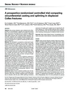
reduced needle (RN) group or the SN group. Randomization was 1:1 with optimal allocation according to Pocock’s minimization technique for sequential randomization (Pocock, 1983). Consideration was given to the woman’s age, number of previous oocyte aspirations, pre-medication and the number of follicles with an external diameter exceeding 10 mm at the last ultrasound before oocyte aspiration. The patient was blinded to which group she had been randomized into. For practical reasons, the doctor and the nurse were not blinded to patient randomization group. The study flow chart of patients randomized is shown in Fig. 1.
Rating of pain Rating of pain was performed by self-assessment by means of visual analogue scale (VAS) consisting of a line oriented vertically on a paper with a range from 0 mm (no pain) to 100 mm (unbearable pain) (McCormack et al., 1988). Immediately after the oocyte aspiration and before the patient had been given the final result of the number of oocytes retrieved, she was asked by a nurse to rate the ‘overall pain for the whole procedure’. This was the primary end-point. The nurse only asked the patient to put a mark on the vertical line on the paper with the VAS. All data were recorded on a case report form, which was transferred manually into an electronic database for analysis. The database was quality-controlled by two individuals who double checked all parameters. They independently measured the marks on the VAS. All data input was done blinded from group allocation.
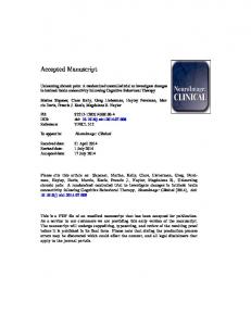
Ovarian stimulation and oocyte aspiration Ovarian stimulation was performed using follicle-stimulating hormone or human menopausal gonadotrophin in combination with GnRH agonists or antagonists according to each clinic’s routines. Dosage was set individually, taking into account age, menstrual cycle length, smoking habits, BMI, the number of pre-antral follicles and, whenever appropriate, the previous response. Ovarian response was monitored by transvaginal ultrasound and serum estradiol when necessary. Oocyte retrieval was carried out 36 – 38 h after hCG injection (Bergh et al., 1997). Standard IVF or ICSI was used according to the indication for the treatment. Transvaginal oocyte retrieval was performed under local anaesthesia (see below) and under guidance of ultrasound with either the RN with an outer diameter of 0.9 mm (20 gauge) and inner diameter of 0.6 mm for the last 50 mm from the tip of the needle and an outer diameter of 1.4 mm (17 gauge) and inner diameter of 1 mm for the remaining length of the needle (Fig. 2) or the SN with an outer diameter of 1.4 mm (17 gauge) and inner diameter of 1 mm for the whole length of the needle (Fig. 2). Both needles were provided by the manufacturer (Vitrolife Sweden AB, Go¨teborg, Sweden). The puncture procedure was performed according to each clinic’s standard routine. Aspiration pressure was kept at a negative pressure between 90 and 120 mmHg. All the physicians (n ¼ 13) performing the oocyte retrievals were skilled, had many years of experience and used the same technique. Laboratory procedures such as culturing and freezing techniques were performed according to each laboratory’s standard technique. Embryo transfer was performed on Day 2, 3 or 5 according to each clinic’s routine.
Sedation and local anaesthesia Pre-operatively, all patients received paracetamol 1 g (Panodil 1 g; Glaco Smith Kline, Ta¨by, Sweden). All patients had a PCB with 10 ml of 1% (10 mg/ml) lidocaine (AstraZeneca, So¨derta¨lje, Sweden) and were given a total dose of 100 mg, using a needle of 0.9 mm diameter and 120 mm length (Mediplast AB, Malmo¨, Sweden). Additionally, all patients received alfentanil (Rapifen 0.5 mg/ml; Janssen-Cilag AB, Sollentuna, Sweden) 0.25 mg i.v. immediately before the PCB was given. If needed (patient
Pain experience at OPU between two different needles
Figure 1 Flow chart of patients for the study.
Figure 2 Illustration of the distal part of the two needles tested. OD, outside diameter; ID, internal diameter.
1379
1380 request), a supplementary dose of 0.25 mg alfentanil was given up to three times, and the total dose was recorded.
Secondary end-points Vaginal bleeding during or after the procedure was graded subjectively by the physician as (i) less than normal, (ii) normal or (iii) more than normal. This was considered the second most important parameter. Aspiration time was measured from the time the first follicle was punctured until the last follicle had been aspirated and the needle withdrawn. The number of follicles aspirated as well as the number of oocytes, the number of fertilized oocytes, the number of embryos cleaved (of normally fertilized), the number of good quality embryos (¼transferred + frozen embryos), the number of embryos transferred and the number of embryos frozen were registered. Clinical pregnancy was defined as gestational sac seen on ultrasound at least 5 weeks after transfer. Furthermore, secondary end-points were also the patients rating of (i) the maximum pain, (ii) pain they experienced directly after the oocyte aspiration, (iii) 30 min and (iv) 60 min after the procedure.
Wikland et al.
Table I Patient characteristics, given as mean + SD (range) per protocol population RN (n 5 126)
SN (n 5 124)
........................................................................................ Age
34.2 + 4.7 (24– 44)
34.4 + 4.2 (24– 43)
BMI
23.2 + 3.6 (17– 35)
23.7 + 3.4 (19– 35)
No. of follicles at the last scan before OPU
10.7 + 5.6 (2–40)
10.3 + 4.6 (2–35)
Previously performed OPU
1.34 + 1.54 (0– 6)
1.37 + 1.81 (0– 12)
No imbalances were observed among the patient characteristics recorded. OPU, oocyte pick-up.
Table II Primary end-point, measurements of VAS, mm; mean + SD (range) RN
SN
P-value
95% CI
........................................................................................
Sample size calculation and statistical analysis The primary end-point was the ‘overall pain experience’, evaluated as described previously. Assuming the mean overall pain in the RN group to be 27 mm (value chosen after analysis of data from a previous study from our group, Cerne et al., 2006) on VAS, with a standard deviation (SD) of 21.5 mm (based on analysis of data from a previous study Cerne et al., 2006), 121 patients in each group would be needed to demonstrate a decrease in overall pain of 8 mm (30%) with 80% power and a significance level of 0.05 (two-tailed tests). Continuous variables were described with mean, SD, median and range and categorical variables as n and percent. For comparison between the two groups, Fisher’s exact test was used for dichotomous variables, Mantel– Haenszel x 2 test for ordered categorical variables and Mann – Whitney U-test for continuous variables. Calculation of confidence interval of differences in means between the two groups for continuous variables was based on the assumption of normality. All significance tests were two-tailed and conducted at the 0.05 significance level. All statistical analyses were performed using SAS 9.2 (SAS Institute Inc., Cary, NC, USA).
Results Two hundred and fifty-seven (n ¼ 257) patients were randomized. Due to practical reasons, 482 patients could not be invited to participate in the study even though they fulfilled the criteria to participate (Fig. 1). Three clinics recruited 70 patients and one clinic 47 patients. One hundred and twenty-nine were allocated to the RN group and 128 to the SN group. Furthermore, in the RN group, three patients were excluded due to protocol violations, resulting in 126 assessed per protocol (PP). In the SN group, one patient was excluded after randomization at her own request, and three were excluded due to protocol violations, resulting in 124 assessed PP (Fig. 1). No patient was lost to follow-up. There were no differences in patient characteristics between the two groups irrespective of whether data were analysed per intention-to-treat (ITT) or PP (Table I).
Intention to treat
n ¼ 129
n ¼ 127
Overall pain during OPU
21.4 + 18.1 (0–78) n ¼ 128a
26.0 + 19.7 (0– 84) n ¼ 126b
Per population
n ¼ 126
n ¼ 124
Overall pain during OPU
21.0 + 17.5 (0–78) n ¼ 125a
26.0 + 19.9 (0– 84) n ¼ 123b
0.047
29.24 to 0.11
0.040
29.72 to 20.36
Mann –Whitney U-test was used to compare differences in means between the two groups. Calculation of confidence interval of differences in means between the two groups was based on the assumption of normality. OPU, oocyte pick-up. a One patient could not register VAS immediately after OPU owing to severe drop in blood pressure. b One patient did not want to register VAS owing to disappointment about the few oocytes retrieved.
Pain rating of OPU The overall pain (primary end-point) for the whole procedure was rated significantly less by means of VAS in the group where the RN was used, irrespective of whether analysed by ITT (mean 21.4 versus 26.0 mm) or PP (mean 21.0 versus 26.0 mm), corresponding to a 19% pain reduction (see Table II). When adjusted for baseline characteristics, such as number of follicles at last ultrasound scan before OPU, previously performed OPU, BMI and age, by use of a multiple linear regression analysis, there is still significantly less overall pain in the RN group when compared with the SN group (P ¼ 0.034). Secondary end-points of pain variables, maximum pain, pain experience directly after 30 and 60 min after the procedure did not differ between the groups (Table III).
Secondary end-points A significant difference was found between the two groups in the assessment of bleeding (see Table IV). Significantly more patients had less vaginal bleeding than expected when the RN was used,
1381
Pain experience at OPU between two different needles
Table III Secondary end-point, measurements of VAS, mm; mean + SD (range) RN (n 5 126)
SN (n 5 124)
P-value
95% CI
............................................................................................................................................................................................. Pain directly after OPU
13.9 + 17.3 (0– 77)
13.3 + 15.0 (0–74)
0.53
23.50 to 4.64
Maximum pain after OPU
33.6 + 26.0 (0– 96)
39.1 + 24.4 (0–96)
0.06
211.81 to 0.79
Pain 30 min after OPU
17.6 + 18.6 (0– 72)
18.0 + 19.7 (0–98)
0.66
25.20 to 4.38
Pain 60 min after OPU
10.8 + 13.7 (0– 61)
11.8 + 14.3 (0–69)
0.27
24.54 to 2.53
% patients who requested more i.v. analgesia
25.4
31.5
0.36
218.3 to 6.40
% patients with ,30 mm on VAS in overall pain during OPU
70.4
61.0
0.14
20.31 to 0.22
Analysis per protocol population. Mann –Whitney U-test was used to compare differences in means between the two groups. Calculation of confidence interval of differences in means between the two groups was based on the assumption of normality. OPU, oocyte pick-up.
Table IV Secondary end-points, treatment and outcome variables, + mean (range) when applicable RN (n 5 126)
SN (n 5 124)
P-value
............................................................................................................................................................................................. Bleeding No. of patients with less than expected bleeding (%)
40 (31.7)
24 (19.4)
No. of patients with normal bleeding (%)
80 (63.5)
93 (75.0)
No. of patients with more than normal bleeding (%)
6 (4.8)
7 (5.6)
0.042
Total OPU time (min)
7.72 + 4.10 (1–22)
7.52 + 3.73 (3–20)
0.85
Oocyte recovery/follicle (%)
77.2 + 24.2
73.4 + 25.4
0.27
No. of mature oocytes
7.57 + 5.31 (0–27)
7.34 + 4.91 (0–27)
0.91
No. of damage oocytes
0.41 + 1.23 (0–8)
0.43 + 1.20 (0–10)
0.67
Fertilization rate (%)
72.7 + 25.8
70.2 + 26.3
0.53
Cleavage ratea (%)
70.8 + 26.6
68.5 + 25.6
0.50
No. of transferred embryos
1.06 + 0.54 (0–2)
1.07 + 0.55 (0–2)
0.89
No. of frozen embryos
1.30 + 2.04 (0–12)
1.28 + 1.89 (0–8)
0.80
Proportion of good quality of embryos (%)
27.9 + 20.2
29.8 + 23.5
0.64
Implantation rate (%)
21.6 + 42.4
25.9 + 44.6
0.40
Clinical pregnancy rate (%)
19.8
24.4
0.48
b
Analysis per protocol population. OPU, oocyte pick-up. a Of normally fertilized. b For definition of good quality of embryos see main text (Materials and Methods).
32% (40 of 126) versus 19% (24 of 124); P ¼ 0.029, 95% CI (1.7 – 23.0%). If also adjusting for the above-mentioned baseline characteristics and vaginal bleeding by use of a multiple logistic regression, there is still significantly more patients with less vaginal bleeding than expected in the RN group when compared with the SN group [P ¼ 0.022, 95% CI (0.28–0.90)]. There were no difference in aspiration time, oocytes recovered, damage to oocytes, fertilization rate, cleavage rate, proportion of good quality embryos, implantation and clinical pregnancy rate between the two groups, as presented in Table IV. Furthermore, there was no difference between the groups with regard to the number of patients who requested more i.v. analgesia (Table III). No clinically important adverse events were observed in either of the groups.
Discussion The overall pain during oocyte aspiration was significantly reduced in the group with the thinner needle when compared with the SN. The reduction in overall pain as measured by self-assessment on a VAS scale was 19%. Furthermore, significantly more patients had less vaginal bleeding than expected. No negative effects were found in relation to the oocyte or the outcome of the treatment. Evaluating pain experience during oocyte retrieval is difficult since there are so many factors that influence pain, fear of pain, anxiety, type of analgesia, physician’s skill and technical factors such as needle diameter and sharpness. These factors have a clear influence on pain threshold (Scott et al., 1983), and are therefore a potential source of bias. In this study, one such factor that could be of importance is the fact that the doctor was not blinded to the type of needle.
1382 Unfortunately, we found it impossible to blind the doctor as to the needle in use, since the needles are easily distinguishable on the ultrasound screen. This is of course a weakness of the study. Another factor that might introduce bias is the lack of blinding of the study nurses. We consider that this is of minor relevance since the nurse’s involvement was limited to asking the patient to mark the VAS. The nurse’s knowledge of which needle was used is thus not believed to influence the patient’s experience of pain. Furthermore, there is no objective method for evaluating pain. Despite those problems, several studies have tried to evaluate the patient’s pain experience during oocyte retrieval, mainly focusing on the method for analgesia. A Cochrane review of such studies concluded that there is insufficient evidence to determine the best method of pain relief for OPU (Kwan et al., 2005). It is interesting to note that technical factors such as needle diameter and type of bevel have largely been neglected with regard to pain related to oocyte retrieval. Almost from the start of human IVF, it has been assumed that an ‘ideal’ follicle aspiration needle should have an inner diameter of at least 0.8 mm, which means that the outer diameter will be between 1.2 and 1.4 mm (16 –18 gauge) depending on the wall thickness of the needle (Lopata et al., 1974). The background for choosing the diameter mentioned above is that such a diameter has, in several studies, proved to result in good recovery rates and a low frequency of damage to oocytes (Renou et al., 1981; Belaisch-Allart et al., 1985). Our results with less overall pain experience in the group where the RN was used is supported by a study by Awonuga et al., who found a significant reduction in how patients perceived pain when oocyte retrieval needle size was decreased from 15 gauge to either 17 or 18 gauge. However, that study evaluated pain using a questionnaire answered by patients post-operatively and not VAS (Awonuga et al., 1996). In another interesting although small study where VAS was used, it was clearly shown that the patient experienced less pain on the side where a thinner needle (16 versus 18 gauge) was used (Aziz et al., 1993). The limit of 30 mm on a VAS is often used for administration of analgesics post-operatively (Rawal and Berggren, 1994). However, there was no statistically significant difference between patients below 30 mm on the VAS scale in the RN group when compared with the SN group (Table III). In conclusion, our results indicate that a needle as thin as 20 gauge does result in a reduction of the overall pain as measured by VAS. Furthermore, even though not objectively measured, we also found a significant difference in the number of patients with less than expected vaginal bleeding between the two needles. The lower proportion of patients with less than expected vaginal bleeding indicate that the needle could be less traumatic. It is reassuring that the thinner needle does not seem to have any negative effects on the cumulus oocyte complex. The reason for this might be that the thinner part of the needle is very short. The low proportion of damaged oocytes in our study is very similar to what has been reported in another prospective randomized controlled study where two different needles were tested (Miller et al., 2004); however, larger studies are needed to confirm this finding. Although, one might expect that a reduction of the inner diameter of the needle by as much as 40% would influence the recovery rate and/or the total aspiration time; however, this was not the case in
Wikland et al.
our study. The reason may be that only a very short part of the needle was reduced. Very few studies have investigated the importance of the needle design for the oocyte recovery and possible damage to the cumulus oocyte complex in clinical IVF. In studies on bovine oocytes, it has been demonstrated that both the diameter and bevel of the needle, as well as the aspiration vacuum, is important for the recovery rate and developmental competence of the oocyte (Bols et al., 1996, 1997). In this study, human oocytes were unaffected by needles as thin as 20 gauge and an aspiration pressure of 90–120 mmHg. In conclusion, oocyte aspiration performed with the RN resulted in significantly less overall pain and less vaginal bleeding without prolonging the retrieval procedure or influencing the oocyte recovery rate, compared with a SN. Furthermore, no adverse effects on the oocyte were found, as evaluated by the outcome of the IVF procedure. The thin tip needle, therefore, seems to be advantageous for the patient.
Authors’ roles All authors played a role in study conception and design. S.B. took part in collection and assembly of data. M.W. and S.B. carried out data analysis, interpreted the findings and drafted the manuscript. All authors critically reviewed and approved the final version of the manuscript.
Acknowledgements Thanks are due to Nils-Gunnar Pehrsson for help with the statistical analyses. The authors also thank Vitrolife Sweden AB for financial support and for providing the needles for the study. Thanks are also due to all the staff at the participating clinics for help with the study.
Funding Vitrolife Sweden AB supported the study financially and provided the needles. Funding to pay the Open Access publication charges for this article was provided by Vitrolife International AB.
References Awonuga A, Waterstone J, Oyesanya O, Curson R, Nargund G, Parsons J. A prospective randomized study comparing needles of different diameters for transvaginal ultrasound-directed follicle aspiration. Fertil Steril 1996;65:109– 113. Aziz N, Biljan M, Taylor C, Manasse P, Kingsland C. Effect of aspirating needle calibre on outcome of in-vitro fertilization. Hum Reprod 1993; 8:1098 – 1100. Belaisch-Allart JC, Hazout A, Guillet-Rosso F, Glissant M, Testart J, Frydman R. Various techniques for oocyte recovery in an in vitro fertilization and embryo transfer program. J In Vitro Fert Embryo Transf 1985;2:99 – 104. Bergh C, Howles CM, Borg K, Hamberger L, Josefsson B, Nilsson L, Wikland M. Recombinant human follicle stimulating hormone (r-hFSH; Gonal-F) versus highly purified urinary FSH (Metrodin HP): results of a randomized comparative study in women undergoing assisted reproductive techniques. Hum Reprod 1997;12:2133 – 2139. Bokhari A, Pollard BJ. Anaesthesia for assisted conception: a survey of UK practice. Eur J Anaesthesiol 1999;6:225 – 230.
Pain experience at OPU between two different needles
Bols PE, Van Soom A, Ysebaert MT, Vandenheede JM, de Kruif A. Effects of aspiration vacuum and needle diameter on cumulus oocyte complex morphology and developmental capacity of bovine oocytes. Theriogenology 1996;45:1001– 1014. Bols PE, Ysebaert MT, Van Soom A, de Kruif A. Effects of needle tip bevel and aspiration procedure on the morphology and developmental capacity of bovine compact cumulus oocyte complexes. Theriogenology 1997;47:1221 – 1236. Cerne A, Bergh C, Borg K, Ek I, Gejervall AL, Hillensjo¨ T, Olofsson JI, Stener-Victorin E, Wood M, Westlander G. Pre-ovarian block versus paracervical block for oocyte retrieval. Hum Reprod 2006; 21:2916– 2921. Cohen J, Avery S, Campbell S, Mason BA, Riddle A, Sharma V. Follicular aspiration using a syringe suction system may damage the zona pellucida. J In Vitro Fert Embryo Transf 1986;3:224 – 226. Corson SL, Batzer FR, Gocial B, Kelly M, Gutmann JN, Go KJ, English ME. Is paracervical block anesthesia for oocyte retrieval effective?. Fertil Steril 1994;62:133 – 136. Ditkoff EC, Plumb J, Selick A, Sauer MV. Anesthesia practices in the United States common to in vitro fertilization (IVF) centers. J Assist Reprod Genet 1997;14:145 – 147. Kwan I, Bhattacharya S, Knox F, McNiel A. Conscious sedation and analgesia for oocyte retrievals during in vitro fertilisation procedures: a Chochrane review. Hum Reprod 2006;21:1672 – 1679. Lopata A, Johnston IW, Leeton JF, Muchnicki D, Talbot JM, Wood C. Collection of human oocytes at laparoscopy and laparotomy. Fertil Steril 1974;25:1030– 1038.
1383 Ludwig AK, Glawatz M, Griesinger G, Diedrich K, Ludwig M. Perioperative and post-operative complications of transvaginal ultrasound-guided oocyte retrieval: prospective study of .1000 oocyte retrievals. Hum Reprod 2006;21:3235– 3240. McCormack HM, Horne DJ, Sheather S. Clinical applications of visual analogue scales: a critical review. Psychol Med 1988; 8:1007 – 1019. Miller KA, Elkind-Hirsch K, Benson M, Pergh P, Drews M, Scott R. A new follicle aspiration needle set is equally effective and as well tolerated as the standard needle when used in a prospective randomized trial in a large in vitro fertilization program. Fertil Steril 2004;8:119 – 193. Ng EH, Tang OS, Chui DK, Ho PC. A prospective, randomized, double-blind and placebo-controlled study to assess the efficacy of paracervical block in the pain relief during egg collection in IVF. Hum Reprod 1999;14:2783 – 2787. Pocock SJ. Clinical Trials: a Practical Approach. Chichester, England: John Wiley, 1983. Rawal N, Berggren L. Organization of acute pain services: a low-cost model. Pain 1994;57:117 – 123. Renou P, Trounson AO, Wood C, Leeton JF. The collection of human oocytes for in vitro fertilization. I. An instrument for maximizing oocyte recovery rate. Fertil Steril 1981;35:409 – 412. Scott LE, Clum GA, Peoples JB. Preoperative predictor of postoperative pain. Pain 1983;15:283– 293. Wikland M. Oocyte retrieval. In: Gardner D. (ed) Vitro Fertilization-a practical approach. USA: Informa Healthcare Inc., 2007.