International Orthopaedics (SICOT) DOI 10.1007/s00264-016-3391-0
ORIGINAL PAPER
A randomized controlled trial demonstrating sustained benefit of Autologous Matrix-Induced Chondrogenesis over microfracture at five years Martin Volz 1 & Jens Schaumburger 2 & Hubert Frick 1 & Joachim Grifka 2 & Sven Anders 2
Received: 9 August 2016 / Accepted: 26 December 2016 # The Author(s) 2017. This article is published with open access at Springerlink.com
Abstract Purpose Autologous Matrix-Induced Chondrogenesis (AMIC®) utilizing a type I/III collagen membrane was compared with microfracture (MFx) alone in focal cartilage lesions of the knee at one, two and five years. Methods Forty-seven patients (aged 37 ± 10 years, mean defect size 3.6 ± 1.6 cm2) were randomized and treated either with MFx, with sutured or glued AMIC® in a prospective multicentre clinical trial. Results After improvement for the first two years in all subgroups, a progressive and significant score degradation was observed in the MFx group, while all functional parameters remained stable for least five years in the AMIC® groups. At two and five years, MRI defect filling was more complete in the AMIC® groups. No treatment-related adverse events were reported. Conclusions AMIC® is an effective cartilage repair procedure in the knee resulting in stable clinical results significantly better than the MFx group at five years.
Keywords Articular cartilage . Autologous Matrix-Induced Chondrogenesis (AMIC®) . Chondro-Gide® . Knee surgery . Microfracture
* Sven Anders
[email protected] 1
Sportklinik Ravensburg, Ravensburg, Germany
2
Department of Orthopedic Surgery, University of Regensburg, Asklepios Clinical Center Bad Abbach, Bad Abbach, Germany
Introduction Cartilage has a low intrinsic regenerative and reparative capacity, and cartilage defects can potentially lead to severe osteoarthritis in the long term. A variety of surgical techniques that aim to resurface and repair the damaged articular cartilage include perforation of the subchondral bone, mosaicplasty, and autologous chondrocyte transplantation. Marrow stimulation methods, such as microfracturing (MFx), involve penetration of the subchondral bone plate to access the bone marrow compartment. The resulting blood clot enriched with bone marrow elements is thought to provide a favourable microenvironment for the development of the cartilage repair tissue. This so-called superclot is capable of further stimulating the migration, proliferation, and chondrogenic differentiation of mesenchymal stem cells from the bone marrow [1]. Currently, microfracturing as well as mosaicplasty is recommended as the treatment of choice for smaller cartilage defects (2 cm2, with a followup of five years. We hypothesize that adding a collagen membrane, fixed either by gluing or sutering, to the conventional microfraturing like in any AMIC® procedure would result in better clinical and morphological results in these patients.
an open procedure (AMIC® glued or AMIC® sutured) neither the patient nor the physicians were blinded. The study was approved by the Ethics Committee (03-088 and 03/173-MZ, ZKS, University of Regensburg) and was conducted according to the declaration of Helsinki and Good Clinical Practice. Sixty-seven patients were initially enrolled (18 MFx, 27 AMIC® glued and 22 AMIC® sutured) in seven centres in Germany. Five centres with a low recruitment number (2 cm2). The hypothesis funding this RCT with AMIC® in focal cartilage defects of the knee was that the use of a covering membrane in an AMIC® procedure would result in sustained benefit compared to microfracturing alone at
International Orthopaedics (SICOT)
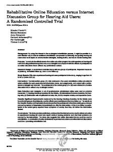
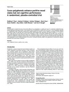
Fig. 5 Example of MRI (1.5 T) follow-up at one, two and five years (glued AMIC®, medial femoral condyle): Proton density weighted sequence (PDw) in sagittal orientation with defect filling almost
complete (20 × 20 mm, see arrow), surface remaining slightly uneven, good marginal integration, and repair tissue nearly isointense to adjacent genuine cartilage
five years. This was confirmed by our results. After improvement for the first two years in all subgroups, a progressive and significant score degradation was observed in the MFx group, while all functional parameters remained stable for at least five years in the AMIC® groups. Both modes of ChondroGide® fixation resulted in similar mid-term clinical benefit. At two years and later, defect filling in MRI was notably more complete in the AMIC® treated groups (at least 60% of the
patients had a defect filling of more than 2/3) compared to the MFx group with only 25% of such a filling. At five years the defect filling was the lowest in the MFx group. Overall, our data are in agreement with the results of Gille et al. reporting a significant improvement in clinical outcomes in patients treated with AMIC®, assessed by five different scores up to 36 months [22]. Similar results were found in a larger multicentre observational study including 57 patients
Table 2
MRI evaluation Glued AMIC®
MFx
Defect filling
Surface
Integration
Signal intensity of defect cover Bone marrow lesion
Sutured AMIC®
1 year (n = 11)
2 year (n = 6)
5 year (n = 9)
1 year (n = 16)
2 year (n = 15)
5 year (n = 11)
1 year (n = 14)
2 year (n = 14)
5 year (n = 12)
none 1/3
0 3
0 2
1 4
1 0
1 3
2 2
2 1
3 2
4 1
1/3-2/3 >2/3 not evaluable
3 5 0
2 2 0
2 2 0
5 9 1
1 10 0
3 4 0
5 6 0
1 8 0
1 6 0
largely uneven partially uneven smooth not evaluable marginal gap up to 50% marginal gap complete not evaluable
4 4 3 0 1
3 1 3 0 2
5 3 0 1 4
2 11 2 1 0
2 6 6 1 1
4 3 2 2 4
4 6 2 2 2
3 4 4 3 1
3 4 1 4 0
8 1 1
1 4 0
4 0 1
10 5 1
9 4 1
3 2 2
5 4 3
3 6 4
2 5 5
inhomogeneous homogenous not evaluable massive (>2 cm) intermediate (1-2 cm) small (