Journal of Geriatric Cardiology (2017) 14: 509514 ©2017 JGC All rights reserved; www.jgc301.com
Research Article
Open Access
A risk prediction score model for predicting occurrence of post-PCI vasovagal reflex syndrome: a single center study in Chinese population Hai-Yan LI, Yu-Tao GUO, Cui TIAN, Chao-Qun SONG, Yang MU, Yang LI, Yun-Dai CHEN Department of Cardiology, Chinese PLA General Hospital, Beijing, China
Abstract Background The vasovagal reflex syndrome (VVRS) is common in the patients undergoing percutaneous coronary intervention (PCI). However, prediction and prevention of the risk for the VVRS have not been completely fulfilled. This study was conducted to develop a Risk Prediction Score Model to identify the determinants of VVRS in a large Chinese population cohort receiving PCI. Methods From the hospital electronic medical database, we identified 3550 patients who received PCI (78.0% males, mean age 60 years) in Chinese PLA General Hospital from January 1, 2000 to August 30, 2016. The multivariate analysis and receiver operating characteristic (ROC) analysis were performed. Results The adverse events of VVRS in the patients were significantly increased after PCI procedure than before the operation (all P < 0.001). The rate of VVRS [95% confidence interval (CI)] in patients receiving PCI was 4.5% (4.1%–5.6%). Compared to the patients suffering no VVRS, incidence of VVRS involved the following factors, namely female gender, primary PCI, hypertension, over two stents implantation in the left anterior descending (LAD), and the femoral puncture site. The multivariate analysis suggested that they were independent risk factors for predicting the incidence of VVRS (all P < 0.001). We developed a risk prediction score model for VVRS. ROC analysis showed that the risk prediction score model was effectively predictive of the incidence of VVRS in patients receiving PCI (c-statistic 0.76, 95% CI: 0.72–0.79, P < 0.001). There were decreased events of VVRS in the patients receiving PCI whose diastolic blood pressure dropped by more than 30 mmHg and heart rate reduced by 10 times per minute (AUC: 0.84, 95% CI: 0.81–0.87, P < 0.001). Conclusion The risk prediction score is quite efficient in predicting the incidence of VVRS in patients receiving PCI. In which, the following factors may be involved, the femoral puncture site, female gender, hypertension, primary PCI, and over 2 stents implanted in LAD. J Geriatr Cardiol 2017; 14: 509514. doi:10.11909/j.issn.1671-5411.2017.08.004 Keywords: Post-percutaneous coronary intervention; Risk prediction score model; Vasovagal reflex syndrome
1 Introduction Nowadays, coronary artery disease (CAD) has become one of the most common lethal diseases with high morbidity and mortality in Chinese population. In 2014, CAD accounted for almost half of all deaths in both rural and urban area in China.[1] Percutaneous coronary intervention (PCI) is an invasive procedure to revascularize narrowed or occluded vessel so that the ischemic area can be reperfused. Although this procedure has saved thousands of lives, some complications may lead to poor prognosis. Vasovagal reflex syndrome (VVRS) is one of the lethal complications post-PCI, which is identified as a complex hemodynamic response characterized by bradycardia, marked hypotension
Correspondence to: Yun–Dai CHEN, MD. General Hospital of People’s Liberation Army, Beijing 100853, China. E-mail:
[email protected] Received: March 24, 2017
Revised: July 2, 2017
Accepted: August 12, 2017
Published online: August 28, 2017
and loss of consciousness.[2] Furthermore, thrombus immediately forms at the stent position if VVR can’t be corrected within 10 min.[3] Several studies have shown that activation of autonomic nerves system, endocrine system, and inflammatory response are all involved in the progression of VVRS. [4,5] In details, vasopressin upon specific stimulation drives hypothalamus to secret IL-6 and TNF-α, which induce contraction of vascular smooth muscle and vasovagal reflex syndrome. Previous researches[6–9] have demonstrated several factors thatare associated with the occurrence of VVRS, including sheath removal, long-time fasting, decreased venous return, and dilation of cavity viscera. However, the sample size was small in these studies and they focused on only one risk factor which failed to effectively predict the vasovagal reflex events, much less prevent. In this study, we aimed to develop a risk prediction score model to identify the determinants for vasovagal reflex events in a large Chinese population cohort who underwent PCI procedure.
http://www.jgc301.com;
[email protected] | Journal of Geriatric Cardiology
LI HY, et al. prediction score model for post-PCI VVRS
510
2 Subjects and methods 2.1 Subjects We retrospectively searched the hospital electronic medical databases of 3550 patients who underwent PCI procedures (78.0% males, mean age 60 years) in Chinese PLA General Hospital from January 1st, 2000 to May 30th, 2016. We identified 161 patients who suffered from vasovagal reflex syndrome (VVRS group). The left patients who suffered no VVRS served as control group. Clinical data of both groups, including demographic characteristics, diagnostic history and PCI associated information, were harvested. 2.2 Symptoms of vasovagal reflex syndrome There are a variety of triggers that can set off the vasovagal reflex. Once a vasovagal reflex has been triggered, a variety of physical symptoms may be experienced. These include chest distress, short of breath, pale complexion, sweating, nausea, and vomiting. It also causes an abrupt dropping of blood pressure (by 15 mmHg) and a sudden reduction in heart rate (by 10 times/min). This group of VVRS patients complained of nausea, vomiting, sweating, dizziness, as well as hematomas, tube removal, anxiety and pain. 2.3 Clinical data collection (1) Demographic data were collected, including age, body mass index (calculated by body weight/height2), gender, cigarette smoking or alcohol drinking. (2) Information on diagnostic work-up included clinical history of hypertension (blood pressure > 140/80 mmHg, or prescribed hypotensor), diabetes, hyperlipidemia, heart failure, atrial fibrillation, prior stroke, cancer, prior cerebral bleeding, anemia and gastrointestinal bleeding. (3) Left ventricular ejection fraction was also concerned. (4) Information on coronary artery disease included unstable angina, stable angina, acute myocardial infarction (MI) , prior MI, prior coronary artery bypass grafting (CABG) or prior PCI. (5) Data associated with PCI itself were also collected, including the number and location of implanted stents, the time duration of PCI, time interval from occurrence of vasovagal relex to PCI manipulation (which is less than six hours in emergent PCI), and the puncture site (coronary sinus or femoral approach).
continuous variables met normal distribution, otherwise the Wilcoxon two-sample test was performed. Categorical variables were compared using chi-square test, and Fishers’ exact test will be performed if estimated cases were less than 5. The multivariate analysis and receiver operating characteristic (ROC) analysis were performed to identify the combination of risk factors which contribute more significantly to VVRS’ incidence and severity. Two-sided P-value < 0.05 was considered statistically significant. Statistical analyses were performed by SPSS 17.0.
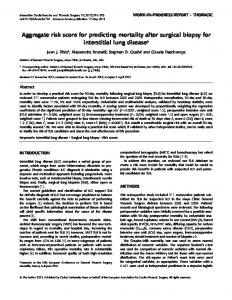
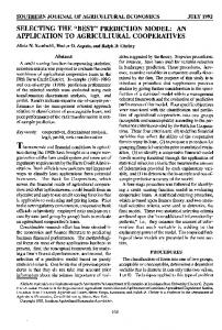
3 Results 3.1 Clinical and demographic characteristics of the two groups Based on the hospital electronic medical database, we identified 3550 patients receiving PCI (78.0% males, mean age 60 years) in Chinese PLA General Hospital from January 1, 2000 to August 30, 2016. Among them, 161 patients (4.5%) suffered from VVRS. Compared with those suffering no VVRS, (n = 3389), the factors involved in VVRS group included female gender, hypertension, over two stents implanted in the left anterior descending (LAD), and the femoral puncture site. In VVRS group, the adverse events of VVRS were significantly increased after PCI manipulation (all P < 0.001, Table 1 and 2). 3.2 Clinical spectrum of VVRS The common presenting symptoms of these 161 VVRS patients included nausea/vomiting (32.9%), sweating (26.7%), palpitation (8.7%), dizziness (6.8%) and confusion (4.3%) in proportional sequence (Figure 1). Hematoma was the leading cause of vasovagal reflex in 161 VVRS patients receiving PCI (28.57%), and other triggers and the following Common causes to include tube draining (21.74%), anxiety (14.91%), puncture site pain (11.18%), and urinary catheterization (4.97%). There were still 30 VVRS patients (18.63%), for whom no any definite cause for VVRS was determined (Figure 2). 3.3 Risk factors of VVRS Regarding all included risk factors, multivariate analysis in the patients revealed that the femoral puncture site, female gender, hypertension, primary PCI, and ≥ 2 stents implanted in LAD, were independent predictors of VVRS (all P < 0.001, Table 3).
2.4 Statistical analysis Continuous data were expressed with mean ± S.D. or median (inter-quartile range). The Student’s t-test or one-way analysis of variance (ANOVA) was used when
3.4 Establishment of a risk prediction score model for VVRS in patients receiving PCI
Journal of Geriatric Cardiology |
[email protected]; http://www.jgc301.com
Based on the results of multivariate analysis, we assigned
LI HY, et al. prediction score model for post-PCI VVRS
511
Table 1. Clinical and demographic characteristics of 3550 patients receiving PCI. Patients without VVRS (n = 3389) Age BMI
60.1 ± 11
Patients with VVRS (n = 161)
P
61.3 ± 11
0.189
25.9 ± 3.3
25.0 ± 3.8
0.024
Female
709 (20.9%)
71 (44.1%)
< 0.001
Smoke
1594 (47.0%)
34 (21.1%)
< 0.001
Alcohol
1085 (32.0%)
25 (15.5%)
< 0.001
194 (5.7%)
4 (2.5%)
0.080
2122 (62.6%)
113 (70.2%)
< 0.001
Discontinuous cigarette smoking/alcohol drinking Medical history Hypertension Diabetes
980 (28.9%)
33 (20.5%)
0.021
Hyperlipidemia
600 (17.7%)
33 (20.5%)
0.366
Heart failure
244 (7.2%)
5 (3.1%)
0.047
56.5±8.8
58.1±9.3
0.054
EF Renal dysfunction
133 (3.9%)
6 (3.7%)
0.899
Atrial fibrillation
87 (2.6%)
2 (1.2%)
0.293
Prior stroke
92 (2.7%)
4 (2.5%)
0.860
Cancer
66 (1.9%)
1 (0.6%)
0.184
Prior brain bleeding
11 (0.3%)
3 (1.9%)
0.023
Anemia
8 (0.2%)
1 (0.6%)
0.342
GI bleeding
6 (0.2%)
0 (0%)
0.757
CAD Unstable angina
1872 (55.2%)
79 (49.1%)
0.124
Stable angina,
2062 (60.8%)
86 (53.4%)
0.060
Acute MI
356 (10.5%)
14 (8.7%)
0.463
Prior MI
232 (6.8%)
11 (6.8%)
0.995
Prior CABG
36 (1.1%)
3 (1.9%)
0.341
413 (12.2%)
6 (3.7%)
0.001
146 (4.3%)
15 (9.3%)
0.003
Femoral
1494 (44.1%)
132 (82.0%)
< 0.001
Radial
1880 (55.5%)
29 (18.0%)
< 0.001
Prior PCI PCI Emergent PCI Puncture site
BMI: body mass index; CABG: coronary artery bypass graft; CAD: coronary artery disease; EF: ejection fraction; GI: gastrointestinal bleeding; MI: myocardial infarction; PCI: percutaneous coronary intervention; VVRS: vasovagal reflex syndrome.
different values for each factor according to their odds ratio (OR) values. Specifically, femoral puncture site weighted 6 points; female gender weighted 2 points; and hypertension, primary PCI and ≥ 2 stents implanted in LAD weighted 1 point, respectively. ROC analysis showed that the area under the ROC curve was 0.76 for the model with the above factors included. When the factor, dropping of systolic blood pressure by more than 30 mmHg, was added, the area under the ROC curve was up to 0.82 (P < 0.01). When the factor, a reduction in heart rate by 10 times/min, was further added, the area under the ROC curve was increased to 0.84 (Table 4). It is indicated that the risk prediction score combined with the dropping of blood pressure plus the reduction in heart rate might predict the incidence of VVRS more
effectively. 3.5 Risk stratification and incidence of vasovagal reflex syndromes in the patients receiving PCI To better predict the prevalence and severity of VVRS, we classified the 161 VVRS patients into four categories based on the established risk prediction score: class I (Score = 0), class II (Score = 1–7), class III (Score = 8–9) and class IV (Score = 10–11). The results showed that the risk prediction score, in parallel to risk category, was closely related to the incidence of VVRS (Table 5 and Figure 3). It is suggested that the risk prediction score model we developed has good value in predicting the incidences of VVRS in patients receiving PCI.
http://www.jgc301.com;
[email protected] | Journal of Geriatric Cardiology
LI HY, et al. prediction score model for post-PCI VVRS
512 Table 2. Number of stents implanted in coronary artery. Patients without
Patients with
VVRS (n = 3389)
VVRS (n = 161)
1 stent
908 (26.8%)
30 (18.6%)
2 stents,
314 (9.3%)
27 (16.8%)
0.002
3 stents
42 (1.2%)
3 (1.9%)
0.489
1 stent
440 (13.0%)
27 (16.8%)
0.165
2 stents
93 (2.7%)
5 (3.1%)
0.784
3 stents
15 (0.4%)
0
0.498
1 stent
514 (15.2%)
22 (13.7%)
0.603
2 stents
203 (6.0%)
14 (8.7%)
0.161
3 stents
54 (1.6%)
4 (2.5%)
0.384
1 stent
67 (2.0%)
3 (1.9%)
0.919
2 stents,
8 (0.2%)
0
0.690
1 stent,
62 (1.8%)
6 (3.7%)
0.086
2 stents
1 (0.0%)
0
0.955
1 stent
10 (0.3%)
2 (1.2%)
0.043
2 stents
1 (0.0%)
0 (0%)
0.955
P
LAD 0.022
LCX
RCA
Figure 1. The proportional distribution of symptoms in 161 patients suffering from vasovagal reflex syndrome.
Diagonal branch
Left main artery
Posterior branch of left ventricle
Posterior branch of right ventricle 1 stent
2 (0.1%)
0
0.911
Obtuse marginal branch 1 stent
35 (1.0%)
3 (1.9%)
0.317
2 stents
6 (0.2%)
0
0.757
Figure 2. Main causes of vasovagal reflex syndrome in the involved 161 patients. VVRS: vasovagal reflex syndrome. Table 3. Multivariate analysis of risk factors for vasovagal reflex syndrome in CAD patients. 95% CI OR
Posterior descending coronary
Lower
Higher
limit
limit
P
1 stents
14 (0.4%)
1 (0.6%)
0.691
Femoral puncture site
6.314
4.136
9.639
< 0.001
2 stents
3 (0.1%)
0
0.170
Female gender
2.874
2.044
4.040
< 0.001
Primary PCI
2.240
1.124
4.466
0.022
0.584
≥ 2 stents implanted in LAD 1.825
1.188
2.803
0.006
0.955
Hypertension
1.445
1.005
2.077
0.047
LAD: left anterior descending artery; LCX: left circumflex coronary artery;
Age
0.996
0.981
1.012
0.647
RCA: right coronary artery; VVRS: vasovagal reflex syndrome.
CAD: coronary artery disease; CI: confidence interval; LAD: left anterior
Ramus intermedius 1 stent 3 stents
12 (0.4%) 1 (0%)
1 (0.6%) 0
descending artery; OR: odds ratio; PCI: percutaneous coronary intervention.
4 Discussion Vasovagal reflex syndrome is an urgent post-PCI complication characterized by marked hypotension and bradycardia. Under normal cardiac rhythm and blood pressure, transitory disturbance followed by spontaneous recovery are usually observed. However, it can sometimes be a serious and even fatal case.[10,11] Our results showed that the incidence of VVRS in patients receiving PCI was 4.5%
(161/3550). All 161 patients with VVRS survived, which were mainly due to the intensive care and timely rescue strategies in our center. Importantly, we established a risk prediction score model, which can well predict the incidence of VVRS following PCI procedure. The following factors were included in the model: femoral puncture site, female gender, hypertension, primary PCI, and ≥ 2 stents implanted in LAD, as well as a
Journal of Geriatric Cardiology |
[email protected]; http://www.jgc301.com
LI HY, et al. prediction score model for post-PCI VVRS
513
Table 4. Predictive ability of VR risk score for vasovagal reflex syndrome in patients receiving PCI. AUC (95% CI)
P
VR risk score
0.76 (0.72–0.79) < 0.001
VR risk score + SBP 30 mmHg
0.82 (0.78–0.85) < 0.001
VR risk score + SBP 30 mmHg + HR 10 bpm
0.84 (0.81–0.87) < 0.001
ROC analysis was performed to determine the predictive ability of Risk Prediction Score Model. AUC: area under ROC curve. The VR risk scores are calculated as following: 6*femoral puncture site + 2*female gender + 1*hypertension + 1* primary PCI + 1* ≥ 2 stents implanted in LAD. SBP 30 mmHg: a dropping of the systolic blood pressure by ≥ 30 mmHg. HR 10 bpm: a reduction in the heart rate by ≥ 10 beats per minute. AUC: area under receiver operating characteristic curve; CI: confidence interval; HR: heart rate; LAD: left anterior descending artery; ROC: receiver operating characteristic; PCI: percutaneous coronary intervention; SBP: systolic blood pressure; VR: vasovagal reflex.
dropping of systolic blood pressure by more than 30 mmHg and a reduction in heart rate by 10 times/min. Interestingly, the latter two in this model have great value in predicting the occurrence of VVRS following PCI. It was previously
reported that VVRS developed secondary to a drop of blood pressure and bradycardia due to the withdrawal of sympathetic tone.[15–17] It has been well known that the drop of blood pressure is attributed to the marked reduction of cardiac output and vascular resistance, while decrease of heart rhythm is related to the vagus nerve excitement. About 20% of nonmyelinated vagal C-fibers were activated during the bradycardia induced by rapid haemorrhage.[18] This shows that drop of blood pressure and decrease of heart rhythm, as important predicting factors, were closely related to VVRS events. In our study, primary PCI, as an independent risk factor, was also added into the model. Our results showed that VVRS events in patients receiving primary PCI were significantly increased compared to those receiving no PCI procedure. To some extent, it may be explained partially by the stressful pre-PCI situation. Repeated PCI procedure stimulates the punctured artery, and is likely to form local hematoma after surgery. Moreover, because of severe coronary artery disease, patients’ cardiac compensatory function, blood volume and stress response are insufficient.
Table 5. Risk categorization and incidence of vasovagal reflex syndrome in the patients receiving PCI (n = 3550). Proportion in risk category
Vasovagal reflex syndrome, %
Class I (score = 0)
Risk category
2.08%
0 (0.00%)
ClassII (score = 1–7)
68.34%
52 (2.14%)
ClassIII (score = 8–9)
22.59%
67 (8.35%)
ClassIV (score = 10–11)
6.99%
42 (16.94%)
C statistic (95% CI)
P
0.72 (0.67–0.76)
< 0.001
*
The risk scores are calculated as following: 6*femoral puncture site + 2*female gender + 1*hypertension + 1* primary PCI + 1* ≥ 2 stents implanted in LAD.
CI: confidence interval; LAD: left anterior descending artery; PCI: percutaneous coronary intervention.
In our study, left ventricular ejection fraction was slightly higher in VVRS group. It was rather confusing that the patients with left ventricular dysfunction could have VVRS. VVRS is a complex process and requires for a vigorous ventricular response with the mechanoreceptors activation, so it is rarely observed in patients with left ventricular dysfunction. It may be explained by some extraventricular and neurohormonal mechanisms.
5 Clinical significance
Figure 3. Cumulative hazard of vasovagal reflex syndrome. VVRS: vasovagal reflex syndrome. VR: vasovagal reflex.
All included factors in this risk prediction score model were easily-available at bedside. So, it can be used conveniently in clinical practice. We considered all involved risk factors in together, instead of evaluating the relevance of single risk factor with incidence of VVRS as in other studies. ROC analysis indicated that this risk prediction score model could well predict the incidence of VVRS, in which, the two predictors, drop of blood pressure and reduc-
http://www.jgc301.com;
[email protected] | Journal of Geriatric Cardiology
LI HY, et al. prediction score model for post-PCI VVRS
514
tion in heart rate play significant roles. Actually, our clinical practice experience has proved that, this model is greatly helpful in warning the secondary cardiac arrest and is critical to successful rescue of VVRS patients after PCI operation.
7
8
6 Limitation Some factors, like NT-proBNP, were not enrolled into the risk prediction score model. NT-proBNP, a serum biomarker for heart failure, may be useful in the diagnosis of VVRS and guiding the treatment procedure.[19–21] Heart rate variability and baroreflex sensitivity could do some help in predicting VVRS, but we failed to evaluate how these factors work due to the retrospective pattern of this study. In addition, the database we used was just with 10 years and only 161 VVRS patients were picked up. More studies, with larger sample size, and longer duration will be worthwhile to confirm the availability of this risk prediction score model.
9
10
11
12
13
7 Conclusions The risk prediction score model we have developed can well predict the incidence of VVRS in patients receiving PCI. Multiple factors may be attributed to the incidence of VVRS in patients receiving PCI, including the femoral puncture site, female gender, hypertension, primary PCI, and over two stents implanted in LAD.
14
15
16
References 1
2
3
4
5
6
Chen WW, Gao RL, Liu LS, et al. China cardiovascular diseases report 2015: a summary. J Geriatr Cardiol 2017; 14: 1–10. Kang XQ, Xu G, Tian HY, et al. An analysis of the intervention effect of perioperative evidence-based nursing on orthopedic trauma patients' vagal reflex. Eur Rev Med Pharmacol Sci 2015; 19: 2537–2543. Jang YE, Do SH, Song IA, et al. Vasovagal cardiac arrest during spinal anesthesia for Cesarean section ––a case report. Korean J Anesthesiol 2013; 64: 77–81. Hiraki T, Baker W, Greenberg JH, et al. Effect of vagus nerve stimulation during transient focal cerebral ischemia on chronic outcome in rats. J Neurosci Res 2012; 90: 887–894. Borovikova LV, Ivanova S, Zhang M, et al. Vagus nerve stimulation attenuates the systemic inflammatory response to endotoxin. Nature 2000; 405: 458–462. Juergens CP, Lo S, French JK, et al. Vaso–vagal reactions during femoral arterial sheath removal after percutaneous coronary intervention and impact on cardiac events. Int J Cardiol 2008; 127: 252–254.
17
18
19
20
21
Journal of Geriatric Cardiology |
[email protected]; http://www.jgc301.com
Raj SR, Faris PD, Semeniuk L, et al. Rationale for the Assessment of Metoprolol in the Prevention of Vasovagal Syncope in Aging Subjects Trial (POST5). Am Heart J 2016; 174: 89–94. Wieling W, Jardine DL, de Lange FJ, et al. Cardiac output and vasodilation in the vasovagal response: An analysis of the classic papers. Heart rhythm 2016; 13: 798–805. Fuca G, Dinelli M, Suzzani P, et al. The venous system is the main determinant of hypotension in patients with vasovagal syncope. Europace 2006; 8: 839–845. Hadden PW, Scott RC. Cardiac arrest during phacoemulsification using topical anesthesia in an unsedated patient. J Cataract Refract Surg 2006; 32: 369. Schrag B, Vaucher P, Bollmann MD, et al. Death caused by cardioinhibitory reflex cardiac arrest-a systematic review of cases. Forensic Sci Int 2011; 207: 77–83. Suarez-Penaranda JM, Cordeiro C, Rodriguez-Calvo M, et al. Cardiac inhibitory reflex as a cause/mechanism of death. J Forensic Sci 2013; 58: 1644–1647. Zysko D, Szewczuk-Boguslawska M, Kaczmarek M, et al. Reflex syncope, anxiety level, and family history of cardiovascular disease in young women: case-control study. Europace 2015; 17: 309–313. Stanton CM, Low PA, Hodge DO, et al. Vasovagal syncope in patients with reduced left ventricular function. Clin Auton Res 2007; 17: 33–38. Dietz NM, Halliwill JR, Spielmann JM, et al. Sympathetic withdrawal and forearm vasodilation during vasovagal syncope in humans. J Appl Physiol (1985) 1997; 82: 1785–1793. Mosqueda-Garcia R, Furlan R, Fernandez-Violante R, et al. Sympathetic and baroreceptor reflex function in neurally mediated syncope evoked by tilt. J Clin Invest 1997; 99: 2736–2744. Jardine DL, Melton IC, Crozier IG, et al. Decrease in cardiac output and muscle sympathetic activity during vasovagal syncope. Am J Physiol Heart Circ Physiol 2002; 282: H1804–1809. Oberg B, Thoren P. Increased activity in left ventricular receptors during hemorrhage or occlusion of caval veins in the cat. A possible cause of the vaso-vagal reaction. Acta Physiol Scand 1972; 85: 164–173. Stryjewski PJ, Nessler B, Kuczaj A, et al. The role of NT–proBNP in the diagnostics and differentiation of cardiac and reflex syncope in adults: relative importance to clinical presentation and medical examinations. J Interv Card Electrophysiol 2014; 41: 1–8. Klemenc M, Strumbelj E. Predicting the outcome of head-up tilt test using heart rate variability and baroreflex sensitivity parameters in patients with vasovagal syncope. Clin Auton Res 2015; 25: 391–398. Virag N, Sutton R, Vetter R, et al. Prediction of vasovagal syncope from heart rate and blood pressure trend and variability: experience in 1,155 patients. Heart rhythm 2007; 4: 1375–1382.