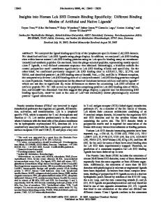
SLP-76 Binding to p56lck: A Role for SLP-76 in CD4-Induced Desensitization of the TCR/CD3 Signaling Complex1 Ralf Sanzenbacher, Dieter Kabelitz, and Ottmar Janssen2 Nonreceptor protein tyrosine kinases and associated substrates play a pivotal role in Ag receptor stimulation of resting cells and in the initiation of activation-induced cell death (AICD) of preactivated T cells. CD4-associated p56lck has been implicated not only in the activation of primary T cells, but also in the inhibition of T cell responses. We have previously shown that CD41 T cell clones can be rescued from AICD when surface CD4 is engaged before the TCR stimulus. In this study, we show that prevention of AICD is associated with a CD4-dependent inhibition of TCR-triggered tyrosine phosphorylation of the Src homology 2 domain-containing leukocyte protein of 76 kDa (SLP-76) and Vav. We provide evidence for a SLP-76 interaction with Src homology 3 domains of p56lck and identify amino acids 185–194 of SLP-76 as relevant docking site. In view of the multiple functions of p56lck and SLP-76/Vav in the initiation of TCR/CD3/CD4 signaling, we propose a model for the CD4-dependent inhibition of TCR signaling and AICD of preactivated T cells. Our data suggest that preformed activation complexes of adapter proteins and enzymes in the vicinity of the CD4/p56lck complex are no longer available for the TCR signal when CD4 receptors are engaged before TCR stimulation. The Journal of Immunology, 1999, 163: 3143–3152.
T
he cell death pathway referred to as activation-induced cell death (AICD3) can be initiated in preactivated mature T lymphocytes by religation of the Ag receptor with Ag, superantigen, or mAb against the TCR/CD3 complex (1–3). AICD is based on a protein tyrosine kinase (PTK)-dependent up-regulation of Fas ligand (FasL, Apo-1L, CD95L) surface expression to subsequently trigger the Fas (CD95, Apo-1) death signaling cascade (4 – 8). Thus, AICD of mature T cells seems to require primary and secondary activation to result in surface expression of both the death receptor and the corresponding death factor. Although AICD has been extensively studied over the past years, only a few reports have analyzed the potential modulation of AICD by coligation of other T cell surface molecules. For example, ligation of CD28 has been demonstrated to inhibit subsequent apoptosis by TCR/CD3 ligation in murine and human T cells (9, 10). We have reported that preligation of CD4 molecules on T cell clones with mAb or gp120 of HIV significantly inhibits AICD (11). The inhibition was associated with a diminished TCR-induced tyrosine phosphorylation, reduced levels of CD3-inducible FasL mRNA and FasL surface expression, and a block of TCRstimulated production of IFN-g and IL-2. In the present study, we obtained evidence for a biochemical link between CD4 engagement and negative regulation of TCR/ CD3 stimulation and AICD. We identified the product of the pro-
Department of Immunology, Paul-Ehrlich-Institute, Langen, Germany Received for publication February 24, 1999. Accepted for publication July 7, 1999. The costs of publication of this article were defrayed in part by the payment of page charges. This article must therefore be hereby marked advertisement in accordance with 18 U.S.C. Section 1734 solely to indicate this fact. 1
This work forms part of the Ph.D. thesis of R.S.
2
Address correspondence and reprint requests to Dr. Ottmar Janssen, Institute for Immunology, Christian-Albrechts-University, Brunswiker Strasse 4, D-24105 Kiel, Germany. E-mail address:
[email protected] 3 Abbreviations used in this paper: AICD, activation-induced cell death; FasL, Fas ligand; PLC, phospholipase C; PTK, protein tyrosine kinase; SEA, staphylococcal enterotoxin A superantigen; SH2 and SH3 domain, Src homology 2 and 3 domain, respectively; SLP-76, SH2 domain-containing leukocyte protein of 76 kDa; TCL, total cell lysate.
Copyright © 1999 by The American Association of Immunologists
tooncogene Vav (12) and the Vav-associated multifunctional SH2 domain-containing leukocyte protein of 76 kDa (SLP-76; Refs. 13–15) as components of TCR/CD3 signaling cascade that are differentially phosphorylated upon TCR ligation in CD4-stimulated vs unstimulated T cell clones. The three functional domains of SLP-76 mediate binding to SH2-containing proteins, including Vav (acidic N terminus when phosphorylated), to SH3-containing molecules such as Grb-2 (proline-rich central part), and tyrosine-phosphorylated molecules such as SLAP-130 (5FYB) and pp62 (C-terminal SH2 domain) (15). More recently, studies with SLP-deficient T cells and SLP-762/2 mice revealed a profound role of this adapter protein in T cell development and activation (16 –18). Thus, SLP-76 couples TCRassociated PTKs to PLC-g1-induced signaling cascades, and is absolutely required for normal T cell development and function. Interestingly, the expression of SLP-76 is restricted to T lymphocytes. In B cells, BLNK or SLP-65, an adapter protein with a very similar overall structure, but a relatively low homology to SLP-76, fulfills the same role in linking the B cell receptor signals to PLC-g1 activation (19, 20). To our knowledge, a direct association of SLP-76 with the TCR/ CD3/CD4-complex or the respective src-related kinases p56lck or p59fyn has not been demonstrated. We show that SLP-76 does not only interact with Grb-2 in a SH3-mediated fashion, but also directly, and selectively binds to SH3 domains of p56lck. With a peptide competition strategy, we map the binding site to a prolinerich stretch located between amino acids 185 and 194 of SLP-76, which is different from the sequence that has been shown to mediate Grb-2 binding. Most important, we are able to coprecipitate substantial amounts of p56lck with SLP-76 from lysates of untransfected Jurkat cells. Our results suggest that upon ligation of CD4, crucial components of the TCR/CD3 signaling cascade (i.e., SLP-76 and Vav) are sequestered to the CD4-p56lck complex via SH3-mediated interactions. As a consequence, we observe a transient TCR/CD3 desensitization of anti-CD4-pretreated cells and provide a model to explain the reduced TCR-dependent cytokine production and AICD described before (11). 0022-1767/99/$02.00
3144
Materials and Methods T cell clones and lines The IL-2-dependent CD31CD41CD51 T cell clone D894/25 has been described (3, 11). Clone cells were restimulated periodically with irradiated PBMC and EBV-transformed lymphoblastoid cell lines in the presence of PHA (0.5 mg/ml; Wellcome, Burgwedel, Germany). Three days after restimulation, the cells were washed extensively and further expanded in the presence of human rIL-2 (10 U/ml; EuroCetus, Frankfurt, Germany) for several days before use in the assays. Dead cells were removed by Ficoll gradient centrifugation when necessary. For biochemical analysis, we also used leukemic Jurkat cells (clone E6.1; American Type Culture Collection (ATCC), Manassas, VA) and PHA-stimulated peripheral blood T lymphocyte populations. To this end, PBMC were cultured with PHA (0.5 mg/ml) for 3 days and further expanded in rIL-2-containing medium. All cells were grown at 37°C in a humidified atmosphere with 6% CO2. Culture medium was RPMI 1640 with 10% (v/v) FBS (Biochrom, Berlin, Germany), antibiotics (penicillin at 100 U/ml and streptomycin at 100 mg/ml), L-glutamine (2 mM), and HEPES buffer solution (10 mM).
Antibodies and reagents For stimulation of the TCR/CD3 complex, we used anti-CD3 mAb OKT3 (mouse IgG2a; Cilag, Sulzbach, Germany) or staphylococcal enterotoxin superantigens from Toxin Technologies (Sarasota, FL). Anti-CD3 mAb were cross-linked with rabbit anti-mouse IgG secondary Abs (Jackson ImmunoResearch, West Grove, PA) when indicated. For ligation or immunoprecipitation of CD4, mAb OKT4 (mouse IgG2b, hybridoma from ATCC) and 5F8 (mouse IgG1, generated in our laboratory) were used. Anti-CD5 mAb UCHT2 (provided by the Sixth International Leukocyte Typing Workshop) was utilized as a control in some experiments. The anti-SLP-76 sheep polyclonal antiserum and anti-pTyr mAb 4G10 were purchased from Upstate Biotechnology (Lake Placid, NY). Anti-c-Cbl (C15, rabbit polyclonal antiserum), anti-Vav (C-12, rabbit polyclonal antiserum), and anti-Sam68 (7-1, mAb) were obtained from Santa Cruz Biotechnology (Santa Cruz, CA). The anti-p56lck antiserum was raised against a lck-specific peptide (residues 39 – 64) (22), and mAb 4/215 (IgG1) against the SH3lck fusion protein (this laboratory). Anti-GST mAb B11F8 (this laboratory) was used for far Western blotting with GST fusion proteins. For Western blotting with ECL detection (Amersham, Braunschweig, Germany), HRP-conjugated secondary reagents (anti-IgG) from Rockland (Gilbertsville, PA; rabbit anti-sheep) and Amersham (donkey anti-rabbit, rabbit anti-mouse) were used. The expression and purification of GST fusion proteins containing the SH2 and/or SH3 domains of p56lck and p59fyn(T) have been described elsewhere (21, 22).
T cell stimulation and preparation of cell lysates To determine effects of CD4 ligation on TCR signals, 1.5 3 106 (total cell lysates) or 20 –50 3 106 (precipitations) clone cells per sample were incubated in the absence or presence of anti-CD4 (or anti-CD5) mAb at 10 mg/ml for 1 h at 37°C. In other experiments, respective numbers of Jurkat cells were used without CD4 engagement. Before TCR stimulation, the cells were washed twice in RPMI with 2% FBS. A total of 50 ml of prewarmed Ab solution with mAb OKT3 at 10 mg/ml and cross-linking Abs at 1 mg/ml were added to the pellet, and incubation was performed at 37°C for the indicated intervals. Clone cells were also activated with SEA at 5 ng/ml. Stimulation was stopped by adding 1 ml of cold PBS, quick spin centrifugation, aspiration of the PBS/Ab supernatant, and immediate lysis in 30 ml of Brij96 (Sigma, Deisenhofen, Germany) or Nonidet P-40 (Fluka Chemie AG, Buchs, Switzerland) lysis buffer (1% (v/v) of detergent in 20 mM Tris-HCl (pH 7.4), 150 mM NaCl, with protease and phosphatase inhibitors aprotinin (10 mg/ml), leupeptin (10 mg/ml), 1 mM PMSF, 1 mM sodium orthovanadate, 1 mM sodium pyrophosphate (all from Sigma), and 10 mM sodium fluoride (Fluka)). Lysates remained on ice for 15–30 min before centrifugation at 4°C and 14,000 rpm for 7 min. Supernatants were then transferred into fresh tubes for further analysis.
Immunoprecipitations, precipitations with fusion proteins, and Western blotting For precipitation with fusion proteins or immunoprecipitation, supernatants were incubated for 90 min rotating at 4°C with 2–5 mg of the respective Ab or 20 –50 mg of GST fusion protein. A total of 60 ml of a 50% slurry of protein A-Sepharose CL4B, protein G-Sepharose, or glutathione Sepharose 4B beads (Pharmacia, Piscataway, NJ) was added, and the samples were rotated for an additional 30 min. The beads were pelleted, washed thrice in cold lysis buffer, and boiled in sample buffer containing 2-ME. Total cell lysates boiled with an equal volume of sample buffer or precipitates were
SLP-76/p56lck INTERACTION REGULATES TCR SIGNALING Table I. Peptides used for competition assays to determine SLP-76/SH3 interaction sitesa
Peptide
SLP-76 peptides A B C D E F G H I K c-Cbl peptides L M a
Corresponding to Amino Acids
Amino Acid Sequence
488–497 84–93 143–152 182–191 185–194 195–204 206–215 283–292 348–357 400–411
G R A T Q A H Q K L
542–551 489–498
L P P P P P P D R P P Q A S L P P V P P
L K D P P A S K P P
R P Y Q P L P P S L
G Q E Q V P L P P P
K V P P P P P L M N
E P P P P P P P N K
D R P V Q P P P P P
F F S P R A Q T L R
L P N P P G T T P P
S E D Q M R N E S P S P
Proline residues characteristic for potential SH3 binding sites are highlighted in
bold.
loaded onto SDS polyacrylamide gels. Separated proteins were transferred to nitrocellulose membranes (Hybond C-Extra; Amersham). Protein loading and efficiency of transfer were monitored with Ponceau S (Sigma). The blots were blocked with 5% BSA (Sigma) for 1 h, and proteins were analyzed with the indicated primary and secondary Abs or with fusion proteins, anti-GST mAb, and HRP-conjugated anti-mouse Ig, and ECL detection reagents.
Peptide competition assay The peptides listed in Table I were synthesized on an AMS 422 peptide synthesizer (Abimed, Langenfeld, Germany). All peptides were dissolved in PBS and used at concentrations specified in Results. For competition experiments, 20 mg of the respective fusion protein on beads was incubated with peptide for 10 –15 min at 4°C with constant rotation. A total of 100 ml of filtered cell lysates corresponding to 25–50 3 106 cells was then added, and incubation was prolonged for 10 min. The beads were then washed extensively and subjected to SDS-PAGE and Western blotting, as described. For ex vivo peptide competition of the SLP-76/lck interactions, filtered lysates of 400 3 106 Jurkat cells were incubated overnight, rotating at 4°C with 2 mM of peptides, followed by SLP-76 immunoprecipitation and anti-lck immunoblot.
Results Inhibition of TCR/CD3-induced tyrosine phosphorylation by preceding CD4 engagement CD41CD31 T cells (clone D894/25) were incubated for 1 h in the presence or absence of anti-CD4 (OKT4) or anti-CD5 (UCHT2) mAbs before stimulation with SEA for 2 or 5 min. As shown in Fig. 1A, a selective reduction of the level of tyrosine phosphorylation of polypeptides with approximate m.w. of 40 – 44, 48 –50, 62– 64, 76, 80, 95, and 145–150 kDa (arrows) was observed in Nonidet P-40 lysates of CD4-treated cells. Preligation of CD4 also inhibited SEA-induced cytokine production and AICD in 894/25 cells (11). Time-course experiments revealed that CD4 ligation was most effective in reducing the subsequent superantigen-induced phosphorylation when CD4 engagement was initiated at least 30 min before the TCR stimulation (not shown). To identify proteins that may be involved in the CD4-mediated down-regulation of the SEA-induced TCR response, we first focused on the polypeptide of 76 kDa. Recently, the SH2 domain-containing leukocyte protein of 76 kDa (SLP-76) has been described as an essential transducer of TCR signals to IL-2 gene activation in concert with Vav, Grb-2, and ZAP-70 (23–25). We therefore reprobed the same blot shown in Fig. 1A with an anti-SLP antiserum and visualized equal amounts of SLP-76 protein loaded in each lane (Fig. 1B). The overlay of the two films strongly suggested that the 76kDa band in fact represented SLP-76. Of note, CD5 ligation before
The Journal of Immunology
3145 strictly dependent on the activation of the CD4 molecule or the CD4/p56lck complex. CD4 ligation blocks TCR/CD3-induced tyrosine phosphorylation of Vav and SLP-76
FIGURE 1. Anti-CD4 mAb treatment inhibits TCR/CD3-induced tyrosine phosphorylation. Cells of T cell clone D894/25 were incubated for 1 h in medium or in the presence of mAbs against CD4 (OKT4) or CD5 (UCHT2), respectively. Stimulation with SEA was performed for 2 or 5 min at 37°C. Clone cells not exposed to SEA served as a control (2). The cells were then lysed in Nonidet P-40 lysis buffer, and proteins were separated by SDS-PAGE. Upon transfer to nitrocellulose membranes, the blots were developed with anti-pTyr mAb (4G10, A) and reprobed with antiSLP-76 antiserum (B). The position of SLP-76 is marked with an asterisk; arrows point to protein bands that were differentially phosphorylated in CD4-untreated and CD4-pretreated cells.
TCR stimulation did not alter TCR-initiated phosphorylation of the indicated proteins, suggesting that the observed inhibition might be
FIGURE 2. CD4 ligation inhibits TCR-triggered phosphorylation of Vav and SLP-76. Clone cells were incubated for 1 h in the absence (A) or presence of anti-CD4 mAb OKT4. The cells were then pelleted, incubated for 3 min in medium without (2) or with (1) SEA, and lysed in Brij96 lysis buffer. Immunoprecipitations of Vav and SLP-76 were performed with 3 mg of the respective Ab and protein A- and protein G-Sepharose beads (beads alone served as a control). Precipitated proteins were separated by SDS-PAGE, and blotted to nitrocellulose. Blots were developed with anti-pTyr mAb 4G10 (A) and reprobed with antiVav (B) and anti-SLP-76 (C) Abs.
With the next set of experiments, the SEA-induced tyrosine phosphorylation of SLP-76 and Vav was analyzed with or without preceding engagement of CD4 by mAbs. Activation of these proteins during TCR-stimulated cytokine production has been shown to be associated with an increased tyrosine phosphorylation mediated by ZAP-70 (23–25). We therefore immunoprecipitated SLP-76 and Vav, respectively, from unstimulated (2) or superantigen-stimulated (1) clone cells that had been exposed or not to anti-CD4 mAb OKT4 for 1 h and analyzed the tyrosine phosphorylation of the blotted proteins with anti-pTyr mAb 4G10 (Fig. 2A). Both proteins were tyrosine phosphorylated during a 3-min stimulation with SEA (see also total cell lysates (TCL)), and the TCR-induced phosphorylation of both proteins was almost completely blocked after exposure to anti-CD4 mAb before SEA stimulation. The protein content in precipitates of untreated or CD4-treated cells was checked by reprobing the blot with anti-Vav (B) or anti-SLP-76 (C) Abs. Preferential, tyrosine phosphorylation-independent association of SLP-76 with SH3 domains of p56lck From the experimental data shown so far, we hypothesized that Vav and/or SLP-76 should be able to interact with both the CD4/ p56lck complex and the TCR/CD3/p59fyn complex in a competitive manner. Also, the respective interaction should be independent of tyrosine phosphorylation. Given the structure of SLP-76 with three protein interaction domains including the proline-rich central portion (26, 27), we favored a potential SH3 association with p56lck and p59fyn(T) in T cells. Therefore, we investigated whether
3146
SLP-76/p56lck INTERACTION REGULATES TCR SIGNALING
FIGURE 3. p56lck SH3 domain binding to SLP-76. Unstimulated (2) or anti-CD3-stimulated (A and B–D, where indicated (1)) Jurkat cells were analyzed. TCL (Nonidet P-40 lysis buffer) and precipitates with anti-SLP-76 antiserum or GST-SH3 fusion proteins were separated by SDS-PAGE. As a control for anti-SLP-76 immunoprecipitates, the same amount of protein A- and G-Sepharose beads was used. Blots were developed with anti-pTyr mAb 4G10 (A and B), and blot B was reprobed with the anti-SLP-76 (C) antiserum and GST-SH3lck fusion protein (D), respectively. The position of SLP-76 is marked with an arrow. Blots A and B–D are from two different experiments.
SLP-76 can be detected in SH3-based precipitates from Nonidet P-40 lysates of T cells. For experimental reasons, these studies were initially done with Jurkat cells. However, similar results were obtained with clone cells and with PHA-stimulated T cell lines. In these experiments, we detected a phosphotyrosine-containing protein band in SH3 precipitates that comigrated with immunoprecipitated SLP-76 and phosphorylated SLP-76 in cell lysates from CD3-stimulated cells (Fig. 3A, arrow). As can also be seen in A, we observed a preferential binding of SLP-76 to isolated SH3 domains of p56lck as compared with p59fyn(T). Again, we detected an increased tyrosine phosphorylation of SLP-76 upon stimulation of Jurkat cells with anti-CD3 mAb OKT3 (Fig. 3B, 1). The presence of equal amounts of protein was tested by reprobing the blot with anti-SLP-76 antiserum (Fig. 3C). In addition, we were able to verify a direct SH3-mediated association with p56lck in a far Western approach utilizing the GST-SH3lck fusion protein and an anti-GST mAb to detect SH3-binding SLP-76 in TCL and anti-SLP-76 immunoprecipitates (Fig. 3D). Kinetics of tyrosine phosphorylation of SLP-76 and coprecipitated proteins SH3 binding is achieved by an interaction of proline-rich motifs of the target protein with a specific binding pocket within the SH3 domain (28, 29). Although SH3 binding occurs independently of tyrosine phosphorylation, it has been suggested that SH3-mediated interactions provide a means to recruit substrates to the receptor/ PTK complexes. In fact, as shown in Fig. 4, a rapid (on after 10 –30 s) and transient (off after 10 –15 min) tyrosine phosphorylation of SLP-76 protein immunoprecipitated from lysates of OKT3-stimulated Jurkat cells is seen (Fig. 4A). GST-SH3lck fusion proteins precipitate phosphorylated and nonphosphorylated SLP-76. The GST part of the fusion protein did not yield any detectable protein (Fig. 4B). As has been reported for other SH3binding proteins, SLP-76 binding to the SH3 domain was not sig-
nificantly influenced by CD3-induced tyrosine phosphorylation. Thus, SLP-76 protein levels were comparable in all GST-SH3lck precipitates (Fig. 4B). Kinetics of SLP-76 phosphorylation in SH3 precipitates was slightly different in that a weak phosphorylation remained detectable even after 30 min of OKT3 stimulation. Of note in this context, SLP-76 coprecipitated with a number of yet unidentified tyrosine-phosphorylated proteins with approximate m.w. of 42– 44, 62– 64, and 120 kDa under these conditions. Preferential SLP-76 binding to SH3 domains of p56lck To further analyze the specificity of SLP-76 SH3 binding to Src kinases, we compared protein levels precipitated from PHA-stimulated PBMC with GST-SH3 fusion proteins of p56lck and of p59fyn(T). As shown in Fig. 5, no SLP-76 protein was detected in controls using protein A and protein G beads (c) or GST and glutathione beads. GST-SH3lck precipitated SLP-76 more efficiently than GST-SH3fyn, pointing to a preferential association of SLP-76 to the CD4-associated PTK. Again, the amount of SLP-76 was unchanged in precipitates from unstimulated (2) vs OKT3-stimulated (1) cells. Identification of the SH3lck interaction site within SLP-76 To identify potential docking regions of SLP-76 binding to SH3 domains of p56lck, we chose a peptide competition strategy. To this end, a series of peptides was synthesized according to the published SLP-76 and c-Cbl sequences (14, 30). SLP-76 peptides B-K contained proline residues characteristic for putative SH3 binding sites (Table I). In addition, we used a control SLP-76 peptide (peptide A) without proline residues and two peptides corresponding to potential SH3-binding regions within c-Cbl (peptides L and M). When SH3 domains of p56lck were preincubated as GST-fusion proteins with the different peptides at a concentration of 1 mM, only peptides E (QPPVPPQRPM, corresponding to amino acids 185–194 of SLP-76) and L (LPPPPPPDRP, corresponding to
The Journal of Immunology
3147
FIGURE 4. CD3-induced tyrosine phosphorylation of SLP-76 does not interfere with SH3 binding. A total of 50 3 106 Jurkat cells was stimulated for the indicated times with cross-linked anti-CD3 mAb OKT3. The cells were lysed in Brij96 lysis buffer, and lysates were subjected to immunoprecipitation with anti-SLP-76 antiserum (A) or precipitation with GST-SH3lck fusion proteins (B). For SLP-76 immunoprecipitations, a mixture of protein A- and protein G-Sepharose beads was used as a control (C) for stimulated cells; for SH3 precipitates, GST served as a control. Blots were developed with anti-pTyr mAb 4G10 and reprobed with the anti-SLP-76 antiserum. TCL from unstimulated (2) or CD3-stimulated (1) cells were also included on the gels.
amino acids 542–551 of c-Cbl) efficiently competed for binding of SLP-76 protein from Jurkat cells (Fig. 6A). Identical results were obtained when clone cells or PHA-stimulated PBMC were used (not shown). Interestingly, under these conditions, peptide E (of SLP-76) also completely blocked SH3 binding to Sam68 (31–34) and c-Cbl, whereas peptide L markedly reduced the SH3/c-Cbl interaction, but only mildly inhibited binding of Sam68 (Fig. 6A). This result was confirmed by titration experiments, as shown for peptides E and L in Fig. 6B. Peptide E inhibited precipitation of Sam68 and c-Cbl with SH3 domains of p56lck at a 4-fold lower dose compared with peptide L. Of note in this context, peptide E effectively competed off SLP-76 binding to SH3lck, but did not
FIGURE 5. Preferential binding of SLP-76 to SH3 domains of p56lck. Nonidet P-40 lysates from 50 3 106 unstimulated (2) or OKT3-stimulated (1) PHA blasts, cross-linked with rabbit anti-mouse Ig, were separated by SDS-PAGE as TCL (corresponding to 1.5 3 106 cells) or upon immunoprecipitation with anti-SLP-76 antiserum or precipitation with GST-SH3 fusion proteins (25 mg/sample). For SLP-76 immunoprecipitations, a mixture of protein A- and protein G-Sepharose beads was used as a control to precipitate unspecifically associated proteins from OKT3-treated cells. Upon transfer to nitrocellulose membranes, the blots were developed with anti-SLP-76 antiserum. The position of SLP-76 is marked with an arrow.
significantly influence the association of SLP-76 with GST-Grb-2 fusion proteins. In addition, peptides E and L, but not peptide A, covalently linked to activated Sepharose beads, in vitro bound GST-SH3lck fusion protein, but not to GST alone (not shown). In vivo association of SLP-76 and p56lck Having established the association of SLP-76 and p56lck in vitro, the next relevant experiment was to show that the two proteins can be coprecipitated from intact cells. Being aware of the difficulties of such experiments due to the instablity of protein-protein complexes even under mild lysis conditions, we chose the following strategy: Brij96 lysates from 50 3 106 unstimulated Jurkat cells were subjected to immunoprecipitation with anti-lck mAb (Fig. 7A, lane 5) or anti-lck antiserum (lane 6). For the anti-SLP-76 immunoprecipitation (Fig. 7A, lane 7), we increased the cell number to 400 3 106. Protein G-bound immunocomplexes were separated by SDS-PAGE on a 8.5% gel. Upon Western blotting of the precipitated proteins, p56lck protein was analyzed first with the anti-lck mAb (upper panel) and reprobed with the polyclonal antiserum (lower panel). As can be seen in Fig. 7, p56lck (and p60lck) could be detected by this type of analysis with both the monoclonal and the polyclonal anti-lck Abs not only in immunoprecipitates formed by anti-lck reagents (lanes 5 and 6), but also in antiSLP-76 immunocomplexes (lane 7). This clearly established that the two proteins, SLP-76 and p56lck, can be coprecipitated as a fairly stable complex from unstimulated intact cells. As shown in Fig. 7B, under similar conditions, peptide E (lane 9), but not control peptide A (lane 8), interfered with the coprecipitation of p56lck (lane 5 or 7). Also, depletion of lysate from CD4/lck by three rounds of anti-CD4 immunoprecipitation almost completely reduced the amount of lck in the subsequent anti-SLP-76 immunoprecipitation.
Discussion Tyrosine phosphorylation mediated by nonreceptor PTKs is required for the initiation of TCR-dependent activation (35, 36). Similarly, in the course of AICD, PTKs such as p56lck provide
3148
SLP-76/p56lck INTERACTION REGULATES TCR SIGNALING
FIGURE 6. Peptide competition reveals aa 185–194 as SH3lck binding site of SLP-76. Peptides corresponding to stretches within the SLP-76 and c-Cbl molecules, as detailed in Table I, were used at 1 mM (A) or at varying concentrations from 0.25–5 mM (B) in PBS. A total of 20 mg of GST-SH3lck fusion protein on glutathione beads was incubated with the respective peptide with constant rotation for 15 min at 4°C. A total of 100 ml of filtered Jurkat cell lysates (25 3 106 cells lysed in 1% Brij96 lysis buffer) was then added, and incubation was prolonged for 10 min. The beads were washed, and precipitated proteins were subjected to SDSPAGE and Western blotting. Blots were developed with the anti-SLP-76 antiserum and subsequently with anti-Sam68 mAb and anti-c-Cbl antiserum. Equal loading of fusion protein was checked in each experiment by Ponceau S staining (data not shown).
necessary and essential signals for the regulation of FasL expression (37–39). Therefore, PTK inhibitors efficiently block CD3/ TCR-triggered activation and AICD (40 – 42). An inhibitory modulation of TCR-dependent kinase signaling cascades by engagement of costimulatory and/or accessory molecules could be explained on the basis of negative signaling, e.g., by phosphatases, or by a recomposition of preformed signaling complexes in the vicinity of Src- or Syk-type PTKs associated with the corresponding surface receptors. We have previously observed that CD4 stimulation of cloned T cells with mAb or HIV-1 gp120 modified subsequent TCR signaling, resulting in a marked reduction of cytokine production and inhibition of AICD (11). This inhibition of TCR signaling was associated with reduced levels of tyrosine phosphorylation of a number of proteins detected in whole cell lysates (11). We now report that two PTK substrates implicated in TCR-mediated signal transduction, the product of the protooncogene vav (12, 43– 45) and the associated SH2 domain-containing leukocyte protein of 76 kDa (SLP-76) (14 –18, 25–27), show this characteristic inhibition of TCR-inducible tyrosine phosphorylation in clone cells pretreated with anti-CD4 mAb. Our results demonstrate that CD4 ligation can prevent the formation of signal-transducing units around the TCR/CD3 complex leading to a desensitization of the Ag receptor. Recently, Jabado and colleagues also pointed to a defective formation of p21ras activity-regulating complexes, including PLC-g1, p120GAP, and other yet unidentified proteins upon CD4 ligand binding (46). In addition, Goldman et al. suggested that CD4 ligation may inhibit TCR signaling by sequestering p56lck to the actin-cytoskeleton (47). When they pretreated cells with gp120
1– 4 h before stimulation, TCR-directed PTK activation was markedly reduced. Also, in gp120/anti-gp120-stimulated cells, p56lck was found predominantly in association with the cytoskeleton in the Nonidet P-40 detergent-insoluble cytoskeletal, but not in the Nonidet P-40 detergent-soluble cytosolic fractions (47). With Vav and SLP-76, we link two other key players in TCR signal transduction to the CD4/p56lck complex. The multifunctional adapter protein SLP-76 was characterized as a molecule that is phosphorylated by ZAP-70 upon TCR stimulation and subsequently associates with SH2 domains of Vav via phosphotyrosylcontaining motifs in the N-terminal portion of SLP-76 (13, 23–25). The physical link between Vav and SLP-76 may also be differentially regulated by associated phosphatases including CD45 (48). In addition, a constitutive association of SLP-76 with Grb-2 SH3 domains has been reported, mediated by the proline-rich central part (14, 15). With its C-terminal SH2 domain, SLP-76 also reacts with phosphoproteins including SLAP-130 (5FYB) and pp62 (27, 49, 50). Recent studies with SLP-76-deficient mice and SLP-76-deficient T cell lines revealed a very profound role of the adapter protein in T cell development and activation (16 –18). It was shown that SLP-76 couples TCR-associated PTKs to PLC-g1-induced signaling cascades and that it is absolutely required for normal T cell development and function. Interestingly, the expression of SLP-76 is restricted to hemopoietic cells of monocyte, granulocyte, and T lymphocyte lineage (51), whereas in B cells, SLP-65 (or BLNK), an adapter protein with a very similar overall domain structure but fairly low homology, fulfills the same role in linking the B cell receptor signals to PLC-g1 activation (19, 20).
The Journal of Immunology
3149
FIGURE 7. The in vivo association of SLP-76 and p56lck can also be blocked by peptide competition. A, Brij96 lysates from unstimulated Jurkat cells (50 3 106 for lck immunoprecipitation (lanes 5 and 6) and 400 3 106 for SLP immunoprecipitation (lane 7)) were incubated overnight with an anti-lck mAb (lane 5), a polyclonal anti-lck antiserum (lane 6), or the polyclonal anti-SLP-76 antiserum (lane 7), respectively. Protein G-bound immunocomplexes were separated by SDS-PAGE on an 8.5% gel. As controls, equivalent amounts of Abs (lanes 1–3) and protein G-Sepharose beads (lane 4) and TCL (lane 8) were loaded on the same gel. B, Brij96 lysates from unstimulated Jurkat cells (50 3 106 for CD4 and lck immunoprecipitation (lanes 2– 4) and 400 3 106 for SLP immunoprecipitation (lane 5)) were incubated overnight with the indicated Abs before protein G-bound immunocomplexes were separated by SDS-PAGE on a 8.5% gel. Anti-CD4 Ab alone was used as control (lane 1). In addition, lysates of 400 3 106 cells were depleted of CD4 and CD4associated lck by three rounds of anti-CD4 immunoprecipitation before SLP-76 was precipitated with the specific anti-SLP-76 antiserum (lane 6). Furthermore, anti-SLP immunoprecipitations were performed from 400 3 106 cells in the absence or presence of 2 mM of peptides A or E, respectively (lanes 7–9). Upon transfer to nitrocellulose, the blots were developed first with the anti-lck mAb (upper panels) and reprobed with the polyclonal anti-lck antiserum (lower panels). The position of the lck band is marked with an arrow.
We show that besides the multiple interactions of SLP-76 that have been reported to date, the adapter protein also binds to SH3 domains of Src kinases, preferentially of p56lck. The amount of SLP-76 precipitated with SH3 domains of p56lck was comparable with the amount of protein precipitated with full-length Grb-2 fusion proteins (not shown). Using a peptide competition strategy, we mapped the putative binding region to a XPPXPPXXP motif corresponding to amino acids 185–194 of SLP-76. Interestingly, the SLP-76 peptide (QPPVPPQRPM) used for the competition experiments also completely blocked the SH3lck interaction with Sam68 and c-Cbl, indicating that in fact it tightly occupies the binding pocket of the SH3 domain. Also of note, in vitro SH3 binding to isolated domains of p59fyn was consistently weaker, indicating a preferential association with the SH3 domain of
p56lck. Furthermore, peptide E effectively competed off SLP-76 binding to SH3lck, but did not significantly influence association with GST-Grb-2. Interestingly, according to the work by Motto et al. (15), the interaction of SLP-76 with either or both Grb-2 SH3 domains occurs via amino acids 225–244 of SLP-76, an area that does not contain an obvious SH3-binding motif. Although a preferential binding of type 1 ligand motifs (R/KxxPxxP) to SH3 domains of Src kinases has been suggested, peptide E (and also peptide L) contains type II SH3-binding motifs (PxxPxR). However, the binding selectivity of Src-SH3 domains for type I or type II motifs seems to be far from exclusive. The reports by Yu et al. (29), Feng and colleagues (52), and Lim et al. (53) showed that the ligands share a common invariable PxxP core motif, and that peptide binding is mainly dependent on the critical
3150 salt bridging of the arginine residue of the peptide and the conserved aspartate at position 99 of the Src-family SH3 domain, with the residues in positions 3, 4, 6, and 7 of the peptide intercalating into the binding site and thus determining the binding orientation of the peptide. This was also confirmed by Morton and colleagues (54), who investigated ligand binding to the SH3 domain of Fyn. Based on the comparison of different peptides (including type I and type II) for SH3 binding, they suggested that SH3 domains bind to polyproline peptides in a promiscuous manner, although the ligand with the closest match to the class I consensus sequence bound with highest affinity and in the predicted orientation. Also, the NMR of the SH3 domain of lck (55) confirmed the previous suggestions that peptide ligands can bind in two different orientations to SH3 domains. This study also points to some unique features within the SH3 domain of Lck, which could help to explain the observed preference of SLP-76 for Lck compared with Fyn. In view of the specific regulation of SH3 binding by the critical arginine residue, it should also be mentioned that peptides D (aa 182–191) and E (aa 185 to 194) are overlapping peptides that share seven amino acids including the PxxP core. However, only the inhibitory peptide E contains an arginine residue in a position that determines the type II-binding motif PxxPxR. Most important, we were able to demonstrate that p56lck can be coprecipitated in an immunocomplex formed by anti-SLP-76 Abs from unstimulated Jurkat cells, and that the coprecipitation can be abrogated by preincubation of the cell lysates with the competitor peptide E. This established that the phosphorylation-independent (SH3-mediated) association detected and analyzed in vitro is also operational in vivo in intact cells. The SH3 domain-mediated interaction of SLP-76 with p56lck may help to explain the observations mentioned above (46, 47). Since we found a reduced tyrosine phosphorylation of SLP-76 upon CD4 pretreatment, it seems likely that CD4 ligation resulted in a spatial dissociation from its kinase, ZAP-70. Thus, if p56lck is not available for the phosphorylation of the z-chains, ZAP-70 could not bind to the TCRz/CD3 complex to get activated. We are presently analyzing whether the dissociation occurs due to sequestering p56lck to the cytoskeleton, as has been suggested by Goldman and colleagues (47), or by capping mechanisms that have been described for other systems. In fact, the influence of the CD4 proximity to the TCR/CD3 complex had been correlated earlier to inhibitory or stimulatory signals provided by CD4 (56). As a functional outcome of negative tuning of TCR responses by CD4 engagement, the selective activation of CD4-positive naive but not memory cells has been suggested as a consequence of MHC class II molecules on APC and CD4 ligation (57). In the context of thymocyte development, it was shown that CD4 engagement inhibited TCR expression and function in immature CD41CD81 thymocytes (58). The effects of other ligands of CD4, including envelope proteins of HIV and the recently cloned IL-16, seem to be manyfold and very much depending on the investigated system. For IL-16 as a natural ligand of CD4 (59, 60), a block of CD3-dependent lymphocyte activation and proliferation has been reported by Cruikshank and coworkers (61). In addition, IL-16 seems to play a role in the regulation of HIV-1 infection and/or replication (62– 64). A protective effect of rIL-16 against AICD has also been described, although the underlying mechanism needs further studies (64). The preparation of IL-16 that we used in our previous study was ineffective in our system. Moreover, CD4 ligation in our hands affected CD95L but not CD95 expression (11), as has been suggested by others (64). The multiple effects of cross-linking of CD4 by anti-CD4 mAb or gp120 of HIV on TCR-induced activation of resting vs preactivated cells have been extensively discussed in our previous report
SLP-76/p56lck INTERACTION REGULATES TCR SIGNALING
FIGURE 8. Model for the involvement of SLP-76 in CD4-induced desensitization of the TCR/CD3 signaling complex. We hypothesize that a preformed TCR/CD3/CD4 signaling complex exists in preactivated T cells. This complex would include PTKs, adapter proteins, and unknown downstream elements associated in a tyrosine phosphorylation-independent manner due to SH3-mediated interactions. A. TCR ligation without CD4 engagement triggers PTKs associated with the TCR/CD3/CD4 complex. The result is a tyrosine phosphorylation-dependent activation and potentially AICD. B, Upon ligation of CD4, components of the preformed complex (i.e., SLP-76 and Vav) are removed from the TCR/CD3 complex. The transient TCR/CD3 desensitization results in an incomplete activation capacity of TCR/CD3-triggered PTK-dependent pathways.
(11). They range from costimulation of resting T cells to proliferate (65, 66), priming of resting cells for AICD (67–70) and direct induction of apoptosis (71–76) to the inhibition of TCR-induced activation (46, 47, 77– 80), and AICD (11). Taken together, these studies suggest that the observed differences in the CD4/TCR cross-talk between resting and activated cells very much depend on the investigated T cell population. Overall, resting cells seem to be coactivated by CD4 engagement, whereas inhibitory effects of CD4 ligation have been mainly observed in preactivated populations. Although OKT4 does not bind to the gp120-binding region, it needs to be addressed in further studies whether the binding of different CD4 ligands or mAb to distinct functional epitopes within the CD4 molecule provokes different signals with regard to the inhibition of AICD (81). In conclusion, SH3-mediated interactions of SLP-76 and associated molecules with the CD4/p56lck complex play a crucial role in negative signaling through the CD4 molecule. In activated T cells, CD4 ligation may lead to a removal of essential components of the TCR signaling complex and to a TCR desensitization with reduced responses in terms of proliferation, cytokine production, and AICD (see model in Fig. 8). Whether the removal of SLP-76associated proteins from the TCR/CD3 complex is physical, e.g., due to a phosphorylation-independent translocation of SLP-76/ p56lck complexes to the cytoskeleton, is presently under investigation.
Acknowledgments We thank Dr. Joachim Denner for peptide synthesis, and EuroCetus and Cilag GmbH for providing essential reagents.
References 1. Russell, J., C. L. White, D. Y. Loh, and P. Meleedy-Rey. 1992. Receptor-stimulated death pathway is opened by antigen in mature T cells. Proc. Natl. Acad. Sci. USA 88:2151.
The Journal of Immunology ¨ streicher, K. Pechold, A. Bender, 2. Janssen, O., S. Wesselborg, B. Heckl-O S. Schondelmaier, G. Moldenhauer, and D. Kabelitz. 1991. T cell receptor/CD3signaling induces death by apoptosis in human T cell receptor gd1 T cells. J. Immunol. 146:35. 3. Kabelitz, D., and S. Wesselborg. 1992. Life and death of a superantigen-reactive human CD41 T cell clone: staphylococcal enterotoxins induce death by apoptosis but simultaneously trigger a proliferative response in the presence of HLA DR1 antigen-presenting cells. Int. Immunol. 4:1381. 4. Nagata, S., and P. Golstein. 1995. The Fas death factor. Science 267:1449. 5. Dhein, J., H. Walczak, C. Baeumler, K. M. Debatin, and P. H. Krammer. 1995. Autocrine T-cell suicide mediated by APO-1/(Fas/CD95). Nature 373:438. 6. Brunner, T., R. J. Mogil, D. LaFace, N. J. Yoo, A. Mahboubi, F. Echeverri, S. J. Martin, W. R. Force, D. H. Lynch, C. F. Ware, and D. R. Green. 1995. Cell-autonomous Fas (CD95)/Fas-ligand interaction mediates activation-induced apoptosis in T-cell hybridomas. Nature 373:441. 7. Ju, S. T., D. J. Panka, H. Cui, R. Ettinger, M. el-Khatib, D. H. Sherr, B. Z. Stanger, and A. Marshak-Rothstein. 1995. Fas(CD95)/FasL interactions required for programmed cell death after T-cell activation. Nature 373:444. 8. Alderson, M. R., T. W. Tough, T. Davis-Smith, S. Braddy, B. Falk, K. A. Schooley, R. G. Goodwin, C. A. Smith, F. Ramsdell, and D. H. Lynch. 1995. Fas ligand mediates activation-induced cell death in human T lymphocytes. J. Exp. Med. 181:71. 9. Radvanyi, L. G., Y. Shi, H. Vaziri, A. Sharma, R. Dhala, G. B. Mills, and R. G. Miller. 1996. CD28 costimulation inhibits TCR-induced apoptosis during a primary T cell response. J. Immunol. 156:1788. 10. Groux, H., G. Torpier, D. Monte´, Y. Mouton, A. Capron, and J. C. Ameisen. 1992. Activation-induced death by apoptosis in CD41 T cells from human immunodeficiency virus-infected asymptomatic individuals. J. Exp. Med. 175:331. 11. Oberg, H.-H., R. Sanzenbacher, B. Lengl-Janben, T. Dobmeyer, S. Flindt, O. Janssen, and D. Kabelitz. 1997. Ligation of cell surface CD4 inhibits activation-induced cell death of human T lymphocytes at the level of Fas-ligand expression. J. Immunol. 159:5742. 12. Katzav, S., D. Martin-Zanca, and M. Barbacid. 1989. Vav, a novel human oncogene derived from a locus ubiquitously expressed in hematopoietic cells. EMBO J. 8:2283. 13. Tuosto, L., F. Michel, and O. Acuto. 1996. p95vav associates with tyrosine-phosphorylated SLP-76 in antigen-stimulated T cells. J. Exp. Med. 184:1161. 14. Jackman, J. K., D. G. Motto, Q. Sun, M. Tanemoto, C. W. Turck, G. A. Peltz, G. A. Koretzky, and P. R. Findell. 1995. Molecular cloning of SLP-76, a 76-kDa tyrosine phosphoprotein associated with Grb2 in T cells. J. Biol. Chem. 270: 7029. 15. Motto, D. G., S. E. Ross, J. Wu, L. R. Hendricks-Taylor, and G. A. Koretzky. 1996. Implication of the Grb2-associated phosphoprotein SLP-76 in T cell receptor-mediated interleukin 2 production. J. Exp. Med. 183:1937. 16. Pivniouk, V., E. Tsitsikov, P. Swinton, G. Rathbun, F. W. Alt, and R. S. Geha. 1998. Impaired viability and profound block in thymocyte development in mice lacking the adaptor protein SLP-76. Cell 94:229. 17. Clements, J. L., B. Yang, S. E. Ross-Barta, S. L. Eliason, R. F. Hrstka, R. A. Williamson, and G. A. Koretzky. 1998. Requirement for the leukocytespecific adapter protein SLP-76 for normal T cell development. Science 281:416. 18. Yablonski, D., M. R. Kuhne, T. Kadlecek, and A. Weiss. 1998. Uncoupling of nonreceptor tyrosine kinases from PLCg1 in a SLP-76-deficient T cell. Science 281:413. 19. Wienands, J., J. Schweikert, B. Wollscheid, H. Jumaa, P. J. Nielsen, and M. Reth. 1998. SLP-65: a new signaling component in B lymphocytes which requires expression of the antigen receptor for phosphorylation. J. Exp. Med. 188:791. 20. Fu, C., C. W. Turck, T. Kurosaki, and A. C. Chan. 1998. BLNK: a central linker protein in B cell activation. Immunity 9:93. 21. Prasad, K. V. S., O. Janssen, R. Kapeller, M. Raab, L. C. Cantley, and C. E. Rudd. 1993. Src-homology 3 domain of protein kinase p59fyn mediates binding to phosphatidylinositol 3-kinase in T cells. Proc. Natl. Acad. Sci. USA 90:7366. 22. Prasad, K. V. S., R. Kapeller, O. Janssen, J. S. Duke-Cohen, L. C. Cantley, and C. E. Rudd. 1993. Phosphatidylinositol 3-kinase and phosphatidylinositol 4-kinase binding to the CD4 –p56lck complex: p56lck SH3 domain mediates binding to PI-3 kinase but not to PI-4 kinase. Mol. Cell. Biol. 13:7708. 23. Wu, J., D. G. Motto, G. A. Koretzky, and A. Weiss. 1996. Vav and SLP-76 interact and functionally cooperate in IL-2 gene activation. Immunity 4:593. 24. Wardenburg, J. B., C. Fu, J. K. Jackman, H. Flotow, S. E. Wilkinson, D. H. Williams, R. Johnson, G. Kong, A. C. Chan, and P. R. Findell. 1996. Phosphorylation of SLP-76 by the ZAP-70 protein tyrosine kinase is required for T-cell receptor function. J. Biol. Chem. 271:19641. 25. Raab, M., A. J. da Silva, P. R. Findell, and C. E. Rudd. 1997. Regulation of Vav-SLP-76 binding by ZAP-70 and its relevance to TCRz/CD3 induction of interleukin-2. Immunity 6:155. 26. Musci, M. A., D. G. Motto, S. E. Ross, N. Fang, and G. A. Koretzky. 1997. Three domains of SLP-76 are required for its optimal function in a T cell line. J. Immunol. 159:1639. 27. Koretzky, G. A. 1997. The role of Grb2-associated proteins in T cell activation. Immunol. Today 8:401. 28. Ren, R., B. J. Mayer, P. Cicchetti, and D. Baltimore. 1993. Identification of a ten-amino acid proline-rich SH3 binding site. Science 259:1157. 29. Yu, H., J. K. Chen, S. Feng, D. C. Dalgarno, A. W. Brauer, and S. L. Schreiber. 1994. Structural basis for the binding of proline-rich peptides to SH3 domains. Cell 76:933. 30. Blake, T. J., M. Shapiro, H. C. Morse III, and W. Y. Langdon. 1991. The sequences of the human and mouse c-cbl protooncogenes show v-cbl was generated
3151
31.
32. 33. 34.
35.
36. 37.
38. 39.
40.
41.
42.
43. 44. 45.
46.
47. 48.
49.
50.
51.
52.
53.
54.
55. 56.
57.
58.
by a large truncation encompassing a proline-rich domain and a leucine zipperlike motif. Oncogene 6:653. Fukazawa, T., K. A. Reedquist, T. Trub, S. Soltoff, G. Panchamoorthy, B. Druker, L. Cantley, S. E. Schoelson, and H. Band. 1995. The SH3 domain-binding T cell tyrosyl phosphoprotein p120: demonstration of its identity with the c-cbl protooncogene product and in vivo complexes with Fyn, Grb-2 and phosphatidylinositol 3-kinase. J. Biol. Chem. 270:19141. Taylor, S. J., and D. Shalloway. 1994. An RNA-binding protein associated with Src through its SH2 and SH3 domains in mitosis. Nature 368:867. Fumagalli, S., N. F. Totty, J. J. Hsuan, and S. A. Courtneidge. 1994. A target for Src in mitosis. Nature 368:871. Lock, P., S. Fumagalli, P. Polakis, F. McCormick, and S. A. Courtneidge. 1996. The human p62 cDNA encodes Sam68 and not the RasGAP-associated p62 protein. Cell 84:23. Rudd, C. E., O. Janssen, Y.-C. Cai, A. J. da Silva, M. Raab, and K. V. S. Prasad. 1994. Two-step TCRz/CD3-CD4 and CD28 signaling in T cells: SH2/SH3 domains, protein-tyrosine kinases and lipid kinases. Immunol. Today 15:225. Alberola-Ila, J., S. Takaki, J. D. Kerner, and R. Perlmutter. 1997. Differential signaling by lymphocyte antigen receptors. Annu. Rev. Immunol. 15:125. Oyaizu, N., S. Than, T. W. McClosey, and S. Pahwa. 1995. Requirement of p56lck in T-cell receptor/CD3-mediated apoptosis and Fas-ligand induction in Jurkat cells. Biochem. Biophys. Res. Commun. 213:994. Di Somma, M. M., S. Nuti, J. L. Telford, and C. T. Baldari. 1995. p56lck plays a key role in transducing apoptotic signals in T cells. FEBS Lett. 363:101. Gonzalez-Garcia, A., L. R.-Borlado, E. Leonardo, I. Me´rida, C. Martinez-A., and A. C. Carrera. 1997. Lck is necessary and sufficient for Fas-ligand expression and apoptotic cell death in mature cycling T cells. J. Immunol. 158:4104. June, C. H., M. C. Fletcher, J. A. Ledbetter, G. L. Schieven, J. N. Siegel, A. F. Phillips, and L. E. Samelson. 1990. Inhibition of tyrosine phosphorylation prevents T-cell receptor-mediated signal transduction. Proc. Natl. Acad. Sci. USA 87:7722. Migita, K., K. Eguchi, Y. Kawabe, A. Mizokami, T. Tsukada, and S. Nagataki. 1994. Prevention of anti-CD3 monoclonal antibody-induced thymic apoptosis by protein tyrosine kinase inhibitors. J. Immunol. 153:3457. Oberg, H.-H., B. Lengl-Janben, M. J. Robertson, D. Kabelitz, and O. Janssen. 1997. Differential role of tyrosine phosphorylation in the induction of apoptosis in T cell clones via CD95 or the TCR/CD3-complex. Cell Death Differ. 4:403. Collins, T. L., M. Deckert, and A. Altman. 1997. Views on Vav. Immunol. Today 18:221. Bustelo, X. R., J. A. Ledbetter, and M. Barbacid. 1992. Product of vav protooncogene defines a new class of tyrosine protein kinase substrates. Nature 356:68. Margolis, B., P. Hu, S. Katzav, W. Li, J. M. Oliver, A. Ullrich, A. Weiss, and J. Schlessinger. 1992. Tyrosine phosphorylation of vav protooncogene product containing SH2 domain and transcription factor motifs. Nature 356:71. Jabado, N., A. Pallier, F. Le Deist, F. Bernard, A. Fischer, and C. Hivroz. 1997. CD4 ligands inhibit the formation of multifunctional transduction complexes involved in T cell activation. J. Immunol. 158:94. Goldman, F., J. Crabtree, C. Hollenback, and G. A. Koretzky. 1997. Sequestration of p56lck by gp120, a model for TCR desensitization. J. Immunol. 158:2017. Onodera, H., D. G. Motto, G. A. Koretzky, and D. M. Rothstein. 1996. Differential regulation of activation-induced tyrosine phosphorylation and recruitment of SLP-76 to Vav by distinct isoforms of the CD45 protein-tyrosine phosphatase. J. Biol. Chem. 271:22225. Musci, M. A., L. R. Hendricks-Taylor, D. G. Motto, M. Paskind, J. Kamens, C. W. Turck, and G. A. Koretzky. 1997. Molecular cloning of SLAP-130, an SLP-76-associated substrate of the T cell antigen receptor-stimulated protein tyrosine kinases. J. Biol. Chem. 272:11674. da Silva, A. J., Z. Li, C. De Vera, E. Canto, P. Findell, and C. E. Rudd. 1997. Cloning of a novel T-cell protein FYB that binds FYN and SH2-domain-containing leukocyte protein 76 and modulates interleukin 2 production. Proc. Natl. Acad. Sci. USA 94:7493. Clements, J. L., S. E. Ross-Barta, L. T. Tygrett, T. J. Waldschmidt, and G. A. Koretzky. 1998. SLP-76 expression is restricted to hemopoietic cells of monocyte, granulocyte, and T lymphocyte lineage and is regulated during T cell maturation and activation. J. Immunol. 161: 3880. Feng, S., J. K. Cheng, H. Yu, J. A. Simon, and S. L. Schreiber. 1994. Two binding orientations for peptides to the Src SH3 domain: development of a general model for SH3-ligand interactions. Science 266:1241. Lim, W. A., F. M. Richards, and R. O. Fox. 1994. Structural determinants of peptide-binding orientation and of sequence specificity in SH3 domains. Nature 372:375. Morton, C. J., D. J. R. Pugh, E. L. J. Brown, J. D. Kahmann, D. A. C. Renzoni, and I. D. Campbell. 1996. Solution structure and peptide binding of the SH3 domain from human fyn. Structure 4:705. Hiroaki, H., W. Klaus, and H. Senn. 1996. Determination of the solution structure of the SH3 domain of human p56lck tyrosine kinase. J. Biomol. NMR 8:105. Ledbetter, J. A., C. H. June, P. S. Rabinovitch, A. Grossmann, T. T. Tsu, and J. B. Imboden. 1988. Signal transduction through CD4 receptors: stimulatory vs inhibitory activity is regulated by CD4 proximity to the CD3/TCR receptor. Eur. J. Immunol. 18:525. Farber, D. L., M. Luqman, O. Acuto, and K. Bottomly. 1995. Control of memory CD4 T cell activation: MHC class II molecules on APCs and CD4 ligation inhibit memory but not naive CD4 T cells. Immunity 2:249. Nakayama, T., C. H. June, T. I. Munitz, M. Sheared, S. A. McCarthy, S. O. Sharrow, L. E. Samelson, and A. Singer. 1990. Inhibition of T cell receptor expression and function in immature CD41CD81 cells by CD4. Science 249: 1558.
3152 59. Cruikshank, W. W., D. M. Center, N. Nisar, M. Wu, B. Natke, A. C. Theodore, and H. Kornfeld. 1994. Molecular and functional analysis of a lymphocyte chemoattractant factor: association of biologic function with CD4 expression. Proc. Natl. Acad. Sci. USA 91:5109. 60. Center, D. M., H. Kornfeld, and W. W. Cruikshank. 1996. Interleukin 16 and its function as a CD4 ligand. Immunol. Today 17:476. 61. Cruikshank, W. W., K. Lim, A. C. Theodore, J. Cook, G. Fine, P. F. Weller, and D. M. Center. 1996. IL-16 inhibition of CD3-dependent lymphocyte activation and proliferation. J. Immunol. 157:5240. 62. Baier, M., A. Werner, N. Bannert, K. Metzner, and R. Kurth. 1995. HIV suppression by interleukin-16. Nature 378:563. 63. Zhou, P., S. Goldstein, K. Devadas, D. Tewari, and A. L. Notkins. 1997. Human CD41 cells transfected with IL-16 cDNA are resistant to HIV-1 infection: inhibition of mRNA expression. Nat. Med. 3:659. 64. Idziorek, T., J. Khalife, O. Billaut-Mulot, E. Hermann, M. Aumencier, Y. Mouton, A. Capron, and G. M. Bahr. 1998. Recombinant human IL-16 inhibits HIV-1 replication and protects against activation-induced cell death (AICD). Clin. Exp. Immunol. 112:84. 65. Anderson, P., M.-L. Blue, C. Morimoto, and S. F. Schlossman. 1987. Crosslinking of T3 (CD3) with T4 (CD4) enhances the proliferation of resting T lymphocytes. J. Immunol. 139:678. 66. Oravecz, T., and M. A. Norcross. 1993. Costimulatory properties of the human CD4 molecule: enhancement of CD3-induced T cell activation by human immunodeficiency virus type 1 through viral envelope glycoprotein gp120. AIDS Res. Hum. Retroviruses 9:945. 67. Newell, M. K., L. J. Haughn, C. R. Maroun, and M. H. Julius. 1990. Death of mature T cells by separate ligation of CD4 and the T cell receptor for antigen. Nature 347:286. 68. Banda, N. K., J. Bernier, D. K. Kurahara, R. Kurrle, N. Haigwood, R.- P. Sekaly, and T. H. Finkel. 1992. Cross-linking CD4 by human immunodeficiency virus gp120 primes T cells for activation-induced apoptosis. J. Exp. Med. 176:1099. 69. Choy, E. H. S., J. Adjave, L. Forrest, G. H. Kingsley, and G. S. Panayi. 1993. Chimeric anti-CD4 monoclonal antibody cross-linked by monocyte Fcg receptor mediates apoptosis of human CD4 lymphocytes. Eur. J. Immunol. 23:2676. 70. Algeciras, A., D. H. Dockrell, D. H. Lynch, and C. V. Paya.1998. CD4 regulates susceptibility to Fas ligand- and tumor necrosis factor-mediated apoptosis. J. Exp. Med. 187:711.
SLP-76/p56lck INTERACTION REGULATES TCR SIGNALING 71. Oyaizu, N., T. W. McCloskey, M. Coronesi, N. Chirmule, V. S. Kalyanaraman, and S. Pahwa. 1993. Accelerated apoptosis in peripheral blood mononuclear cells (PBMCs) from human immunodeficiency virus type-1 infected patients and in CD4 cross-linked PBMCs from normal individuals. Blood 82:3392. 72. Wang, Z.-Q., A. Dudhane, T. Orlikowsky, K. Clarke, X. Li, Z. Darzynkiewicz, and M. K. Hoffmann. 1994. CD4 engagement induces Fas antigen-dependent apoptosis of T cells in vivo. Eur. J. Immunol. 24:1549. 73. Desbarats, J., J. H. Freed, P. A. Campbell, and M. K. Newell. 1996. Fas (CD95) expression and death-mediating function are induced by CD4 cross-linking on CD41 T cells. Proc. Natl. Acad. Sci. USA 93:11014. 74. Howie, S. E. M., A. J. Sommerfield, E. Gray, and D. J. Harrison. 1993. Peripheral T lymphocyte depletion by apoptosis after CD4 ligation in vivo: selective loss of CD442 and ‘activating’ memory T cells. Clin. Exp. Immunol. 95:195. 75. Tian, H., R. Lempicki, L. King, E. Donoghue, L. E. Samelson, and D. I. Cohen. 1996. HIV envelope-directed signaling aberrancies and cell death of CD41 T cells in the absence of TCR co-stimulation. Int. Immunol. 8:65. 76. Corbeil, J., M. Tremblay, and D. D. Richman. 1996. HIV-induced apoptosis requires the CD4 receptor cytoplasmic tail and is accelerated by interaction of CD4 with p56lck. J. Exp. Med. 183:39. 77. Bank, I., and L. Chess. 1985. Perturbation of the T4 molecule transmits a negative signal to T cells. J. Exp. Med. 162:1294. 78. Mittler, R. S., and M. K. Hoffmann. 1989. Synergism between HIV gp120 and gp120-specific antibody in blocking human T cell activation. Science 245: 1380. 79. Dianzani, U., A. Shaw, M. Fernandez-Cabezuda, and C. A. Janeway Jr. 1992. Extensive CD4 cross-linking inhibits T cell activation by anti-receptor antibody but not by antigen. Int. Immunol. 4:995. 80. Liegler, T. J., and D. P. Stites. 1994. HIV-1 gp120 and anti-gp120 induce reversible unresponsiveness in peripheral CD4 T lymphocytes. J. Acquired Immune Defic. Syndr. 7:340. 81. Baldari, C. T., E. Milia, M. M. Di Somma, F. Baldoni, S. Valitutti, and J. L. Telford. 1995. Distinct signaling properties identify functionally different CD4 epitopes. Eur. J. Immunol. 25:1843.