identification of growth factor receptors (for which the ligands have yet to be identified) that are expressed specifi- cally on hematopoietic cells. For example, the ...
MOLECULAR AND CELLULAR BIOLOGY, Feb. 1994, p. 880-887
Vol. 14, No. 2
0270-7306/94/$04.00+0 Copyright X 1994, American Society for Microbiology
A Simple and Efficient Procedure for Generating Stable Expression Libraries by cDNA Cloning in a Retroviral Vector JOHN R. RAYNER AND THOMAS J. GONDA*
Hanson Centre for Cancer Research and Division of Human Immunology, Institute of Medical and Veterinary Science, Adelaide 5000, South Australia, Australia Received 1 July 1993/Returned for modification 29 September 1993/Accepted 3 November 1993
cDNA expression cloning is a powerful method for the rescue and identification of genes that are able to confer a readily identifiable phenotpe on specific cell types. Retroviral vectors provide several advantages over DNA-mediated gene transfer for the introduction of expression libraries into eukaryotic cells since they can be used to express genes in a wide range of cell types, including those that form important experimental systems such as the hemopoietic system. We describe here a straightforward and efficient method for generating expression libraries by using a murine retroviral vector. Essentially, the method involves the directional cloning of cDNA into the retroviral vector and the generation of pools of stable ecotropic virus producing cells from this DNA. The cells so derived constitute the library, and the virus they yield is used to infect appropriate target cells for subsequent functional screening. We have demonstrated the feasibility of this procedure by constructing several large retroviral libraries (105 to 106 individual clones) and then using one of these libraries to isolate cDNAs for interleukin-3 and granulocyte-macrophage colony-stimulating factor on the basis of the ability of these factors to confer autonomous growth on the factor-dependent hemopoietic cell line FDC-P1. Moreover, the frequency at which these factor-independent clones were isolated approximated the frequency at which they were represented in the original plasmid library. These results suggest that expression cloning with retroviruses is a practical and efficient procedure and should be a valuable method for the isolation of important regulatory genes. master genes that encode transcription factors responsible
The hematopoietic system provides us with an exceptional experimental system for studying the control of cellular proliferation and differentiation. During the past several years our understanding of this complex system has improved considerably, largely because of the isolation of growth factors and their cognate receptors, as well as the genes which encode them. Despite this progress, the manner in which such genes and their products interact to maintain normal hematopoiesis in the bone marrow environment is yet to be clearly elucidated. Although several hematopoietic growth factors are known to be produced by marrow-derived stromal cells in vitro (14, 22), the existence of a number of presently unknown growth factors can be imputed from the identification of growth factor receptors (for which the ligands have yet to be identified) that are expressed specifically on hematopoietic cells. For example, the tyrosine kinase class receptor flk-2, which is related to c-fins (the colony-stimulating factor 1 receptor), is expressed in populations enriched for hematopoietic stem and early progenitor cells but not in mature cells (30), which suggests that this receptor, and thus its ligand, might have a specific regulatory role in hematopoiesis. Similarly, the human homolog of the v-mpl oncogene, c-mpl, has recently been cloned and shown to be highly homologous to the hematopoietin receptor superfamily (42). c-mpl is expressed in a variety of cells of hematopoietic origin, but, as for the flk-2 gene, no function can be ascribed to it, nor is the ligand known. Additionally, relatively little is known about the role of genes whose products function within the cell to regulate the processes of proliferation and differentiation. Such genes might include
for lineage-specific gene expression. We have chosen to approach the isolation of potential hematopoietic regulatory genes by the use of cDNA expression cloning. Expression cloning techniques, notably those developed by Seed and Aruffo (2, 38), have proven successful for the isolation of genes encoding adhesion molecules, growth factor receptors, and a variety of other putative regulatory molecules, but the techniques so far devised have a number of limitations when applied to hematopoietic cells and the cell lines derived from them. Hematopoietic cells, and especially primary cells, can be transfected only at relatively low frequency. This makes adequate representation of genes in a complex cDNA library difficult, particularly if a rare cell type is being targeted for selection. Another disadvantage of this technology is that the use of transient gene expression for phenotypic selection of target cells limits the assay and selection period to 2 to 3 days, so that assays requiring longer periods are not practical. We describe here a retroviral expression cloning system that we have developed to address many of what we perceive to be the shortcomings of conventional expression cloning systems. In contrast to transfection, retroviruses can efficiently infect and transfer genes to a wide range of cell types, including primary hematopoietic cells (25, 27). Moreover, the viral DNA is stably integrated, in a predictable configuration, in the infected cells at one or a few copies per cell. This allows for expansion of individual infected cells displaying a particular phenotype and facilitates recovery of sequences inserted in the provirus. We have tested this system by creating a retroviral cDNA library from activated T cells and isolating the cDNAs for the hematopoietic growth factors interleukin-3 (IL-3) and granulocyte-macrophage colony-stimulating factor (GM-CSF) by using a functional assay in a factor-dependent cell line.
* Corresponding author. Mailing address: Hanson Centre for Cancer Research and Division of Human Immunology, Institute of Medical and Veterinaiy Science, Frome Rd., Adelaide 5000, South Australia, Australia. Phone: 618-228-7305. Fax: 618-232-4092.
880
GENERATION OF RETROVIRAL EXPRESSION LIBRARIES
VOL. 14, 1994
MATERIALS AND METHODS Vector construction. pRUFneo (see Fig. 2) was derived from the MPZen vector described by Johnson et al. (19) and a rearranged M3Neo(myb) provirus present in the U22.4 cell line (12). Briefly, the multicloning site shown in Fig. 2 was inserted into the unique XhoI site of MPZen, and the sequence from the SacI site in the 5' long terminal repeat (LTR) to the BamHI site in the multiple-cloning site (MCS) was replaced by a 1,570-bp fragment that encompasses a portion of the LTR (5' of the SacI site), 5' untranslated sequences, and the indicated (see Fig. 2) gag sequences all derived originally from the myeloproliferative sarcoma virus-based M3Neo retroviral vector (23). The rearrangement resulted in a partial deletion of sequences from the proviral gag and pol genes and a complete loss of the neo gene (12). The sequence between the BglII site in the MCS and the ClaI site in MPZen was replaced by the 1,090-bp XhoI-DdeI neo fragment of pMClneo (41). A complete description of the construction and properties of this vector will be presented elsewhere. cDNA synthesis and cloning. cDNA was synthesized essentially as described by Huse and Hansen (16) with the following modifications. (i) For first-strand synthesis, 2,ug of poly(A)+ mRNA (isolated as described by Gonda et al. [13]) from lectin-stimulated cells of the murine T-cell line LB3 was incubated for 1 h at 37°C in a 25-,u reaction mixture containing 50 mM Tris-HCl (pH 8.3), 75 mM KCI, 3 mM MgCl2, 8 mM dithiothreitol, 4 mM sodium PPi, 36 U of RNA Guard (Pharmacia), 400 ,uM dATP, 400 ,uM dTITP, 400 ,uM dGTP, 200 ,uM 5-methyl-dCTP (Boehringer), and 200 U of Superscript Reverse Transcriptase (GIBCO). The reaction was primed with the synthetic oligonucleotide (GA)10 CTC GAG CGG CCG CiT (T)16. (ii) For second-strand synthesis, the reaction from the first-strand synthesis was made up to a final volume of 160,ul by the addition of 32 RIu of 5 x reaction buffer [94 mM Tris-HCl, 453 mM KCl, 23 mM MgCl2, 50 mM (NH4)2SO4], 4 ,ul of second-strand deoxynucleoside triphosphates (dNTPs) (10 mM each dATP, dTI?, and dGTP and 26 mM dCT7P), 6 p.l of 100 mM dithiothreitol, and water to 160 p.1. The reaction was started by adding 32 U of Escherichia coli DNA polymerase I (Pharmacia) and 0.8 U of E. coli RNase H (Pharmacia), and incubation was carried out at 16°C for 2 h, at which time the double-stranded cDNA was ethanol precipitated. To blunt the ends of the cDNA, the pellet was resuspended in 50 p.l of T4 polymerase buffer (33 mM Tris-acetate [pH 8.5], 66 mM potassium acetate, 10 mM magnesium acetate, 0.5 mM dithiothreitol, 100 p.g of bovine serum albumin per ml) and the mixture was made up to 0.2 mM with respect to dNTP; the reaction was initiated by the addition of 8 U of T4 DNA polymerase I (Promega). Incubation was carried out for 10 min at 37°C, and then the enzyme was heat inactivated at 75°C for 30 min. After being cooled on ice, the reaction mixture was supplemented with ATP to a final concentration of 1 mM; 0.1 optical density at 260 nm unit of a BamHI-NotI adaptor (Pharmacia) and 8 U of T4 DNA ligase were added, and the mixture was incubated overnight at 16°C. The ligase was heat inactivated at 65°C for 30 min, and the cDNA was phosphorylated with 15 to 20 U of T4 polynucleotide kinase at 37°C for 30 min. The cDNA was then digested for 2 h with XhoI after the total salt concentration had been adjusted to 150 mM. The digest was phenol extracted, and the cDNA was passed through a Sephacryl S-400 spin column (Pharmacia) to select for cDNA fragments greater than 500 bp. Cloning into pRUFneo. pRUFneo containing a 1-kb stuffer
881
sequence cloned into the unique BamHI and XhoI sites (see Fig. 2) was cut with these two enzymes. The vector was separated from the stuffer fragment on a 0.8% low-meltingpoint agarose gel (FMC Corp.) and recovered from the agarose by digestion of the melted gel with Agarase (New England Biolabs). A 40-ng portion of the size-selected cDNA (average size, approximately 1,500 bp) was ligated into 30 ng of the gel-purified vector in a 20-p,l reaction consisting of 1 mM ATP, 1 x One-Phor-All Plus buffer (Pharmacia), and 0.8 U of T4 DNA ligase (Pharmacia). After ligation, the reaction mixture was made up to 100 p.l with TE (10 mM Tris [pH 7.5], 1 mM EDTA), phenol extracted, and ethanol precipitated in the presence of 20p,g of glycogen (Boehringer). The pellet was washed in 70% ethanol and resuspended in 10 RI of deionized water in preparation for electroporation. Amplification of the library. Aliquots of 1 p.l (ca. 5 ng) of the resuspended ligation mix were electroporated into E. coli DH1OB (GIBCO) by using a Gene Pulser apparatus (BioRad). The electroporated cells were grown for 1 h at 37°C in 1 ml of SOC medium (37a), plated out on 150-mm LBampicillin plates (100 p.g of ampicillin per ml) and grown overnight at 37°C. The resulting colonies were scraped from the plates into LB medium, and the total cells from all the plates for a particular experiment were pooled. The cells were pelleted, and supercoiled plasmid DNA was prepared from the pellet by alkaline lysis followed by purification on a CsCl gradient. When necessary, more DNA was generated by reelectroporation of E. coli DH1OB with aliquots of this DNA. Cell lines. PA317 (31), T2 (28), and Tcrip cells (8) were maintained in Dulbecco's modified Eagle's medium (DMEM) supplemented with 10% (vol/vol) heat-inactivated fetal calf serum (FCS), 2 mM L-glutamine, and antibiotics. Infected cells were selected in DMEM-FCS containing G418 at 400 ,ug/ml and thereafter were maintained in DMEM-FCS containing G418 at 200 ,ug/ml. FDC-P1 cells (10) were maintained in DMEM-FCS supplemented with 80 U of murine GM-CSF per ml (FD medium). Lectin-stimulated T cells (LB3) (20, 21) were a generous gift from Anne Kelso (Walter and Eliza Hall Institute, Melbourne, Australia). Transfection and infection procedures. Amphotropic cell lines (PA317, Tcrip) were transfected by a standard calcium phosphate transfection procedure essentially as described by Miller et al. (32) with 40 p.g of retroviral plasmid per 10-cm dish (seeded with 106 cells the previous day). After overnight incubation, the medium containing the calcium phosphateDNA coprecipitate was removed, and the cells were shocked with 2.5 ml of 15% glycerol in DMEM for 4 min. The glycerol was removed by aspiration and gentle rinsing with DMEM and replaced with 10 ml of DMEM-10% FCS. Following a further 24-h incubation, the virus-containing supernatant was harvested from the culture dishes, filtered through an NML filter (pore size, 0.45 p.m; Sartorius), and stored at -70°C. Aliquots of these supernatants, supplemented with 5 jig of Polybrene per ml, were used to infect T2 cells plated the previous day at 106/10-cm dish. After 24 h, infected cells were transferred to 225-cm2 tissue culture flasks, selected in G418 at 400 p.g/ml, and used to infect FDC-P1 cells by cocultivation. Briefly, pools of 106 infected T2 cells were irradiated (25 Gy) and cocultivated with 5 x 10⁵ FDC-P1 cells in FD medium (see above) for 2 days in 25-cm2 flasks. The FDC-P1 cells were then separated from the adherent T2 cells and selected for factor independence either as pools in liquid culture (by growth in factor-free DMEM-10% FCS) or as clones by plating in soft agar as described by Johnson (18) in the absence of GM-CSF.
882
MOL. CELL. BIOL.
RAYNER AND GONDA
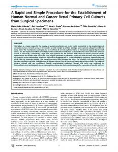
Additionally, infected cells were selected in FD medium containing 1 mg of G418 per ml and maintained in this medium at a reduced G418 concentration (200 ,g/ml). Genomic DNA isolation. Genomic DNA was isolated from cells by using a proteinase K-sodium dodecyl sulfate procedure essentially as described by Hughes et al. (15). PCR of genomic DNA. PCRs with 1 p,g of genomic DNA were performed essentially as described by Saiki (37). The primers used for amplification were RCFI (TTGGGGACTC TGCTGACCAC), which corresponds to the vector gag sequence approximately 80 bp 5' of the MCS, and RCR1 (CTTGCAAAACCACACTGCTCG), which corresponds to the MClneo sequence immediately adjacent to the 3' end of the MCS. The reactions were performed in a Perkin-Elmer Thermocycler for 35 cycles, and the cycling parameters were denaturation at 94°C for 1 min, annealing at 60°C for 2 min, and extension at 72°C, initially for 2.5 min and then increasing by 5 s per cycle. The reaction mixtures were denatured at 94°C for 4 min before cycling commenced, and a final 7-min extension was included after cycle 35. Southern blots of genomic DNA. Genomic DNA, digested with either BamHI or SacI, was fractionated on a 0.7% agarose gel, transferred to Hybond N+, UV cross-linked at 0.75 J/cm2, and probed with a 32P-labeled 1,090-bp XhoIDdeI neo fragment from pMClneo as recommended by the manufacturer. Southern blots of PCR-generated DNA. DNA from PCR of genomic DNA (see above) was fractionated on a 1.2% agarose gel and prepared for probing as described in the preceding section. Blots were probed as described by Mason and Williams (29) with 32P-labeled oligonucleotides specific for either IL-3 (GATAACGTATCTGTCCTCAGGATC) or GMCSF (ATCTfCAGGCGGGTCTGCACACATG). Northern (RNA) blots. Poly(A)+ RNA was isolated from factor-independent clones as described by Gonda et al. (13). A 1-,ug sample of this RNA was fractionated on a formaldehyde-agarose gel and blotted to a Hybond N membrane (Amersham) as specified by the manufacturer. The blot was dried, UV cross-linked in at 0.4 J/cm2, and probed with the neo probe described above for genomic DNA. RESULTS Outline and rationale of the protocol. An outline of the protocol is presented in Fig. 1. Briefly, it begins with the generation of cDNA from a source that is appropriate for the isolation of the gene(s) in question. The cDNA is directionally cloned into the retroviral vector (see below) and amplified in E. coli. The vector DNA thus obtained is used to generate a representative pool of virus-producing cells. This is done by first transfecting the library into an amphotropic packaging cell line ('crip or PA317) and then using the transiently generated virus (48 h posttransfection) to infect an ecotropic packaging cell line (T2). The infected ecotropic packaging cells are selected for the expression of a drug resistance gene (Neor) carried by the retroviral vector and are then used to infect a suitable target cell population. Target cells displaying the desired phenotype are isolated, and the gene is subsequently recovered by PCR from the retroviral DNA integrated in those cells. The retroviral vector that we have constructed (Fig. 2; pRUFneo) will be described in more detail elsewhere, but the salient features of the vector are (i) an MCS to allow directional cloning; (ii) the myeloproliferative sarcoma virus LTR, which is known to function well in hematopoietic cells (5, 40); (iii) the MClneo cassette containing the Neor gene
Directional cDNA synthesis Size selection Ligate into pRUFNeo and transform E. coli by electroporation Isolate DNA
Transfect amphotropic packaging cells Collect transient supernatant Infect ecotropic packaging cells Select with G418 Use pool to infect target cells
Functional screen
FIG. 1. Outline of the procedure used to generate cDNA expression library (see text for details).
a
retroviral
driven by the f9 polyomavirus enhancer (41) (MClneo was chosen in preference to tkneo because our preliminary experiments (data not shown) showed that it was efficiently expressed in a variety of cell types, including fibroblasts, primary hematopoietic cells [from fetal liver], and hematopoietic cell lines); and (iv) sequences from the rearranged gag-pol genes of the M3Neo(myb) provirus integrated in the U22.4 cell line described by Gonda et al. (12). This rearrangement resulted in increased expression of the myb gene carried by the provirus, and our experiments indicated that it functions similarly in the RUFneo vector. Expression of myb from the U22.4 provirus and of cDNAs inserted into the MCS of pRUFneo occurs via a subgenomic mRNA generated by splicing between the normal retroviral splice donor (at nucleotide 206) and a cryptic splice acceptor (at nucleotide 1353) described in references 11 and 12 (Fig. 2; see also Fig. 4B). In addition, the presence of gag sequences has been shown to substantially increase retroviral titers (1, 3). Generation of cDNA libraries. We electroporated the retroviral vector containing the cDNA into E. coli and grew the cells overnight on ampicillin plates to amplify the library. By this method, we were able to obtain a library of 1.5 x 10⁶ colonies from approximately 40 ng of LB3 cDNA, an efficiency of about 3.75 x 10⁷ clones per ,ug of cDNA. In other experiments, we also generated a bone marrow stromal cell library of 1.3 x 10⁶ clones from 130 ng of cDNA (44). Both libraries contained cDNAs ranging in size from 0.4 to 6 kb (data not shown). A major concern for the generation of cDNA libraries is the need for adequate representation, in the final library, of
GENERATION OF RETROVIRAL EXPRESSION LIBRARIES
VOL. 14, 1994
883
Polylinker 0
co
0
0
0
C4
ts 2
CV
~~~~~~CC,
~~~-
-
0
U9
la,
O CC,
1l)
~a
0
a -
-,
C)
;23X
-~~~~~~U %
J.
~AMPR ORI
'q
FIG. 2. The structure of the RUFneo retroviral plasmid showing landmark restriction endonuclease cleavage sites, the cloning sites in the polylinker (MCS), and other major features including the splice donor (SD) and splice acceptor (SA) sites used to generate the subgenomic mRNA (see text and Fig. 4B; see also Materials and Methods for details of construction). The nucleotide sequence numbers of the retroviral portions of the plasmid are derived from the sequence of Moloney murine leukemia virus (39).
all the genes expressed in the source. Estimates vary widely, but there are probably 30,000 to 120,000 different mRNA species present in the cytoplasm of a normal mammalian cell (4, 7, 36). In fact, adequate representation in a cDNA expression library is likely to require at least 1 order of magnitude more clones than this since synthesis of large, full-length cDNAs is relatively inefficient. Although it is fairly easy to generate libraries of this complexity as plasmids (as we have done [see above]) or phages in E. coli, the generation of similarly complex libraries in eukaryotic cells is generally more difficult. The protocol that we describe here is designed to circumvent this problem. The initial steps involve (i) transfection of the DNA obtained from amplification of the library (see above) into an amphotropic packaging cell line (PA317) and (ii) use of the transiently generated retrovirus (48 h posttransfection) to infect an ecotropic packaging cell line. Infection is a more desirable way to transfer genes into the cells that will constitute the final library of (ecotropic) virus-producing cells, since it has been shown to yield substantially higher viral titers from these cells (17, 26, 32). Moreover, infection generally results in a smaller number of proviral integrations per cell (i.e., low copy number), which means that each infected cell in the total pool represents a single (or at most a only a few) cDNA species in the library. In this manner, we have derived several populations (i.e., libraries) of virus-producing cells expressing a complement of retroviruses that should represent all the mRNA species present in the original cells from which the cDNA was derived. The results from several experiments suggest that the generation of sufficient numbers of virus-producing cells is dependent on at least two factors: the particular amphotropic cell line used to produce the transient retrovirus and the volume of virus-containing supernatant used to infect the ecotropic packaging cells (Table 1). In pilot experiments we found that the 'Ic' cell line generated approximately onequarter of the number of infectious units as did PA317 in the same experiment (data not shown, but compare Table 1, experiments 1 and 2), so we chose PA317 for our subsequent experiments. Furthermore, we determined the titer of the
transient supernatants obtained from the PA317 cells and established that under limiting-dilution assay conditions, titers in the vicinity of 1 x 10⁴ to 3 x 10⁴/ml could be obtained (results not shown). This suggested that a T2 library of a complexity of approximately 106 could be generated from 100 ml of viral supematant. However, when larger-scale experiments were performed, it appeared that the actual number of colonies obtained was strongly influenced by the volume of the supernatant used to carry out the infection. Table 1, experiment 3, shows that when 2 ml of supernatant was used to infect one dish of T2 cells, approximately 46,000 colonies were recovered whereas 8 ml of the same supernatant yielded only 32,000 colonies. This suggests that the infection frequency under the conditions used here is more a function of virus concentration than of the absolute number of infectious units. Table 1, experiments 4 and 5, shows the numbers of infected cells obtained during the generation of a retroviral library from a different source, TABLE 1. Numbers of clones derived by infection of P2 cells
with amphotropic supernatants
Expte
No. of dishes'
1 2 3 3 4 4 5
12 (T'm,) 10 3 (PA317) 1 (PA317) 1 (PA317) 1 (PA317) 12 (PA317)
(PA317)
Amt of
No. of clones/
(mI)C 10 10 8 2 10 2 2
dishd
Total no. of clones
3,270 7,000
39,200 70,000
superatant/dish
32,500 46,000 360,000 360,000 45,000
97,500 46,000 360,000 360,000 540,000
a Experiments 1 to 3 were carried out with the Lb-3 library, and experiment 4 was carried out with a stromal cell library. b Total number of dishes of l2 cells used in each experiment. The amphotropic cell line used to derive the transient supernatant is shown in
parentheses.
c Volume of transient amphotropic supematant used to infect each 10-cm petri dish of 12 cells. d Number of infected W2 clones derived from each dish; these were maintained as separate pools for subsequent use.
884
RAYNER AND GONDA
MOL. CELL. BIOL.
TABLE 2. Occurrence of factor-independent FDC-P1 cells following infection with the retroviral LB3 cDNA library
A
---
No factor GM-CSF +G418 GM-CSF
Ratio of factorindependent to total infected cells
0 0 17,500
T2Bpool b
AI2Epool b
15 6,167 19,417
18 7,875 15,500
11 2,000 5,624
1:411
1:437
1:182
a Infected FDC-P1 cells were plated in soft agar (as described in Materials and Methods), and colonies (>50 cells) were counted 1 week later. Numbers represent colonies per 50,000 cells plated. In practice, fewer cells were plated in GM-CSF than in the absence of factor to ensure that the cells could be counted. b The pools used contained approximately 32,500 independently infected %I2 clones and were those obtained from Table 1, experiment 3.
3.2kb
X.
Plating conditions
'T2Abpool
-
---
cDNA
No. of coloniesa generated following cocultivation with:
'IP2%2
-
N
B B
C L,
< C:
-