Sep 20, 2016 - Collie Miller, Larry Filipow, Stuart Jackson et al. A comparison of ... The effect of couch attenuation is incorporated in the more complex ...
Home
Search
Collections
Journals
About
Contact us
My IOPscience
A simple method for the correction of photon attenuation caused by patient couches in SPECT
This content has been downloaded from IOPscience. Please scroll down to see the full text. 1991 Phys. Med. Biol. 36 1693 (http://iopscience.iop.org/0031-9155/36/12/013) View the table of contents for this issue, or go to the journal homepage for more
Download details: IP Address: 185.58.164.42 This content was downloaded on 20/09/2016 at 10:13
Please note that terms and conditions apply.
You may also be interested in: Planar imaging quantification using 3D attenuation correction data and Monte Carlo simulated buildup factors Collie Miller, Larry Filipow, Stuart Jackson et al. A comparison of attenuation correction methods for quantitative single photon emission computed tomography S Webb, M A Flower, R J Ott et al. Improved cone-beam SPECT via an accurate correction for non-uniform photon attenuation A Welch, S Webb and M Flower Truncation artifact suppression in cone-beam radionuclide transmission CT using maximum likelihood techniques: evaluation with human subjects S H Manglos The effect of non-uniform attenuation compensation on myocardial SPECT defect analysis S H Manglos, F D Thomas and B J Hellwig Evaluation of attenuation corrections using Monte Carlo simulated lung SPECT Agnetha Gustafsson, Björn Bake, Lars Jacobsson et al.
Phys. Med. Eiol., 1991, Vol. 36, No 12, 1693-1697. Printed i n the U K
Technical note
A simple method for the correction of photon attenuation caused by patient couches in SPECT R T Staff and H G Gemmell Department of Eio-Medical Physics and Eio-Engineering, Grampian Health Board and the University of Aberdeen, Foresterhill, Aberdeen AB9 ZZD, U K Received 13 May 1991. i n final form I5 July 1991
1. Introduction
Single photon emission computed tomography (SPECT)has the potential for absolute quantitation (Murphy 1987) although there are still a number of factors which prevent this goal from being achieved. I n recent years several different methods of attenuation correction have been investigated (Budinger and Gullburg 1977, Murase 1987, Fleming 1990) but, understandably, these have been concerned with correcting for tissue attenuation within the patient. It has been pointed out, however, that the couch supporting the patient can produce significant distortion of the reconstructed image in the form of a drop-off in count density towards the couch (Khan and Ell 1982). This is due to additional photon attenuation in views acquired with the couch wholly or partly between the detector of the SPECT imager and the patient. The image distortion caused by the photon attenuation will be considerable in all SPECT images of the body, e.g., the liver, heart, lung and skeleton. It is likely that this factor will be less important in brain imaging when a head-rest, which is usually made from material of lower attenuation, is generally used. The effect of couch attenuation is incorporated in the more complex attenuation correction methods such as the iterative attenuation compensation technique proposed by Moore et a1 (1982) and the transmission attenuation correction technique proposed by Bailey et al (1987), but this paper describes a simple method of resolving the problem using the first order Chang attenuation correction method (Chang 1978) to create a standard array of correction factors to compensate for the patient couch. This technique is then tested on a phantom for two different SPECT systems and couches. 2. Materials and methods
2.1. Data acquisition Two different rotating gamma camera systems were used for data acquisition in this investigation, a Siemens Orbiter 7500 ZLC with a Maxdelta data processor and an ICE 400 ACT with a Link Analytical MAPS 5050 data processor. For both systems, images were collected using a 360 degree circular orbit to obtain 64 views in a 6 4 x 6 4 word array. Image reconstruction and attenuation correction for data acquired on both systems was carried out on the Maxdelta computer which included a digital Microvax 11. The patient couches investigated in this study were the standard ones supplied by Siemens and ICE for use with their SPECT gamma cameras. The two couches were quite different in construction. The Siemens couch was made of PVC foam sandwiched 003l-9155/9l/l21693+05$03.50 0 1991 IOP Publishing Ltd
1693
1694
K 7 SrafTnnd H G Gemmell
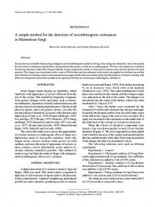
Figure I. Siemr.n\ couch. l a I l h e ('hang l ' # r \ r M r d u correction arr.i): I h l the Siemenr couch crosi~seclion: l c l a slice i h i o u s h the comscrim srr;ii \ho* inp rhr \ o r i i i i i o n o f the correction facrors.
hetween two aluminium sheets; the PVC foam was assumed to he air equivalent and, therefore, the bed was considered to he a hollow shell. T h e I C E couch was solid a n d made of carbon fibre. Their cross-sections are shown in figures l ( h ) a n d 2 ( h ) ,respectively. The effective attenuation coefficients ofthe couches were measured by supporting
Fixure 2. IGI- couch / t i l '111s('h;inR l i r r r ~ x d r rcomecriun nrril:: 1 h i i l i e IGF couch crosi~secrion: ( c l \Iicc through rhe c o r ~ c c i i o n.trr.'i 'hoiiin8 rhc i d r i a l i t m i o f c n r r ~ c t i o nfactors.
it
Correction ofphoton attenuation in
SPECT
1695
a source of 99Tcmat a fixed distance (30 cm) above the gamma camera face. The source was placed in a collimated container to produce 'narrow heam' conditions. The count rate was recorded with and without the couch between the source and the gamma camera and the effective attenuation coefficient calculated. A general purpose collimator was used for all data acquisition. All the projected images were uniformity corrected using the appropriate flood image. The phantom studies were carried out using a water filled Perspex elliptical phantom of major axis 33.3 cm and minor axis 27.4 cm into which approximately 500 MBq of 99Tcmwas introduced. This was placed on the patient couch in a central position. Images were reconstructed with a ramp Hanning filter and were corrected for tissue attenuation using a first order Chang correction with an effective attenuation coefficient of 0.095 cm-'. This value produced a uniform cross-sectional image of the elliptical phantom suspended in air, i.e., not supported by the couch.
2.2. The correction method The correction method consists of two parts. The first involves the calculation of an array of Chang correction factors to compensate for couch attenuation; this need only be done once for a particular combination of couch and radionuclide. Cross-sectional outlines for the couches used in this study, described above, were computed on the Microvax. Then, for every pixel exterior to the couch cross-section, the Chang correction factor was calculated using the following equation from Chang (1978): c(X,y)=
[
M
( 1 / ,I =~I
ex pi^,^,,)]
-,
(1)
where C ( x , y ) is the Chang correction factor for pixel ( x , y ) , M is the number of projections taken, U,, is the effective attenuation coefficient and le, is the path length in the attenuating medium in direction Bi from point (x, y ) . I n this way an array of Chang correction factors is produced for each couch as shown in figures l ( a ) and 2 ( . a ) . The second part of the correction method concerns the modification of the reconstructed images with this array of Chang correction factors. This involves spatially registering the couch correction array with the image to he modified, i.e., overlaying the correction array on the image array such that the outline of the couch is in its true spatial position in the reconstructed image. This is done by first locating the bottom edge of the reconstructed image of the phantom. Determination of the source boundary is required for source attenuation correction which then sets all pixels outside the boundary to zero. Establishing the position of the bottom edge of the image i s therefore simple. The spatial registration is accomplished by calculating the lowest point of this bottom edge and translating the correction array until it coincides with the lowest point on the upper edge of the couch outline. The same translation is applied to all other points in the array. This technique is successful because of the concave shape of both couches, i.e., the lowest point in the source will always be in the centre of the couch. After registration the correction process is a simple multiplication of the two arrays. The second p a n of the correction method can be summarized by the following equation: A'(;,j ) = A ( ; ,j ) C (i + x -x', j + y - y ' ) (2) where (x, y ) is the lowest point of the image to be corrected, (x', y ' ) is the lowest point of the top edge of the couch outline, A(i,j ) is the image array to be corrected, A'(i,j ) is the corrected image array and C ( i ,j ) is the array of Chang correction factors. These algorithms were written in Fortran on the Microvax.
R T Stuff and H G Gemmell
1696
-Lae 0
Figure 3. Phantom cross-sections. (ai A venical slice through rhe phanram imaged on the Siemens couch, before and after the correction technique has been applied: ( h ) a vertical slice through the phantom imaged on the IGE couch, before and after application of the correction technique.
3. Results
The effective attenuation coefficient was found to be 0.0223 mm-' for the Siemens couch and 0.00796 mm-' for the IGE couch. The distorting effects of couch attsniiathn czn be seen in fgures ? ( G ) and ( 6 ) A~ vertical section through the image of the phantom shows that for the Siemens system there was a 23.7% decrease in count density from the mean due fa couch attenuation and a 9.9% decrease for the IGE couch. Figures 3 ( u )and ( b ) also show vertical sections through the phantom images after correction. 4. Discussion
This method exploits the fact that the size and shape of the couch used with a particular SPECT imager remains constant and that its effective attenuation coefficient is easily measured. This approach has some similarities to that adopted by Fleming (1990) for correction of patient attenuation, but differs in that a standard correction array is used. The technique for locating the couch correction array relative to the reconstructed image was found to work effectivcly for both phantom and clinical studies. Of course. if the position of the couch relative to the centre of rotation was fixed for all studies, then no spatial registering of the two arrays would be required since the couch would always be in the same position relative to the reconstructed image. However, the flexibility of most modern tomographic gamma cameras, including the two used in this study, means that this is not usually the case and spatial registration is necessary. Initially, the computational requirements of this technique are similar to those required to perform a Chang correction algorithm, but, as has been pointed out, this need only be done once for each couch. The correction of image arrays is much less demanding, simply requiring the translation and multiplication of two arrays. These results show that the distortion due to couch attenuation is greater for the Siemens couch than for the one supplied with the IGE system, when imaging homogeneous phantoms. This study has also shown that when quantitative SPECT images are required, the distortion caused by couch attenuation must be considered.
Correction of photon attenuation in
SPECT
1691
In particular, it is inconsistent to correct for tissue attenuation hut to neglect the effect of couch attenuation. This effect is likely to be significant in SPECT imaging of the lungs, liver or skeleton. The main clinical use of quantitative SPECT at present is probably the analysis of myocardial perfusion images acquired using TI-201, but correction for couch attenuation is probably less important in these studies as many centres employ 180 degree, mainly antierior, acquisition (Hoffman (1982)). It is likely, however, that this investigation will he superseded by the new 99Tc" agents using 360 degree SPECT in the near future (Lahiri 1989) in which case couch attenuation correction will soon become important in cardiac imaging. Acknowledgments One of the authors (RTS) was supported by the Medical Research Council for part of this work. The authors would like to thank Professor P F Sharp for his helpful comments and criticisms of this manuscript. References Bailey D I, Hutlon B F and Walker P J 1987 Improved SPECT using simullaneous emission and transmission tomography J, Nuel. Med. 28 844-51 Budinger T F and Gullberg G T 1977 Transverse section reconstruction o f gamma-ray emitting radionuclides i n patients Reconstrucrion Tomography in Diagnorric Radiolog>, and Nuclear medicine ed M M TerPogosiian (Baltimore, MD: University Park Press) pp 315-42 Chang L T 1978 A method for attenuation correction in radionuclide computed tomography IEEE Trans. Nuel. Sci. NS-231) 638-9 Fleming J S 1990 Technique for the use of standard outlines for attenuation correction and quantification i n SECI N u d Med. C m m . I I 685.96 Hoffman E J 1982 180 degree compared with 360 degree sampling i n SPECT. Teaching editorial J. Nucl. Med. 23 745-7 Khan 0 and Ell P J 1982 Liver and Spleen computed emission tomography Computed Emkriun Tomography ed P J Ell and B L Holman (Oxford: Oxford Medical Publicationi) p 449 Lahiri A, Higley B, Kelly J D, Webbon P, Edwards B, Archer C, Crawley J C W and Raftery E B 1989 Myocardial perfusion imaging in man using new Tc-YYm functionalised Uiphosphine Complexes (Ab) Eur J. Nuel. Med. 15 p 425 Moore S C. Brunelle J A and Kirsh C M 1982 Qualitative multi-detector emission computerized tomography using iterative attenuation compensation J. Nucl. Med. 23 706-14 Murase K, ltoh H, Mogami H, lshine M, Kawamura M, lio A and Hamamolo K 1987 A comparative study ofattenuation corvxtioii algorithms in single photon emission tomography Eur. J. Nurl. Med. 13 55-62 Murphy P H 1987 Quantitative Emission Tomography (Editorial) J. N~tcl.Med. 28 922-33