U.P.B. Sci. Bull., Series B, Vol. 75, Iss. 1, 2013
ISSN 1454-2331
A SIMPLE METHOD FOR THE QUANTIFICATION OF ISOTHIOCYANATES FROM MUSTARD Melinda-Rita MARTON1, Vasile LAVRIC2 Lucrarea de faţă prezintă o metodă simplă de analiză pentru cuantificarea din muştar a iizotiocinaţilor din legumele cruciferoase. The present work describes a simple method for the analysis of iosthiocyanates from mustard in cruciferous vegetables.
Keywords: ITC, mustard, GC-MS. 1. Introduction Isothiocyanates (ITCs) received much attention over the past years especially because of their potent anticarcinogenic properties which were proved in in vitro models as well as in in vivo models. ITCs are the hydolysis products of glucosinolates (GLSs), secondary plant metabolites present in members of the Cruciferous plants, including the Brassica crops. From a chemical point of view, GLSs are substituted β-thioglucoside N-hydroxysulfates with a chemical structure of a β-D-glucopyranose residue linked via a sulfur atom to a (Z)-Nhydroximinosulfate ester, plus a variable group derived from amino acids. Based on the chemical structure of the precursor amino acid, GLSs can be classified into three categories: aliphatic GLSs derived from alanine, leucine, isoleucine, methionine and valine; aromatic GLSs derived from phenylalanine or tyrozine and indole GLSs from tryptophan [1]. Under normal physiological conditions GLSs are stable, the presence of the plants own enzyme, myrosinase is required to the hydrolysis. The myrosinase enzyme in the plant is separated from GLSs; it is stored in so called myrosinase cells. When the integrity of the plant is compromised (chewing, chopping) the enzyme and GLSs come into contact and the hydrolysis reaction takes place upon the cleavage of the glucosid bond. The reaction yields glucose and an unstable aglycone, thiohydroxamate-O-sulfonate. The aglycone depending on the reaction
1
PhD Student, Chemical Engineering Department, University POLITEHNICA of Bucharest, Romania, e-mail:
[email protected] 2 Prof., Chemical Engineering Department, University POLITEHNICA of Bucharest, Romania, email:
[email protected]
64
Melinda-Rita Marton, Vasile Lavric
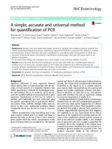
pH conditions undergoes spontaneous rearrangement and different end products are formed. At nearly neutral pH the reaction yields mostly ITCs (Fig.1.).
Fig. 1.Scheme of hydrolysis of GLS to ITC. GLSs under nearly neutral pH are enzymatically hydrolyzed by myrosinase to ITCs.
ITCs are characterized by the presence of the N=C=S group, which central carbon atom is highly electrophilic. The chemopreventive activity and their toxic effect as well probably can be attributed to this characteristic. The biological activities of the ITCs are mediated through the reaction of this carbon atom with nucleophilic reagents in the cells [2]. Although the presence of myrosinase enzyme is needed for the hydrolysis of GLSs into ITC, it was shown in many studies that the human intestinal microflora is capable of hydrolysing GLSs, even when the enzyme was destroyed by heat inactivation [3, 4]. It was shown in many studies that GLSs and the end products of the hydrolysis reaction have a role in the plant-insect interactions, for example GLSs can serve as general poison and deterrent for harmful insects [5]. ITCs are well known for their chemoprotective effects, the mechanisms behind these effects involve modulation of xenobiotic metabolism, protection from oxidative stress, inhibition of tumor growth, induction of apoptosis, inhibition of cell cycle. It was shown that ITCs also have anti-inflammatory properties through regulation of Nuclear factor-kappa B and its downstream signalling and inhibition of TNF-ά and lipopolysaccharide stimulated inflammatory response; anti-bacterial properties against Helicobacter pylori and cardioprotective effects [6, 7]. In the human body ITCs are metabolized by the mercapturic acid pathway and eliminated through the urine in the form of N-acetylcysteine (NAC)conjugates. As the research on the biological activity of the ITC advanced there was a growing need for accurate analytical methods for identification and quantification of ITCs and their different metabolites which are formed during the metabolism in the human body. Thin layer chromatography like analytical method for the quantitation of ITCs was used in the early years of ITC research when more
A simple method for the quantification of isothiocyanates from mustard
65
advanced methods were not available. Although its limitation, it made still possible to quantify ITC-NAC conjugates from urine samples [8, 9]. Gas chromatography was the most rarely used advanced analytical method for the measurement of ITCs, probably because of the limit of detection which can be achieved using this method. With a GC method the limit of detection is in the µg range, which does not allow direct quantification of ITC content from urine or blood samples with low ITC concentration. Although this limitation of the GC method was observed, it can be still used for the measurement of samples with high ITC content for example samples of vegetable origin [10, 11, 12]. The cyclocondensation assay was widely used for the quantification of total ITC content from various types of samples, it was first published by Zhang et al in 1992 and used for the quantification of ITCs in vegetables. The assay is based on the characteristic of ITCs to react quantitatively with an excess of vicinal dithiols, the end product of the reaction being a five-membered cyclic condensation product (cyclic thiocarbonyl) with a broad ultraviolet absorption band at 270 nm [13]. During the years the sensitivity of the method was greatly [14-17]. HPLC was the most frequently used analytical method used for the quantitation of ITCs and their derivates. Most of the methods were using RP-LC with C18 columns and aqueous mobile phases [11, 18- 28]. Lamy et al. [29] demonstrated that a single consumption of mustard, containing approximately 25 mg total ITC can strongly reduce cell sensitivity towards direct-acting genotoxins. They also showed that a three day human intervention with mustard significantly reduced the DNA damage and micronucleusformation induced by hydrogen peroxide or benzo(a)pyrene diolepoxide [30]. Based on our results the ITC concentration required for inducing the beneficial biological effects can be reached by consuming approximately three tablespoon of mustard. Mustard (Sinapis spp.) seeds contain high concentrations of ITC precursor compounds. During the preparation of mustard, the GLSs are hydrolysed into ITCs, so mustard contains already the released ITCs. Mustard is widely consumed in the world like a condiment and is a good dietary source of allyl-, benzyl- and phenethyl isothiocyanates (AITC, BITC and PEITC). Mustard consumption can have beneficial effects on human health. For the estimation of human bioavailability of ITC from mustard it is necessary to know the exact ITC concentrations in the mustard. The aim of this study was to establish a simple and rapid method for the quantificationof ITCs from mustard. Human bioavailability of ITCs from plant products is higher when the product contains the free ITCs and not the precursor compounds. If the plant was heat-treated and because of this reason the myrosinase is heat inactivated, the bioavailability is even lower. During the mustard preparation the seeds come into
66
Melinda-Rita Marton, Vasile Lavric
contact with water and the ITCs are released, so the bioavailability is maximal [30]. 2. Material and Methods 2.1 Chemicals and Standards ITC standards were purchased from Fluka (Germany), acetonitrile and methanol (HPLC degree) were obtained from J. T. Baker (Germany), n-hexane, butyl-benzene were purchased from Merck (Darmstadt, Germany). All solvents were of HPLC grade and water was of Milli-Q quality. 2.2 Equipment and Chromatographic Conditions for the determination of Isothiocyanates A Varian CP-3800 gas chromatograph equipped with a Varian 1200 Quadropule MS/MS was used for the analysis. The applied column was a Varian VF-5ms 0.25 mm x 30 m x 0.25 μm. The oven temperature was programmed from 50ºC for 5 min, for 110ºC with a rate increase of 5ºC/min and 300ºC with a rate increase of 20ºC/min for 3.5 min. The temperature of the ion source was set to 255ºC, for the transfer line at 270ºC. We used positive ion modus and an e-energy of 70 eV. The injection was splitless, the injector temperature was set to 250ºC, the volume of the injected sample was 1 µL. The flow rate of the helium carrier gas was 1.5 mL/min. Mass spectra were obtained by electron ionisation by Extended Dynamic Range (EDR) and quantification was made by selective ion monitoring of the 99 m/z fragment for AITC, 149 m/z for BITC, 163 m/z for PEITC and 134 m/z for the internal standard butyl-benzene. 2.3 Standard Solutions and Quantification Prior to analysis, standard solutions were prepared with n-hexane containing all three ITC. Three concentration ranges were analyzed using butylbenzene as internal standard (IS) for the calibration. The analyzed concentration ranges were the following: 10-100 µg/mL, 1-10 µg/mL and 0.1-1 µg/mL ITC solutions, each containing the IS at 50 µg/mL. The calibration curve was derived by three independent measurements, each conducted in triplicate and was linear (r2 = 0.99) over the analyzed concentrations ranges. 2.4 Sample Preparation 2 mL of mustard was extracted with 1 mL of n-hexan using a 15 mL plastic tube (conical bottom Cellstar PP tubes, 15 mL, Greiner bio-one, Germany).
A simple method for the quantification of isothiocyanates from mustard
67
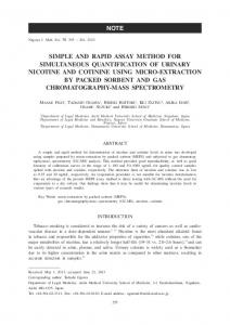
The mixture was stirred for 1 min at 2600 rpm and centrifuged at 5000 rpm for 5 min, at 20ºC. The eliminated supernatant was collected in glass vials. The nhexane extraction was repeated with the remained material, the two supernatants were combined and filtered trough a 0.45 µM syringe driven filter unit (Millipore, U.S.A.) and quantified with the described GC-MS method. 2.5 Data analysis Results were analysed using MS Excel. Data represent mean values ± standard deviation (SD) of three independent experiments, conducted in triplicate. 3. Results The calibration curve was linear over the analyzed concentrations ranges as shown in Fig.1. Calibration AITC (10 - 100 µg/mL)
Calibration BITC (10 - 100 µg/mL)
40
40
y = 0,3262x - 0,3835 2
Counts Quotient
20
y = 0,3254x - 1,0887
30
2
R = 0,9978
20
10
10
0
0 0
20 40 60 80 100 Analyte concentration (µg/mL)
0
120
20
40
60
Calibration PEITC (10 - 100 µg/mL)
y = 0,3506x - 1,1931 2
R = 0,9974
30
20
10
0 0
20
40
80
100
Analyte concentration (µg/mL)
40
Counts Quotient
Counts Quotient
R = 0,9994 30
60
80
100
120
Analyte concentration (µg/mL)
Fig. 1. Calibration curves for AITC, BITC and PEITC.
120
68
Melinda-Rita Marton, Vasile Lavric
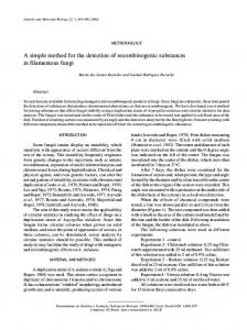
The limits of detection (LOD) and limit of quantification (LOQ) under the present chromatographic conditions were determined with the formula (SD/Slope)*3.3; (SD/Slope)*10, respectively. The LOD was 12.8 ng/mL (0.13 µM) for AITC, 27.5 ng/mL (0.185 µM) for BITC and 10.6 ng/mL (0.065 µM) for PEITC. The LOQ was 38.9 ng/mL (0.393 µM) for AITC, 83.4 ng/mL (0.56 µM) for BITC and 32 ng/mL (0.196 µM) for PEITC. The GC-MS chromatograms of the analyzed ITCs and of the ITSD are shown in Fig.2. The recorded retention times were in the range of 2 minutes for AITC, 14 minutes for the internal standard, 19 minutes for BITC and 21 minutes for PEITC. Because of the large time differences between the recorded retention times of the ITCs and internal standard the overlapping of the chromatographic peaks can be excluded.
.
Fig. 2. Chromatograms of AITC, ITSD, BITC and PEITC of 100 µg/mL standard solutions.
A simple method for the quantification of isothiocyanates from mustard
69
The measured ITC concentrations are shown in Table 1. As expected the highest concentration was detected for AITC, because mustard seeds are a very rich source of sinigrin, the precursor compound of AITC. The lowest concentration was recorded for BITC. The total ITC content was found to be 670.68 µg in 1 g mustard. In the total ITC content AITC has the higher concentration: 731.9 µg corresponding to 99.11 % of the total ITC content. BITC and PEITC were found in significantly lower concentrations: 0.07 µg BITC in 1 g mustard (0.009 % from to total ITC content) and 6.45µg PEITC in 1 g mustard (0.87 % from the total ITC content). Table 1 ITC concentration in mustard samples Nr. 1 Nr. 2 Nr. 3 725.31 762.92 707.47 Conc AITC (µg/mL) 0.074 0.077 0.071 Conc BITC (µg/mL) Conc PEITC (µg/mL) 6.47 7.09 6.99 2.09 2.09 1.95 Conc AITC (g/kg DW) 0.21 0.21 0.20 Conc BITC (mg/kg DW) Conc PEITC (mg/kg DW) 18.67 19.44 19.25
Mean 731.90 0.07 6.85 2.04 0.21
SD 28.31 0.00 0.33 0.08 0.01
RSD 3.87% 3.78% 4.87% 4.07% 4.35%
19.12
0.40
2.07%
4. Conclusions In the present paper a simple GC-MS method was used for the quantitation of ITCs from mustard samples. Determination of the exact concentration in ITC containing samples is essential for the assessment of bioavailability of ITCs in the human body. The advantages of the method presented in this paper are: no requirement for time consuming extraction method, time efficiency and simplicity. Acknowledgements Melinda-Rita Márton is partly funded by the Sectorial Operational Programme “Human Resources Development 2007-2013” of the Romanian Ministry of Labour, Family and Social Protection through the Financial Agreement POSDRU/88/1.5/S/60203
70
Melinda-Rita Marton, Vasile Lavric
REFERENCES [1]. B.A.Halkier and J.Gershenzon, “Biology and Biochemistry of Glucosinolates”, in Annual Review of Plant Biology, vol. 57, 2006, pp. 303–33 [2]. Y.Zhang, “Cancer-preventive isothiocyanates: measurement of human exposure and mechanism of action”, in Mutation Research, vol. 555, 2004, pp.173–190 [3]. C.Krul, C.Humblot, C.Philippe, M.Vermeuulen, M.van Nuenen, R.Havenaar, and S.Rabot, “Metabolism of sinigrin (2-propenyl glucosinolate) by the human colonic microflora in a dynamic in vitro large-intestinal model”, in Carcinogenesis, vol. 23, no. 6, 2002, pp. 10091016 [4]. L.Elfoul, S.Rabot, N.Khelifa, A.Quinsac, A.Duguay, and A.Rimbault, “Formation of allyl isothiocyanate from sinigrin in the digestive tractof rats monoassociated with a human colonic strainof Bacteroides thetaiotaomicron”, in FEMS Microbiology Letters, vol. 197, 2001, pp. 99-103 [5]. I.R.Redovnikov, T.Gliveti, K.Delonga, and J.Vorkapi-Fura, “Glucosinolates and their potential role in plant”, in Periodicum Biologicum, vol. 110, no. 4, 2008, pp. 297-309 [6]. Y.-S.Keuma, W.-S.Jeong, and A.N.Kong, “Chemoprevention by isothiocyanates and their underlying molecular signaling mechanisms”, in Mutation Research, vol. 555, 2004, pp. 191-202 [7]. Y.Nakamura, “Chemoprevention by Isothiocyanates:Molecular Basis of Apoptosis Induction”, in Food Factors for Health Promotion, Forum Nutrition Basel, Karger, vol. 61, 2009, pp. 170–181 [8]. B.D.Brüsewitz, B.D.Cameron, I.L.Chasseaud, K.Gorler, D.R.Hawkins, H.Kock, and W.H.Mennicke, “The Metabolism of Benzyl Isothiocyanate and its Cysteine Conjugate”, in Biochemical. Journal, vol. 162, 1977, pp. 99-107 [9]. W. H.Mennicke, K.Gorler, and G.Krumbiegel, “Metabolism of some naturally occurring isothiocyanates in the rat”, in Xenobiotica, vol. 13, 1983, pp. 203–207 [10]. G.P.Slater and J.F.Manwille, “Analysis of thiocyanates and isothiocyanates by ammonia chemical ionization gas chromatography-mass spectrometry and gas chromatographyFourier transform infrared spectroscopy”, in Journal of Chromatography, vol. 648, 1993, pp. 433-443 [11]. D.Jiao, C.-T.Ho, P.Foiles, and F.-LChung, “Identification and quantification of the Nacetylcysteine conjugate of allyl isothiocyanate in human urine after ingestion of mustard”, in Cancer Epidemiolgy, Biomarkers and Prevention, vol. 3, 1994, pp. 487-492 [12]. Y.Uematsu, K.Hirata, K.Suzuki, K.Iida, T.Ueta and K.Kamata, “Determination of isothiocyanates and related compounds in mustard extract and horseradish extract used as natural food additives”, in Shokuhin Eiseigaku Zasshi, vol. 43, no. 1, 2002, pp. 10-17 [13]. Y.Zhang, C.-G.Cho, G.H.Posner and P.Talalay, “Spectroscopic Quantitation of Organic Isothiocyanates by Cyclocondensation with Vicinal Dithiols”, in Analytical Biochemistry, vol. 205, 1992, pp. 100-107 [14]. Y.Zhang, K.L.Wade, T.Prestera, and P.Talalay, “Quantitative Determination of Isothiocyanates, Dithiocarbamates, Carbon Disulfide, and Related Thiocarbonyl Compounds by Cyclocondensation with 1,2-Benzenedithiol”, in Analytical Biochemistry, vol. 239, 1996, pp. 160–167
A simple method for the quantification of isothiocyanates from mustard
71
[15]. F.-L.Chung, D.Jiao, S.M.Getahun, M.C. Yu, “An urinary biomarker for uptake of dietary isothiocyanates in humans”, in Cancer Epidemiolgy, Biomarkers and Prevention, vol. 7, 1998, pp. 103-108 [16]. L.Liebes, C.C.Conaway, H.Hochster, S.Mendoza, S.S.Hecht, and J.Crowell, “Highperformance liquid chromatographybased determination of total isothiocyanate levels in human plasma: application to studies with 2-phenethyl isothiocyanate”, in Analytical Biochemistry, vol. 291, 2001, pp. 279–289 [17]. L.Ye, A.T.Dinkova-Kostova, K.L.Wade, Y.Zhang, T.A.Shapiro and P.Talalay, “Quantitative determination of dithiocarbamates in human plasma, serum, erythrocytes, and urine: pharmacokinetics of broccoli sprout isothiocyanates in humans”, in Clinica Chimica Acta, vol. 316, 2002, pp. 43–53 [18]. M.Bollard, S.Stribbling, S.Mitchell, and J.Caldwell, “The disposition of allyl isothiocyanate in the rat and mouse”, in Food and Chemical Toxicology, vol. 35, 1997, pp. 933–943 [19]. P.N.Maheshwari, D.W.Stanley, J.I. Gray and F.R. Van de Voort, “An HPLC method for simnultaneous Quantitation of individual isothiocyanates and oxazolidinethione in myrosinase digests of rapeseed meal”, in Jornal of the American oil chemists’ society, vol. 56, Sept. 1979, pp. 837-841 [20]. Y.M.Ioannou, L.T.Burra and H.B Mattews, “Ally1 Isothiocyanate: Comparative Disposition in Rats and Mice”, in Toxicology and Applied Pharmacology, vol.75, 1984, pp. 73-181 [21]. W.H.Mennicke, K.Gorler, G.Krumbiegel, D.Lorenz, and N.Rittmann, “Studies on the metabolism and excretion of benzyl isothiocyanate in man” in Xenobiotica, vol.18, 1988, pp. 441–447 [22]. K.I.Eklind, M.A.Morse and F.-L.Chung, “Distribution and metabolism of the natural anticarcinogen phenethyl isothiocyanate in A/J mice”, in Carcinogenesis, vol. 11, no. 11, 1990, pp. 2033-2036 [23]. F.-L.Chung, M.A.Morse, K.I.Eklind, and J.Lewis, “Quantitation of Human uptake of the anticarcinogen phenetyl isothiocyanate after a watercress meal”, in Cancer Epidemiology, Biomarkers and Prevention, vol. 11, no. 992, pp. 383-388 [24]. F.-L.Chung, D.Jiao, S. M.Getahun, and M.C.Yu, “A urinary biomarker for uptake of dietary isothiocyanates in humans”, in Cancer Epidemiology, Biomarkers and Prevention, vol. 7, 1998, pp. 103–108 [25]. Y.Ji, and M.E.Morris, “Determination of phenethyl isothiocyanate in human plasma and urine by ammonia derivatization and liquid chromatography-tandem mass spectrometry”, in Analytical Biochemistry, vol. 323, 2003, pp. 39–47 [26]. M.Vermeulen, H.J.M.van Rooijen and W.H.J.Vaes, “Analysis of isothiocyanate mercapturic acids in urine: a biomarker for cruciferous vegetable intake”, in Jornal of Agricultural and Food Chemistry, vol. 51, 2003, pp. 3554–3559 [27]. L.Song, J.J.Morrison, N.P.Botting and P.J.Thornalley, “Analysis of glucosinolates, isothiocyanates, and amine degradation products in vegetable extracts and blood plasma by LC–MS/MS”, in Analytical Biochemistry, vol. 347, 2005, pp. 234–243
72
Melinda-Rita Marton, Vasile Lavric
[28]. A.A.Al Janobi, R.F.Mithen, A.V.Gasper, P.N.Shaw, R.J Middleton and C.A.Ortori, “Quantitative measurement of sulforaphane, iberin, and their mercapturic acid pathway metabolites in human plasma and urine using liquid chromatography-tandem electrospray ionisation mass spectrometry”, in Journal of Chromatography B Analytical Technologies in the Biomedical and Life Sciences, vol. 844, 2006, pp. 223–234 [29]. E Lamy, S Schmitz, A Krumbein, V Mersch-Sundermann, “Isothiocyanate-containing mustard protects human cells against genotoxins in nitro and in vivo, in Mutation research, vol. 726, 2011, pp. 146-150. [30]. E Lamy, M Garcia-Käufer, J Prinzhorn, V Mersch-Sundermann, “Antigenotoxic action of isothiocyanate-containing mustard as determined by two cancer biomarkers in a human intervention trial”, in European Journal of Cancer Prevention, vol 21, no 4 , 2012, 400406.