Processing nematodes and other small biological specimens for electron micros- copy is simplified by the use of a chamber to contain the specimens dttring ...
A Simple Method of Processing Nematodes for Electron Microscopy ~ MICHAEL
A. McCLURE and L. J. STOWELL ~
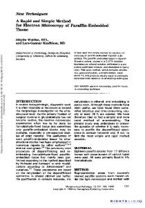
Processing nematodes and other small biological specimens for electron microscopy is simplified by the use of a chamber to contain the specimens dttring fixation, dehydration, and critical-point drying or impregnation with embedding resin. Beem capsules (1) and metallic tubing connectors (3) have been modified for the purpose, but those devices either permit very small specimens to escape or react adversely with fixatives (3). Flat chambers made of nylon mesh and glass tubing (2) depend upon diffusion through the mesh for exchange of solvents. We have constructed a chamber (Fig. IA) which overcomes those limitations. It is inexpensive and easy to assemble, and permits observation of the specimens during processing. Short lengths of glass tubing (4.5-mm inside diameter) are cut and the ends flared slightly with heat. Teflon tubing (4.8 mm outside diameter) covered with nylon mesh, pore size 10 /,m (Tobler, Ernst and Traber, Inc., 420 Saw Mill River Rd., Elmsford, N.Y. 10523) is then forced into the ttared ends. T h e chamber is loaded with specilnens by removing the seal at one end and inserting the other end into a rubber serum-bottle stopper to prevent drainage. After the chamber is filled with water or buffer solution, the specimens are introduced either by hand-picking or pipetting. Contamination can be reduced by handpicking. T h e open end is then sealed and the specimens are ready for processing. Liquid exchange and removal of air bubbles is facilitated by connecting one end of the chamber to a syringe by a long piece of teflon tubing (Fig. 1B). If the tubing is long enough, the chamber can be completely flushed without drawing liquids into tile barrel of the syringe. After fixation and dehydration, the long tubing can be removed and the entire chamber placed in a critical-point drying apparatus. Received for publication 15 March 1978. *Journal Paper No. 2839 of the Arizon~ Agricultural Experiment Station. aProfessor and Teaching Assistant respectivelT¢, l)epartment of Plant Pathology, University of'Arizona, Tucson, Arizona 85721.
376
Since the chamber is constructed only of glass, nylon, and teflon, it is compatible with most fixatives and solvents used in electron microscopy. Nematodes are fixed for scanning-electron microscopy by phmging the chamber into hot formalin:propionic acid (4) in a scintillation vial and rapid exchange of this material with the syringe. For transmission electron inicroscopy, nematodes are fixed in phosphate-buffered cold gluteraldehyde. Specimens can be held in fixative or other solutions by disconnecting tile long teflon tube and capping the vial. T h e y can be viewed (with some optical distortion) by placing the vial under a stereo-microscope. Postfixation with cold phosphate-buffered osmium tetroxide is accomplished in a similar fashion, which causes some darkening of the nylon without affecting its performance. Dehydration can be done with a graded series of ethanol or acetone, but ethanol is less hazardous. Both are miscible with Spurr's low-viscoscity embedding medium. Specilnens in acetone, however, can be critical-point dried without tile use of intermediate solvents. Dried specimens are removed from the chamber with a bristle mounted on a dissecting needle. The bristle is made slightly adhesive by rubbing it on the sticky side of plastic tape. Nematodes prepared by this method for scanning-electron microscopy show little distortion and are relatively free of adheringdebris (Fig. 1C). LITERATURE
CITED
I. DAY, J. W. 1974. A b e e m capsule c h a m b e r pipette for h a n d l i n g small s p e c i m e n s for e l e c t r o n microscopy. Stain T e e h n o l . 49:408410. 2. DeGRISSE, A. T . 1973. A m e t h o d fc~r p r e p a r i n g n e m a t o d e s a n d o t h e r soft tissues for s c a n n i n g electron microscopy. Meded Fac. L a n d blouwet. R i j k s n n i v . Gent. 38:1685-1702. 3. H O G G E R , C. H., a n d R. H. ESTEY. 1976. C h a m h e r for critical-point d r y i n g of n e m a todes anti o t h e r biological specimens. J. Nematol. 8:357-358. 4. N E T S C H E R , C., a n d J. ~,4,:. S E I N H O R S T . 1969. Propitmic acid better t h a n acetic acid for killing n e m a t o d e s . N e m a t o l o g i c a 15:286.
EM Technique: McClure, Stowell 377 i
-i LA
1
i t t FIG. 1. Chamber for processing nematodes. A) construction details; B) chamber attached to syringe; C)
Pralylenchus brachyurus processed in chamber by the critical-point method.