[Downloaded free from http://www.ijccm.org on Wednesday, September 28, 2016, IP: 109.152.41.98] Indian Journal of Critical Care Medicine April-Jun 2012 Vol 16 Issue 2
References 1. 2. 3.
Sodhi K, Single MK, Shrivastava A. Impact of advanced cardiac life support training program on the outcome of cardiopulmonary resuscitation in a tertiary care hospital. Indian J Crit Care Med 2011;15:209-12. Olasveengen TM, Vik E, Kuzovlev A, Sunde K. Effect of implementation of new resuscitation guidelines on quality of cardiopulmonary resuscitation and survival. Resuscitation 2009;80:407-11. Thiagarajan RR, Laussen PC, Rycus PT, Bartlett RH, Bratton SL. Extracorporeal Membrane Oxygenation to aid cardiopulmonary resuscitation in infants and children. Pediatr Cardiol 2007; 116:1693-700. Access this article online Quick Response Code:
Website: www.ijccm.org
DOI: 10.4103/0972-5229.99143
A simple method to prevent devastating complications Sir, We read with interest the letter to editor by Soni et al.[1] in the October – December 2011 issue, on the problem of air leak around endotracheal tube due to malposition of nasogastric tube (NGT) in the trachea. As an intensive care physician, we routinely place gastric tube via oral or nasal route blindly in ventilated or non-ventilated patients. It is common for the nasogastric or orogastric tube (NGT/OGT) to slide in the trachea, also by the side of endotracheal or tracheostomy tube.[2] Malpositioning of NGT/OGT feeding tube in the trachea results in devastating complications, which are usually preventable.[3] As NGT/OGT tube insertion is a routine procedure in ICUs, operation theatres, wards and emergency area, it does not seem practical to check for NGT/OGT position with fiberoptic bronchoscopy in all the patients. We are using a simple 4-step method for a long time now to prevent this avoidable complication: • Insert NGT/OGT with recommended method. • Check by auscultation of air insufflation with 20 ml syringe in the epigastrium. • Keep the proximal end open for one minute to allow injected air to come out. 118
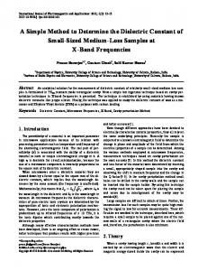
Figure 1: ICU nurse feeling for expiration over her cheek in spontaneously breathing patient
• Bring the proximal end of NGT/OGT close to the cheek and feel for the movement of air [Figure 1]. In spontaneously breathing patient movement of air will be felt on the cheek during expiration while in patients on mechanical ventilation or non-invasive ventilation continuous flow of air is felt on cheek. We always follow these simple steps while inserting NGT/OGT blindly, thereby avoiding the most devastating complications in a simple way. Moreover, if this final step is not fulfilled then we remove and reposition the NGT/OGT. Since, this is an usual practice in our department we have rarely encountered any complication or ventilator malfunction in our ICUs and wards during insertion of NGT/OGT.
Rajesh Chawla, Rakesh Sharma, Sudha Kansal Department of Critical Care Medicine, Indraprastha Apollo Hospital, New Delhi, India
Correspondence: Dr. Rakesh Sharma, Raj Niwas, 1-A, Swatanter Nagar, Narela, Delhi – 110 040, India. E-mail:
[email protected]
References 1. 2. 3.
Soni KD, Gupta B, Agrawal P, D’souza N, Sinha C. An uncommon cause of intraoperative airleak. Indian J Crit Care Med 2011;15:237. Zausig YA, Graf BM, Gust R. Occurrence of a pneumothorax secondary to malpositioned nasogastric tube: A case report. Minerva Anestesiol 2008;74:735-8. Metheny NA, Meert KL, Clouse RE. Complications related to feeding tube placement. Curr Opin Gastroenterol 2007;23:178-82. Access this article online Quick Response Code:
Website: www.ijccm.org
DOI: 10.4103/0972-5229.99144