A simple spot screening test for galactosemia. J. Lab. Clin. NIed. 68:137,. 1966. For personal use only. on September 18, 2017. by guest www.bloodjournal.org.
From www.bloodjournal.org by guest on September 18, 2017. For personal use only.
A Simple
Spot
Red By
Screening
Cell
JEAN-CLAUDE
Test
KAPLAN,
rapid
spot
ANNE-MARIE
screening
diaphorase tilted ture
blood freshly
reagents. filter
Spots
are
and
ONGENITAL deficiency a relatively rare diagnosis of this of the
for
NICOLAS,
mixstock
made
at intervals for
estimation
cause
they
of
can
be
method of Hegesh,4 a relatively fresh
although preparation
prompted
us
to
described
for
fast
capacity
performed
by
measuring
or
of
\Vhole
In
the
the
blood
NADH,
NAD are
washings
of
an
easy
It is based
on
From
the
Center,
itut
Inst
February
Supported
in
man, of
330
rate
of
.
cells;2
the The
reduction
of
The methemoglobin time-consuming
he-
red
The
nitrite-treated
for
rapid
ultraviolet
cells.
diagnosis
spot
red
cell
test
of
NADH-
prmciple
enzyme
already
deficiencies.#{176}’7
METHODS
containing
dye
illuminated
by
Moleculaire,
1970;
a hemolyzing is reduced long
agent, by
France,
NADII
NADH.
wavelength
Paris,
de of
lIE
M.D.: Paris,
from
Associate
Paris,
20, the
ultraviolet
and
and
During
the
the light,
City
of
DCIP. reaction is
trans-
Medical
Hope
France.
Southern
of
Hope
Professor
of
Medical
NicoLAs:
ALENA
Biochemistry, Research
HANZLICKOVA-LEROUX:
Medical
California,
1970.
N1H.
ANNE-MARIE
Mol#{233}culaire, Paris, City
of
March
07449
France.
Pathologic
Medicine,
University
accepted
Grant
Mol#{233}culaire, institut
Medicine,
23, by
KAPLAN,
Division
the
red hemolysates.
specific, requires as a substrate hemoglobin. These drawbacks
other
the
when
Mol#{233}culaire,
Pathologic
INSERM
a mixture
de Pathologic
part
JEAN-CLAUDE
Pathologic
in
Calif.
Submitted
de
to
fluoresces
Duarte,
heterozy-
methemodobin-ferrocyanide
the
AND
nitrited
itself
( NADH ) tedious and
method the
various
of NADH-diaphorase,
which
of
Test
is added
presence
the
reduced of Scott
MATERIALS
Principle
of
simpler and more of enzyme-free
of
six defi-
correctly identibut it is not
detection
in intact activity
detection
of
to NADH-diaphorase rediictase deficiency” ) is by lifelong cyanosis.’ The several methods : the explora-
enzyme
devise
deficiency.
the
All
diaphorase
due
( DCIP)3
repeated
diaphorase
for
the
the presence of and the method
is oxidized. NADH
gotes.
reduction
require
have
suitable
methemoglobin characterized established by
disorder may be
dichiorophenol-indophenol in test
NADH with
METHEMOGLOBINEMIA clinical disorder
HANZLICKOVA-LEROUX
ciency studied have been fled using this technique,
on
( “NADH-dependent
determination
complex4’5 reduction
as
patients
defluores-
methemoglobin
quantitative
ALENA
cence
to a reaction from stable
of
BEUTLER
NADHNi-
Detection
Deficiency
ERNEST
described.
examined
C latter
is
is added prepared
paper
tion
test
deficiency
Fast
NADH-Diaphorase
AND
A
for
France.
ERNEST
Center, Los
BLooc,
Angeles,
VOL.
Calif.,
de
Institut
Research
Assistant
BEUTLER,
Duarte,
Institut
Technician,
M.D.: Clinical
ChairProfessor
Calif.
36,
No.
3
(SEPTEMBER),
1970
From www.bloodjournal.org by guest on September 18, 2017. For personal use only.
SCREENING
TEST
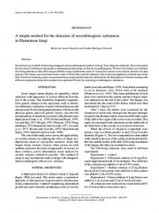
Fig.
1.-UV
without
blood
DCIP.
3.
FOR
formed blood
Normal
2. Nitrited
blood blood
NADH-DIAPHORASE
without plus
NAD,
which
is not
with
sodium
nitrite
effect
upon
normal
normal
nitrite
complete
plus
plus
reaction
1. Reaction
reaction
mixture
mixture
mixture
without
without
DCIP.
4.
mixture.
under
oxidizes
blood.
blood
reaction
fluorescent
331
DEFICIENCY
NADH-diaphorase:
into
the
such
conditions.
hemoglobin,
Pretreatment
thus
preventing
of
its
the
direct
re-
DCIP.
Procedure
Five added
gil. of to
sample
ml.
blood
ml.
a freshly
0.1
of
is allowed
whole of
and
NADII
(Sigma).
10
of 10
examined
test
of
a drop
is
wave
a long
30
NADH,
on
with
a
jd.
of
cent
saponin
mM
DCIP
and
0.27
is freshly
to
blood
spotted
Twenty
Whatman
No.
of
0.2
sodium
Stal)le
mM
sodium
0.5
mixture
1 filter
nitrited
from
vial
reaction
the
and mM
0.27
preweighed
the
the
prepared
containing
are
mixed,
1 per
buffer
mg./ml.)
the
solution
thoroughly
of
mixture
Tris-HCI
aqueous
being
minutes. ml.
0.19
This
NI
ultraviolet
After
0.C4
7.6.
0.06
mM DCIP (6.25 tube containing
minutes,
under
mM
pH
ml.
for
nitrite
sodium
blood.
containing
0.70
buffer, 1.0
19
The
at 37#{176}C. Every
tube
)
( 1.24% whole
temperature
test
containing
adding
NI
ACD
room
a
NI Tris-HCI by
EDTA
at to
mixture
solutions
0.18 or
stand
added
a 0.06
in
.tock
prepared
heparinized
to
are
a reaction
EDTA
are
for added).
sample
ducing
Test
CELL
spot-test (nitrite
nitrited
Normal
RED
mg.
dry
is incubated
paper.
Dry
spots
lamp. RESULTS
Normal
is
blood
omitted,
or
hemolyzed reaction
mLxture, of
the
fluorescence
red
the
blood
a delay cells
defluoresces
in
in
cells
1’4luorescence
are
is heated there
at
is no
the
the
suspended time
30
no
56#{176}Cfor time.
Plasma
mixture. same
in various is
than
blood,
defluorescence.
reaction
gives
less
of
defluorescence of
saline
in
absence
proportional
result
hours
1). being
The
omission does
suspension unwashed
of plasma dilution
When
is
before
alone
A
the
(Fig.
defluorescence
as
amounts to
2
minutes
added
of not
of
DCIP
observed.
If to
nitrite
results
produce
any
washed
nitrited
nitrited whole
or
saline,
factor.
The
the same
the in dered
blood.
increased propor-
If
From www.bloodjournal.org by guest on September 18, 2017. For personal use only.
332
KAPLAN
Fig.
2.-UV
perimental
plus
spot
the
for see
blood.
3 and
normal
tionality
test
conditions,
is observed
usefulness
diaphorase
than
defluorescence
the
other
clearly
for
of the
by
abnormal
less was
It
defluorescence
red
cell
subjects
tively,
is
time
gave
high
normal
cent.
hematocrit
reduction
This
spot
be
was
than
10 per to be
done
from
not
do
in
the
significantly
as compared
to
In
cent
did
24
per
an
hour,
On give
procedure
without infants
2). not
this
spite of blood. The and
subjects,
(Fig.
subjects
young
to six
it
NADH-
these
in
normal
11 per
removing
appropriate
cell
increased
samples, few
increased
counts,
normal.
recommend
to 70 blood that
reticulocyte
not
an
red
number
to prolong to normal
by applying
heterozygous
we
state. applied
of
influences cell
by
in
Their
considerably
two
red
either
cells
verified
cent
(For exmixture
value of
contributing the PCV
packed
test
found
noteworthy was
can
the
Therefore
NADH-diaphorase with
per
samples
results.
results.
The
combined
AL.
blood.
methemoglobinemia.
detection of the carrier The technique has been
positive
the
and normal blood. 2 and 4. Reaction
deficient
hemolysates.
ultraviolet
was blood
plus
suspending
congenital time
hand,
35
or
activity
the
deficient alone.
effect are additive, both We recommend readjusting
time.
with
mixture
mixture
because
the excess of plasma amount of saline.
The
Reaction
diluted
time
and hemoglobin-quenching the defluorescence whenever it is lower
diaphorase:
1.
5. Reaction
with
defluorescence
homozygotes
NADH
text).
ET
any
false
tested,
the
their blood
lowered of two
cent,
respec-
results. DISCuSSION
Since readily test
the
whole
available a simple
procedure equipment
and
fast
method
can and for
be
carried
reagents, diagnosis
out we of
within consider
the
NADH-diaphorase
using
only
fluorescent deficiency.
spot
From www.bloodjournal.org by guest on September 18, 2017. For personal use only.
TEST
SCREENING
The
FOR
stability
of
the average NADH-diaphorase prepared,
the
are
for
is only deficiency.
this
can
be
for
many
stable
simply
it it
can quickly should be
cyanotic
the
of
as
in
in
a
the
an for
well
as
particular
importance,
to make solution
few
the must
state
and
the occasion presents itself.
of the
can
for
since
diagnosis of be freshly
All
minutes.
frozen
NADH when deficiency
provide valuable
infants,
is of
called upon the nitrite
months
333
DEFICIENCY
test
rarely Only
accomplished
added to a preweighed vial examination for NADH-diaphorase
Since samples,
NADH-DIAPHORASE
reagents
laboratory
and
solutions
CELL
RED
with
be
performing
all or none answer on capillary screening methemoglobineniic,
adults
other
merely
an
blood even
or
methemoglobinemia.
REFERENCES 1. Jaff, globinemia
E. R., and Heller, P.: in man. In: Moore,
Brown, E. B. (Eds.) Progress ogy, Vol. IV. New York, Grune 1964,
p.
2. in
The
reduction
erythrocytes
of
of
a patient
methemoglobinemia,
subjects
glucose-6-phosphate
deficiency
and
21:561, 3. of
in
Hematol-
human 1960.
4.
Hegesh,
5.
Scott,
72:339, E.
with
globin
reductase.
congenital erythro-
individuals.
for determining (NADH-methein erythrocytes. J. Lab. 1968.
M.:
A
NI.:
The
relation
erythrocytes
to
J.
Clin.
6. Beutler, procedures
of diaphorase Invest.
Clin. E.:
for
of 39:
ficiency
and
ficiency.
Blood
7. screening
E.,
Calmanovici,
N.,
of
two
DPNH-methemo-
Chim.
Acta,
23:49,
1969.
Blood
inheritance
comparison
determining
A series
and
NIed.
-,
and test
68:137,
of new
pyruvate
glucose-6-phosphate
E.
methemoglobinemia. 1176,
Med. of
with
method reductase
reductase)
Clin. methods
1963. Scott,
New
methemoglobin
dehydrogenase
normal
Avron, M.: fern-hemoglobin moglobin
& Stratton,
48.
-:
cyte
MethemoC. V., and
kinase
screening deficiency,
dehydrogenase glutathione 28:553, Baluda, for
de-
1966.
NI. C.:
galactosemia.
1966.
de-
reductase A
simple
spot
J. Lab.
Clin.
From www.bloodjournal.org by guest on September 18, 2017. For personal use only.
1970 36: 330-333
A Simple Spot Screening Test for Fast Detection of Red Cell NADH-Diaphorase Deficiency JEAN-CLAUDE KAPLAN, ANNE-MARIE NICOLAS, ALENA HANZLICKOVA-LEROUX and ERNEST BEUTLER
Updated information and services can be found at: http://www.bloodjournal.org/content/36/3/330.full.html Articles on similar topics can be found in the following Blood collections Information about reproducing this article in parts or in its entirety may be found online at: http://www.bloodjournal.org/site/misc/rights.xhtml#repub_requests Information about ordering reprints may be found online at: http://www.bloodjournal.org/site/misc/rights.xhtml#reprints Information about subscriptions and ASH membership may be found online at: http://www.bloodjournal.org/site/subscriptions/index.xhtml
Blood (print ISSN 0006-4971, online ISSN 1528-0020), is published weekly by the American Society of Hematology, 2021 L St, NW, Suite 900, Washington DC 20036. Copyright 2011 by The American Society of Hematology; all rights reserved.