technical section
A simple technique to help avoid varus malreduction of reverse oblique proximal femoral fractures DJ Westacott, S Bhattacharaya Heart of England NHS Foundation Trust, UK CORRESPONDENCE TO Daniel Westacott, E:
[email protected]
BACKGROUND
Clinical guidelines from the National Institute for Health and Clinical Excellence recommend the use of an intramedullary nail for the treatment of subtrochanteric fractures.1 Reverse oblique fractures are associated with varus malunion,2 which defunctions the abductors by placing them at a mechanical disadvantage and increases the lever arm across the fracture. Most modern nails have been designed with a lateral bend to accommodate a trochanteric entry point. This simple technique helps to avoid varus malreduction without the need for excessive traction or open reduction.
Figure 2 A more medial entry point has corrected the neck shaft angle but some lateral translation of the proximal fragment has occurred.
References 1. National Clinical Guideline Centre. Hip Fracture. London: NICE; 2011. 2. Shukla S, Johnston P, Ahmad MA et al. Outcome of traumatic subtrochanteric femoral fractures fixed using cephalo-medullary nails. Injury 2007; 38: 1,286–1,293.
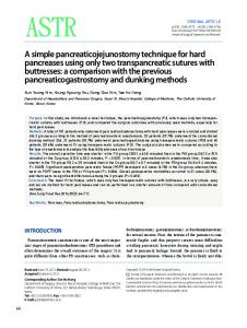
Figure 1 Reverse oblique fractures are prone to varus angulation (A). A medialised entry point (B) corrects the varus when the nail engages the distal fragment (C).
TECHNIQUE
A routine preparation and approach to the greater trochanter is performed. The entry point is positioned medial to the tip of the trochanter. The cortex is breached and the guidewire passed in a direction aiming slightly from medial to lateral (Fig 1). This means that when the nail is inserted, it will engage with the lateral cortex of the distal fragment and rotate the proximal fragment into valgus, closing the fracture gap and restoring anatomy (Fig 2). DISCUSSION
Anatomical closed reduction of a reverse oblique fracture is difficult due to the pull of the abductors. Abducting the leg is often attempted to restore the normal neck shaft angle but this makes access for the procedure more difficult. We describe a simple alteration of surgical technique that can prove very useful in preventing varus malunion, improving functional outcome and speed of union. Readers should be aware that medialising the entry point without angulating the direction of entry leaves the potential for lateral translation, rather than rotation, of the proximal fragment (Fig 2).
74
Preventing tibial and talar component contact during implantation of a total ankle replacement AJ Roche, JD Calder Chelsea and Westminster Hospital NHS Foundation Trust, UK CORRESPONDENCE TO Andrew Roche, E:
[email protected] BACKGROUND
The MOBILITY™ ankle replacement (DePuy, Leeds, UK) is the most frequently used implant in the UK.1 After bony preparation of articulating surfaces, the arthroplasty implants must be manoeuvred carefully into definitive position while preventing inadvertent scratching of bearing surfaces that could lead to wear and early failure.2,3 We describe an easy way of protecting the talar component while implanting the tibial component. TECHNIQUE
We use the anterior approach to the ankle.4 Once the correct components are chosen following trial insertions, they are removed from their sterile packaging. The talus is implanted first as per the manu-
Ann R Coll Surg Engl 2013; 95: 73–81
Technical Section January 2013 95_1.indd 74
04/12/2012 13:31:03
technical section
determine whether the talus component displaces during tibial implantation. This is a simple and safe method to carefully insert the tibial component of a commonly used ankle replacement.
References 1. National Joint Registry for England and Wales. 8th Annual Report 2011. Hemel Hempstead: NJR; 2011. 2. Alaia MJ, Dayan AJ. Catastrophic failure of a metal-on-metal total hip arthroplasty secondary to metal inlay dissociation. J Arthroplasty 2011; 26: 976.e1–976.e5. 3. Chang CB, Yoo JJ, Song WS et al. Transfer of metallic debris from the metal surface of an acetabular cup to artificial femoral heads by scraping: comparison between alumina and cobalt-chrome heads. J Biomed Mater Res B Appl Biomater 2008; 85: 204–209. 4. Wood PL, Karski MT, Watmough P. Total ankle replacement: the results of 100 Mobility total ankle replacements. J Bone Joint Surg Br 2010; 92: 958–962.
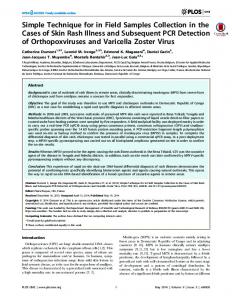
Figure 1 The sterile packaging accompanying the components can be safely cut to size and used to cover the talus component.
facturer’s technique. We use the thin plastic covering accompanying the sterile tibial implant (Fig 1) to protect the talus during tibial insertion. The dome-shaped plastic covering can be trimmed to a smaller talar-sized piece to cover the talus, leaving an unobstructed view during tibial component insertion in the standard fashion (Fig 2). With both implants seated, the plastic can be exchanged for the implant bearing. DISCUSSION
The trial bearing can be used to protect the talus, as suggested by the manufacturer. However, this could dislodge or partially obstruct tibial component insertion, potentially increasing the risk of contact between metal bearings. The plastic covering we use is partially transparent, which can assist the surgeon in viewing the talus and
Figure 2 The cut plastic packaging is a perfect contour to fit neatly over the talus component without risking abrasive damage to the tibia.
Simple method of fluid resuscitation in patients requiring emergency thoracotomy through direct cardiac cannulation M Zakkar, I Hunt St George’s Healthcare NHS Trust, UK CORRESPONDENCE TO Mustafa Zakkar, E:
[email protected] Patients who are critically injured with imminent cardiac arrest may require immediate thoracotomy as an integral component of their initial resuscitation in the emergency department. Fluid resuscitation can be difficult owing to a shutdown state. A simple method for fluid delivery in patients requiring emergency department clamshell thoracotomy entails the insertion of a large bore venous catheter directly into the right atrium at its appendage and using it for fluid resuscitation (Fig 1). This method is quick, safe and easily reproducible. Once the patient is stable, the catheter can be removed and a simple purse string suture used to close the atrium.
Figure 1 Clamshell thoracotomy with a large bore venous catheter in the right atrium (arrow)
Ann R Coll Surg Engl 2013; 95: 73–81
Technical Section January 2013 95_1.indd 75
75
04/12/2012 13:31:05