King, 1970, for a complete description of egg chamber formation and the ensuing 14 stages of egg chamber matura- tion). At stage 8, transported mRNAs are ...
Development 121, 3809-3818 (1995) Printed in Great Britain © The Company of Biologists Limited 1995 DEV5009
3809
A small predicted stem-loop structure mediates oocyte localization of
Drosophila K10 mRNA Thomas L. Serano1,2,† and Robert S. Cohen1,* 1Department 2Department
of Biochemistry, University of Kansas, Lawrence, KS 66045, USA of Biochemistry and Molecular Biophysics, Columbia University College of Physicians and Surgeons, 630 West 168th Street, New York, NY 10032 USA *Author for correspondence †Present address: Department of Molecular and Cell Biology, Howard Hughes Medical Institute, University of California, Berkeley, CA 94720, USA
SUMMARY The establishment of dorsoventral polarity in the Drosophila oocyte and future embryo is dependent on the efficient transport of K10 mRNA from nurse cells into the oocyte. To investigate the cis-requirements of K10 mRNA transport, we used a transgenic fly assay to analyze the expression patterns of a series of K10 deletion variants. Such studies identify a 44 nucleotide sequence within the K10 3′ untranslated region that is required and sufficient for K10 mRNA transport and subsequent localization to the oocyte’s anterior cortex. An inspection of the 44 nucleotide transport/localization sequence (TLS) reveals a strong potential for the formation of a stem-loop secondary structure. Nucleotide substitutions that interfere with the predicted base-pairing of the TLS block mRNA transport and anterior localization. Conversely, mutations that alter the base composition of the TLS while maintaining predicted base-pairing do not block mRNA transport or anterior localization. We conclude that K10 mRNA transport and anterior localization is mediated by a 44
nucleotide stem-loop structure. A similar putative stemloop structure is found in the 3′ untranslated region of the Drosophila orb mRNA, suggesting that the same factors mediate the transport and anterior localization of both K10 and orb mRNAs. Apart from orb, the K10 TLS is not found in any other localized mRNA, raising the possibility that the transport and localization of other mRNAs, e.g., bicoid, oskar and gurken, are mediated by novel sets of cis- and trans-acting factors. Moreover, we find that the K10 TLS overrides the activity of oskar cis-regulatory elements that mediate the late stage movement of the mRNA to the posterior pole. We propose the existence of a family of cisregulatory elements that mediate mRNA transport into the oocyte, only some of which are compatible with the elements that mediate late stage movements.
INTRODUCTION
Lipshitz, 1993). The localization of these mRNAs is a dynamic process that begins with mRNA synthesis in nurse cells and transport to the oocyte’s posterior pole. mRNA synthesis and transport are tightly coupled events inasmuch as mRNAs do not accumulate in nurse cells to detectable amounts. In most cases, mRNA synthesis and transport extend from just prior to egg chamber formation though ~stage 7 of oogenesis (see King, 1970, for a complete description of egg chamber formation and the ensuing 14 stages of egg chamber maturation). At stage 8, transported mRNAs are moved from the oocyte’s posterior pole to the oocyte’s anterior cortex, where mRNA-specific sorting events occur. For example, oskar (osk) and gurken (grk) mRNAs are moved to the oocyte’s posterior pole and anterodorsal corner, respectively, during late stage 8/early stage 9 (Kim-Ha et al., 1991; Ephrussi et al., 1991; Neuman-Silberberg and Schüpbach, 1993). Other mRNAs, e.g., fs(1)K10 (K10), bicoid (bcd) and orb, remain at the oocyte’s anterior cortex until stage 10B or later (Cheung et al., 1992; Berleth et al., 1988; Lantz et al., 1992).
Examples of mRNA localization as a means for protein targeting have increased greatly in recent years (reviewed in Macdonald, 1992; Ding and Lipshitz, 1993; St Johnston, 1995). It is a particularly common mechanism in developmental systems, where it is often desirable to store positional information in an inactive form. It is also widely used in large cells, e.g., eggs and neurons, possibly because it is more energy efficient to localize a relatively small number of mRNAs and translate them many times than it is to independently localize a large number of protein molecules. Despite its widespread occurrence and biological importance, the mechanism of mRNA localization is poorly understood compared to our understanding of other protein targeting mechanisms (e.g., see Pelham and Munro, 1993). The richest identified source of localized mRNA is the Drosophila oocyte, in which over a dozen localized mRNAs have been found (reviewed in Macdonald, 1992; Ding and
Key words: Drosophila K10 gene, mRNA localization, mRNA transport, Drosophila oogenesis, RNA secondary structure
3810 T. L. Serano and R. S. Cohen Pharmacological studies indicate that mRNA transport and subsequent localization events within the oocyte are mediated by microtubules (Pokrywka and Stephenson, 1991, 1995; Theurkauf et al., 1993). Each nurse cell-oocyte complex is connected by a single microtubule array (Theurkauf et al., 1992). Through stage 6, the array’s organizing center (minusend) is positioned at the oocyte’s posterior pole, and its plusends extend into nurse cells. During stages 6-8, this array is dismantled and a new one, nucleated along the oocyte’s anterior cortex and terminated at the oocyte’s posterior pole, is formed (Theurkauf et al., 1992). The dynamics and polarity of microtubules suggest that mRNA transport and anterior localization are both powered by a minus end-directed motor, e.g., a member of the dynein superfamily, while late stage (i.e., stage 9) movement of certain mRNAs to the oocyte’s posterior pole is powered by a plus-end, kinesin-like, motor. Consistent with this idea, kinesin’s motor subunit, when fused to β-galactosidase (β-gal), targets β-gal enzyme activity to the posterior pole of stage 9 and older oocytes (Clark et al., 1994). The observed overlap between the localization patterns of different Drosophila mRNAs is suggestive of a modular design of cis-regulatory elements. Specifically, one might expect all localized mRNAs to contain an element that mediates association with a minus-end motor. Some mRNAs, like osk, that move to the oocyte’s posterior pole at stage 9, might be expected to also contain an element that mediates association with a plus-end motor. Additional elements that mediate the association of mRNAs to non-microtubule components of the cytoskeleton might be required to maintain localization of some mRNAs, particularly those that remain localized during cytoplasmic streaming stages (i.e., stages 11-12), when microtubules are reorganized yet again (Theurkauf et al., 1992). In direct support of a modular design of mRNA localization elements, Kim-Ha et al. (1993) have identified distinct elements within the osk 3′ untranslated region (3′UTR) for mRNA transport and for movement to the oocyte’s posterior pole. Distinct regulatory elements have also been identified in the bcd 3′UTR; the 53 nucleotide BLE1 mediates bcd mRNA transport and localization to the oocyte’s anterior cortex, while three non-contiguous sequences mediate late-stage staufendependent localization to the oocyte’s anterior cytoplasm (Macdonald et al., 1993; Ferrandon et al., 1994). In the most streamlined version of a modular regulatory logic for mRNA localization, all similar localization steps would be mediated by the same set of cis- and trans-regulatory factors. However, this is apparently not the case. For example, bcd mRNA transport, but not the transport of other examined mRNAs, is mediated by exuperantia gene activity (Berleth et al., 1988; St Johnston et al., 1989). Furthermore, there are no obvious sequence homologies between the localization control regions of similarly localized mRNAs. The lack of such sequence homology could, however, simply reflect the physical nature of mRNA localization elements. For example, they may specify RNA secondary structures, which could be difficult to detect by conventional searching algorithms – both because many different sequence combinations can specify a particular structure and because a single structure could be composed of widely separated sequences. Thus, the degree to which the similar movements of different mRNAs are controlled by the same elements remains unknown. In cases where different mRNAs do use different elements for similar
movements, it is of interest to know if the different elements have evolved for trivial or functionally significant reasons. In this paper, we identify and characterize the cis-regulatory sequences of K10 mRNA localization. K10 mRNA is synthesized in nurse cells, transported to the oocyte’s posterior pole and moved to the oocyte’s anterior cortex at stage 8, where it persists through stage 10B (Cheung et al., 1992). Consistent with K10’s relatively simple mRNA localization pattern, we find that a single 44 nucleotide sequence located within the K10 3′UTR is necessary and sufficient for all K10 mRNA localization steps. The activity of this sequence, called the transport/localization sequence (TLS), is apparently dependent on its folding into a stem-loop secondary structure. All mutations that disrupt TLS’s predicted base-pairing pattern also disrupt its mRNA transport and anterior localization activity. Conversely, all mutations that alter TLS’s primary sequence, while maintaining its predicted base-pairing pattern, have little or no adverse affect on mRNA transport and anterior localization. The small size of the TLS, coupled with the observation that all mutations that decrease the TLS’s transport activity similarly decrease its anterior localization efficiency, suggests that the TLS is a single regulatory element. That is, the TLS appears to interact with a single regulatory protein or protein complex, presumably one that facilitates association of the mRNA with a minus end-directed microtubule-based motor protein (see above). A sequence structurally similar to TLS is also found in orb mRNA, which exhibits an mRNA distribution pattern similar to that of K10 (Lantz et al., 1992), suggesting that TLS is a conserved element mediating mRNA association with a minus-end motor. Curiously, the TLS apparently overrides the posterior localization control sequences of osk mRNA: the substitution of the TLS for osk’s putative transport control sequences renders osk’s posterior localization control sequences inactive. We suggest that mRNAs that have different final destinations within the oocyte require functionally different transport control elements. MATERIALS AND METHODS Fly stocks The wild-type stock is Oregon R. The K10 allele is K10LM00 which was provided by T. Schüpbach and is described in Cheung et al. (1992). All markers and balancers are described in Lindsley and Zimm (1992). Transformation constructs All of the constructs described in the text are derived from the same ~4.4 kilobase K10 minigene fragment. The fragment starts at nucleotide (nt) 1 and extends to nt 5363, except the intron has been omitted. Nucleotide positions are from Prost et al. (1988), with sequence corrections described in Cohen and Serano (1995). For further reference, the K10 3′UTR begins at nt 3048 and extends to ~nt 4499, where the poly(A)-addition recognition sequence (AATAAA) starts at nt 4474. Internal deletions of the 3′UTR The following constructs contain deletions (deleted segment indicated by numbers) of the 3′UTR: K∆SSt, 3112-3661; K∆SH, 3112-3970; K∆SD, 3112-4366; K∆HD, 3970-4366; K∆StH, 3661-3970; K∆A, 3661-3749; K∆B, 3750-3824; K∆C, 3825-3895; K∆D, 3896-3970; K∆AB, 3661-3824; K∆CD, 3825-3970; K∆BCD, 3750-3970. The construction of K∆SSt, K∆SH, K∆SD, K∆HD and K∆StH was facilitated
K10 mRNA localization 3811 by the presence of naturally occurring SalI (S), StuI (St), HpaI (H) and DraI (D) restriction sites at nts., 3112, 3661, 3970 and 4366, respectively. The K∆A, K∆B, K∆C, K∆D, K∆AB, K∆CD and K∆BCD constructs were made by joining appropriate PCR-amplified fragments (details available upon request).
with the exception of Ko∆, in which only one line was analyzed. While there were line to line variations in the amount of mRNA produced by a given construct, no differences were seen in the mRNA distribution patterns. The transgenes were introduced into a K10LM00 mutant background by standard genetic crosses.
3′ truncations The K26, KSSt26, KSH26, KStH26 and KSD26 constructs were made by truncating the K10 minigene fragment (described above) at SalI, StuI, HpaI, HpaI and DraI, respectively (coordinates listed above), and thus, lack K10 poly(A)-addition and transcription termination control sequences. Poly(A)-addition and transcription termination control sequences from the hsp26 gene were then spliced onto the truncated minigene fragments. The hsp26 sequences are defined by a 623 nt SacI-ClaI fragment and extend from 317 nt upstream of the hsp26 translational stop codon to 193 nt downstream of the hsp26 poly(A)-addition recognition sequence (Southgate et al., 1983). Endogenous hsp26 mRNA is produced in nurse cells, but not transported or localized (Zimmerman et al., 1983). In addition to the 3′ truncation, KStH26 is internally deleted for the K10 3′UTR SalI-StuI region (nts 3112-3661).
In situ hybridization In situ hybridization to whole-mount ovaries was carried out according to Tautz and Pfeifle (1989) with modifications described in Cheung et al. (1992). Digoxigenin-labeled DNA probes were produced by random-priming according to Feinberg and Vogelstein (1983). The K10 probe corresponds to nucleotides 757-1763 (Prost et al., 1988). The lacZ probe was derived from sequences in the pMC1871 plasmid (Pharmacia; see also Serano et al., 1994). In situ hybridization to mRNA produced by K10 transgenes was carried out in a K10LM00 background, which produces no detectable endogenous K10 mRNA (Cheung et al., 1992). Photography was carried out as previously described (Serano et al., 1995).
TLS substitution and deletion mutants Kstem5′, Kstem3′, Kstem5′3′, Kloop and Kbub differ from the 4.4 kb K10 minigene fragment (described above) only in the TLS, as shown in Fig. 7. These mutations were introduced by PCR-mediated, sitedirected mutagenesis (details available upon request) and verified by dideoxy-sequencing.
K10-oskar fusion constructs The K-osk, Ko∆ and Ko∆+T constructs were made by truncating the K10 minigene fragment at the SalI site, nt 3112 (see above). Following truncation, osk 3′UTR sequences, with or without the K10 TLS, were added to the 3′ end of the minigene. For reference, the osk 3′UTR corresponds to the region 2551 to ~3593 [nt numbering from Kim-Ha et al., (1991)]. K-osk contains the osk DraI-XbaI fragment (osk nts. 2796-3658). The osk 3′UTR sequences of Ko∆ and Ko∆+T were generated by deleting the SphI-SacII region (nts 3083-3231) from the ~4 kb StyI-AspI fragment, which extends from nucleotide 2471 to ~3 kb downstream of the osk poly(A)-addition site. In Ko∆+T, the osk SphI-SacII fragment is replaced by the K10 TLS (sequence shown in shaded region of Fig. 5A). Other constructs K∆SD+A and K∆SD+T were made by substituting a single PCRamplified copy of subregion A and a synthetic TLS (sequences given in Fig. 5A), respectively, for the SalI-DraI region (nt 3112-4366) of the K10 minigene. K∆StH+T was constructed by substituting a single synthetic copy of the TLS for the StuI-HpaI region (nt 3661-3970) of the K10 minigene. KA26 is identical to K26 (described above), except that the former contains a PCR-amplified copy of subregion A (described above) immediately upstream of the hsp26 sequence. 70zStH26 consists, from 5′ to 3′, of a hsp26-sgs3 nurse cell-specific promoter fragment (Frank et al., 1992), all but the first six amino acids of the protein coding region of the E. coli lacZ gene, the K10 StuIHpaI fragment (nucleotides 3661-3970) and the hsp26 SacI-ClaI fragment (described above). Transformations All constructs described above were cloned into the pCaSpeR4 transformation vector (Pirrotta, 1988), with the exception of 70zStH26, which was cloned into the Germ70 transformation vector (Serano et al., 1994). Constructs were introduced into w1118 flies by P elementmediated transformation (Rubin and Spradling, 1982; Spradling and Rubin, 1982). Transposase activity was provided by the p13pwc helper plasmid (Cohen and Meselson, 1985). Several independently transformed lines were generated and analyzed for each construct,
RESULTS In previous studies, we mapped K10 mRNA transport/localization control sequences to the ~1400 nucleotide (nt) 3′UTR of the mRNA (Cheung et al., 1992). To delimit further the K10 mRNA transport/localization control sequences, we constructed a series of K10 deletion mutants lacking various portions of the K10 3′UTR. Unless otherwise noted, each deletion was made within the context of the same 4.4 kilobase K10 minigene that we and others have previously shown possesses wild-type K10 gene activity in transgenic flies (Haenlin et al., 1987; Serano and Cohen, 1995). The transgenes were introduced into a K10LM00 mutant background which produces no endogenous K10 mRNA (Cheung et al., 1992), and tested for their ability to produce transported/localized mRNA by in situ hybridization. The K10 poly(A)-addition and transcriptional termination control regions are not required for K10 mRNA transport/localization We first tested whether the K10 poly(A)-addition and putative transcription termination control regions are required for mRNA transport/localization. We made a K10 construct, called KSD26, in which the 3′ end of the K10 gene, starting at a site 108 nt upstream of the poly(A)-addition control site (AATAAA), was replaced with the poly(A)-addition and transcription termination control sequences from the 3′ end of the hsp26 gene. As seen in Fig. 2B, the KSD26 mRNA distribution pattern is indistinguishable from that of wild-type K10 mRNA. The mRNA is transported to the posterior pole of the oocyte during stages 1-7 and subsequently localized to the oocyte’s anterior cortex during stages 8-10B. In contrast, a control transgene, called K26, which contains the same hsp26 3′ end fragment, but lacks all but the first 65 nt of the K10 3′UTR (Fig. 1), produces mRNA that is retained in nurse cells until nurse cell regression, i.e., until the indiscriminate transfer of nurse cell cytoplasm to the oocyte at stage 11 (Fig. 2C). We conclude that the K10 poly(A)-addition site and putative transcription termination control region are not required for K10 mRNA transport/localization. While K10 transgenes containing hsp26 poly(A)-addition and putative transcriptional termination control sequences
3812 T. L. Serano and R. S. Cohen produce efficiently transported and anteriorly localized mRNA, they exhibit reduced gene activity compared to their wild-type counterparts; K10 mutants carrying one copy of the KSD26 transgene produce eggs that hatch with a frequency of ~20-80%, while K10 mutants that carry one copy of a K10 transgene containing an intact 3′ end typically produce eggs that hatch at a frequency of ≥95% (data not shown). Based on immunocytochemical analyses (data not shown), we attribute the reduced activity of the KSD26 transgene to inefficient
TAG
WT
S
St H
D pA
P
transport/ localization yes
K∆SSt
yes
K∆SH
no
K∆SD
no
K∆HD
yes
K∆StH
no
KSD26
hsp26
yes
KSH26
hsp26
yes
KSSt26
hsp26
no
KStH26
hsp26
yes
70ZStH26
hsp26
yes
K26
hsp26
no
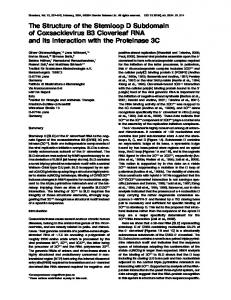
translation of its mRNA. Importantly, the distribution pattern of protein produced from KSD26 mRNA is indistinguishable from that of endogenous K10 mRNA (data not shown). The 309 nt StuI-HpaI region is necessary and sufficient for K10 mRNA transport/localization We next analyzed a series of transgenes lacking different internal portions of the K10 3′UTR. As summarized in Fig. 1, these studies identify a 309 nt StuI-HpaI fragment that is both necessary and sufficient for K10 mRNA transport/localization. This is best illustrated by two constructs: K∆StH and KStH26. K∆StH specifically lacks the StuI-HpaI region and produces transcripts that are retained in nurse cells until nurse cell regression (Fig. 2D). In contrast, KStH26, which contains the Fig. 1. Low-resolution mapping of K10 mRNA transport/localization control sequences. All deletions (with the exception of 70zStH26) were created in the context of a K10 minigene fragment possessing wild-type K10 gene activity (see Materials and Methods). The constructs differ only in the region shown. The black lines represent K10-derived sequences, where deletions are represented by gaps in the line. The approximate positions of the K10 translation stop codon (TAG) and poly(A)-addition site (pA) are indicated. The bottom six constructs contain hsp26-derived poly(A)-addition and putative transcription termination control sequences (labeled rectangle). The ability of the transgenes to produce transported/localized mRNA, as determined by in situ hybridization, is summarized to the right of the constructs (see also Fig. 2). The transcriptional control and protein coding regions of the 70zStH26 construct consists of the hsp26 nurse cell enhancer with an adjoining Sgs3 promoter followed by the E. coli lacZ protein coding region. S, SalI; St, StuI; H, HpaI; D, DraI; P, PstI; WT, wild type.
A
B
C
D
E
F
Fig. 2. Whole-mount in situ hybridization of K10-deletion and K10-hsp26 fusion genes. Egg chambers are arranged from left to right in order of increasing stage of development following whole-mount in situ hybridization with a digoxigenin-labeled K10 (A-E) or lacZ (F) probe. Egg chambers carry the following transgenes: (A) none (wild-type control); (B) KSD26; (C) K26; (D) K∆StH; (E) KStH26; (F) 70zStH26. Transgenes listed in B-E are in a K10LM00 background, which does not produce any detectable endogenous K10 mRNA (Cheung et al., 1992). In this and all other figures, anterior is to the left.
K10 mRNA localization 3813 StuI-HpaI region but lacks all other portions of the 3′UTR (except the first 65 nt), produces transcripts that are transported and anteriorly localized within the oocyte (Fig. 2E). We conclude that most, if not all, of K10’s transport/localization control sequences are contained within the 309 nt StuI-HpaI region. To test whether the StuI-HpaI region is sufficient to direct the transport/localization of a heterologous mRNA, we constructed 70zStH26. The only K10 sequence contained in this lacZ-tagged transgene is the 309 nt StuI-HpaI region (Fig. 1). As seen in Fig. 2F, 70zStH26 mRNA is efficiently transported and localized. We conclude that the 309 nt StuI-HpaI region is sufficient to direct the transport and anterior localization of a heterologous mRNA. A 92 nt subregion of StuI-HpaI is necessary and sufficient for K10 mRNA transport/localization To map more precisely K10 mRNA transport/localization control sequences, we conceptually subdivided the 309 nt StuIHpaI region into four non-overlapping subregions, denoted AD. We deleted each subregion individually, or in combination, within the context of an otherwise intact K10 3′UTR. As summarized in Fig. 3A, our analysis of these constructs indicates that K10 mRNA transport/localization control sequences are A St
WT
H A
B
C
D
transport/ localization yes
K∆A
no
K∆B
yes
K∆C
yes
K∆D
yes
K∆AB
no
K∆CD
yes
K∆BCD
yes
B
transport/ localization TAG
K∆SD+A TAG KA26
A
A
fully contained within subregion A. When we delete subregion A (see e.g., K∆A), only non-transported/non-localized mRNA is produced (Fig. 4A). Conversely, K10 transgenes lacking subregions B, C and/or D (see e.g., K∆BCD) produce efficiently transported/localized mRNA (Fig. 4B). These results show that subregion A is required for K10 mRNA transport/localization and that subregions B-D are dispensable. To test whether subregion A is sufficient for mRNA transport/localization, we constructed K∆SD+A and KA26 (Fig. 3B). The 3′UTRs of these transgenes contain the first 65 nt of the K10 3′UTR followed by a single copy of subregion A. The two constructs differ in that the poly(A)-addition site and putative transcription termination control regions of K∆SD+A and KA26 are derived from the K10 and hsp26 genes, respectively. Constructs identical to K∆SD+A and KA26, but lacking subregion A, produce transcripts that are not transported or localized (see K∆SD and K26 in Fig. 1). As seen in Fig. 4C (and data not shown), K∆SD+A and KA26 produce mRNA that is transported and anteriorly localized. We conclude that most, if not all, of K10’s mRNA transport/localization control sequences are contained within subregion A. We cannot, however, rule out the possibility that there are redundant copies of a second essential sequence, one or more in the region extending from the first nt of K10 mRNA to 65 nt downstream of TAG, and one or more within subregions B-D. The identification of the 44 nt K10 mRNA transport and localization sequence (TLS) Upon visual inspection of the 92 nt subregion A, we identified a 44 nt sequence that has a potential to form a stem-loop
A
B
pA
yes pA
yes
Fig. 3. High-resolution mapping of K10 mRNA transport/localization control sequences. All deletion constructs were created in the context of the same K10 minigene described in Fig. 1 and differ only in the region shown. (A) The black lines represent K10-derived sequences, where deletions are represented by gaps in the line. The 309 nt StuI-HpaI region (see Fig. 1) was subdivided into four non-overlapping subregions, named A, B, C and D, which were deleted individually or in combination as shown. (B) Subregion A (thin line) was inserted between the K10 transcriptional control/protein coding region (thick black line), and poly(A)-addition site-containing region from the 3′ end of the K10 (thick black line) or hsp26 (thick gray line) genes. The ability of the transgenes to produce transported/localized mRNA, as determined by in situ hybridization, is summarized to the right (see also Fig. 4). Symbols are as in Fig. 1.
C
Fig. 4. Whole-mount in situ hybridization of K10-deletion transgenes. Egg chambers are arranged from left to right in order of increasing stage of development following whole-mount in situ hybridization with a digoxigenin-labeled K10 probe. Egg chambers carry the following transgenes in a K10LM00 background: (A) K∆A; (B) K∆BCD; (C) K∆SD+A.
3814 T. L. Serano and R. S. Cohen secondary structure (Fig. 5A,B). We decided to test whether this sequence, called the K10 transport and localization sequence (TLS), is sufficient to direct mRNA transport and localization. We constructed two transgenes, K∆StH+T and K∆SD+T, in which the TLS replaces the 309 nt StuI-HpaI region and the ~1250 nt SalI-DraI region of the K10 3′UTR, respectively (Fig. 5C). As seen in Fig. 5C (and data not shown), both K∆StH+T and K∆SD+T produce transported/localized mRNA. In addition, K10 mutants carrying one copy of either K∆StH+T or K∆SD+T produce eggs that typically hatch at a frequency of 80-95%. These results, together with those of the experiments described below, indicate that the 44 nt TLS contains most, if not all, of the mRNA transport/localization control information present in the entire K10 mRNA.
Kim-Ha et al., 1993). Ko∆ transcripts are detected in nurse cells through early stage 8. Beginning at the end of stage 8, and extending through the end of stage 10B, low amounts of Ko∆ transcripts are also seen at the oocyte’s posterior pole. Presumably, low amounts of Ko∆ transcripts diffuse from nurse cells into the oocyte during early stages of oogenesis and become detectable in the oocyte after stage 8 as they concentrate at the posterior pole via the intact posterior localization control sequences. In contrast to Ko∆ mRNA, Ko∆+T mRNA is efficiently transported into the oocyte during early stages of oogenesis (Fig. 6B). However, while K-osk and wild-type osk mRNAs accumulate only transiently at the oocyte’s anterior cortex during stage 8, Ko∆+T mRNA remains there through at least stage 10B (Fig. 6B; detection becomes problematic after stage 10B due to the secretion of the egg chorion). We conclude that the K10 TLS can direct the transport of a heterologous mRNA. In addition, the K10 TLS apparently overrides the activity of osk mRNA posterior localization control sequences.
The TLS is sufficient for the transport and anterior localization of recombinant osk transcripts, but overrides osk posterior localization control sequences We next wanted to see if the TLS could substitute for the The stem-loop secondary structure of the K10 TLS transport/anterior localization control region of a mRNA is apparently essential for TLS function whose final destination within the oocyte is different than K10’s. We chose osk, since its mRNA transport and localizaAs mentioned above, the K10 TLS was originally identified by tion control sequences had been previously defined. Specifiits striking potential to form a stem-loop secondary structure. cally, Kim-Ha et al. (1993) identified a ~150 nt SphI-SacII To determine whether TLS activity is dependent on such a region that mediates osk mRNA transport and a separate region that A A A B U U mediates late stage posterior localizaU U 5'AGGCCUUAGAUUACACCACUUGA A C tion. We first replaced K10’s 3′UTR A U UUGUAUUUUUAAAUUAAUUCUUA with osk 3′UTR sequences to determine A U U A whether osk transport and localization AAAACUACAAAUUAAGAUCACUC U A U A control sequences retain activity when UCGUGAACGUGUGCUCGAUGGUG U A U A fused to K10 protein coding sequences C and produced under the transcriptional A U U A control of the K10 promoter/enhancer. C G C U A This control construct, called K-osk, S St H D P A pA TAG U A produces mRNA that, like wild-type WT A U osk, is transported to the oocyte’s G U K∆StH+T U A posterior pole during stages 1-7, tranU A 5'C G siently localizes to the oocyte’s anterior K∆SD+T cortex during early stage 8, and moves K∆SD+T back to the oocyte’s posterior pole during late stage 8/early stage 9, where it persists through the remainder of oogenesis (Fig. 6A,B and data not shown; Kim-Ha et al., 1991; Ephrussi et al., 1991). We then constructed two K10-oskar hybrid transgenes, called Ko∆ and Ko∆+T, that lack osk mRNA transport sequences, but retain osk posterior localization sequences (Fig. 6A). In Ko∆+T, the deleted osk transport Fig. 5. The sequence, putative secondary-structure and transport/localization activity of the control sequences are replaced with the K10 TLS. (A) The sequence of subregion A corresponds to nucleotides 3658-3749 of Prost et K10 TLS, while in Ko∆ there is no such al. (1988) with corrections described in Cohen and Serano (1995). The shaded region replacement. As seen in Fig. 6B, Ko∆ corresponds to the 44 nt TLS. (B) The predicted stem-loop secondary structure of the TLS. mRNA is distributed in a manner (C) Schematic diagram and representative mRNA distribution pattern of constructs containing similar to that reported for other osk the TLS. The TLS (represented by the small stem-loop) was added to K∆StH and K∆SD (see mRNAs that lack osk transport control Fig. 1) to give K∆StH+T and K∆SD+T, respectively (symbols are as in Fig. 1). Both sequence, but retain posterior localizatransgenes produce transported/localized mRNA, as shown here for K∆SD+T (see Fig. 2 tion control sequences (see olc15 in legend for in situ hybridization details).
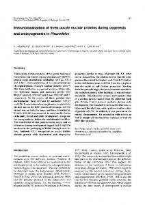
K10 mRNA localization 3815 structure, we introduced small mutations into the TLS, (Alden and Kim, 1979; Seeman et al., 1976), we thought it designed to disrupt its predicted secondary structure and likely that base-specific interactions would be restricted to the primary sequence, or only its primary sequence. single-stranded regions of the predicted stem-loop secondary The first two constructs, Kstem5′ and Kstem3′, contain structure. To look for such interactions, we made Kloop and mutations that disrupt the TLS primary sequence as well as its Kbub. In Kloop, we introduced transition mutations into all putative stem-loop structure. In Kstem5′, we replaced five eight positions of the predicted TLS loop (Fig. 7). In Kbub, internal uracil residues on the 5′ side of the stem with five we removed the two nucleotides that bubble out from the 3′ adenine residues (Fig. 7). Reciprocally, in Kstem3′, we side of the predicted stem (Fig. 7). As seen in Fig. 7, Kloop replaced five internal adenine residues on the 3′ side of the and Kbub both produce transported/localized mRNA, stem with five uracil residues (Fig. 7). As seen in Fig. 7, although like Kstem5′3′, the transport/localization and rescue Kstem5′ and Kstem3′ mRNAs are neither transported nor activities of Kloop and Kbub mRNA are slightly less efficient localized. In addition, K10 mutants carrying one copy of either than wild type (Fig. 7 and data not shown). These results Kstem5′ or Kstem3′ produce eggs that typically hatch with a indicate that the predicted single-stranded regions of the TLS frequency of ≤10% (data not shown). Thus, two separate five do not make essential base-specific contacts with the nucleotide substitutions in the TLS block its ability to direct transport/localization machinery. The possibility that other mRNA transport/localization, even in the context of an regions of the predicted stem-loop structure, viz., the stem otherwise wild-type K10 transgene. These results show that the termini, are engaged in such base-specific recognition events TLS is essential for K10 function and mRNA transport/localis discussed below. ization. The uracil-to-adenine and adenine-toA uracil substitutions described above could destination block transport/localization because they transport within oocyte alter the TLS primary sequence or because pA TAG they alter the predicted stem-loop structure. Kosk yes posterior To distinguish between these possibilities, Ko∆ weak posterior we combined the uracil-to-adenine substituKo∆+T yes anterior tions of Kstem5′ with the compensatory adenine-to-uracil substitutions of Kstem3′ to B make Kstem5′3′ (Fig. 7). Kstem5′3′ thus carries the primary sequence lesions of both Kstem5′ and Kstem3, but has the potential to form the same stem-loop structure predicted for wild-type TLS. As seen in Fig. 7, Kstem5′3′ mRNA is transported/localized almost as efficiently as is wild-type K10 mRNA. Moreover, Kstem5′3′ transgenes Kosk restore almost complete fertility to K10 mutant stocks. K10 mutants that carry one copy of the Kstem5′3′ transgene produce eggs that typically hatch at a frequency of ≥80% (data not shown). These data indicate that extensive alterations of the TLS primary sequence have little or no adverse affect on TLS transport/localization activity, provided Ko∆ that that they do not disrupt the predicted stem-loop secondary structure. We conclude that TLS activity is likely to be dependent on the formation of the predicted stem-loop secondary structure. While the above experiments provide Ko∆+T strong evidence that TLS activity is dependent on an encoded stem-loop Fig. 6. Schematic diagrams and mRNA distribution patterns of K10-oskar hybrid secondary structure, they do not provide transgenes. (A) The thick black lines represent K10-derived sequences. The thick gray much insight into the nature of the interaclines represent osk-derived sequences. The interruption in the line represents the deletion tions between the TLS and the of the ~150 nt SphI-SacII region, which contains osk mRNA transport control sequences transport/localization machinery. Given the (Kim-Ha et al., 1993). The small stem-loop represents the 44 nt K10 TLS (sequence selectivity of mRNA transport and localizashown in Fig. 5). The ability of the transgenes to produce transported mRNA (transport) tion, it is likely that such interactions and the final destination of the mRNA within the oocyte (destination within oocyte) are include base-specific recognition events (St indicated to the right of the diagrams. (B) In situ hybridization montages showing the Johnston, 1995). Because the major groove distribution patterns of K-osk, Ko∆ and Ko∆+T mRNA (see Fig. 2 legend for in situ of double-stranded RNA is deep and narrow hybridization details).
3816 T. L. Serano and R. S. Cohen AA U U U U A C AU AU UA UA UA UA UA C AU UA GC UA A UA AU GU UA UA CG
Wild type
AA U U U U A C AU AU a A a A a A a A a A C AU UA GC UA A UA AU GU UA UA CG
Kstem5'
AA U U U U A C AU AU U u U u U u U u U u C AU UA GC UA A UA AU GU UA UA CG
Kstem3'
AA U U U U A C AU AU au au au au au C AU UA GC UA A UA AU GU UA UA CG gg
c c
c c
Kstem5'3'
Kloop
g u AU AU UA UA UA UA UA C AU UA GC UA A UA AU GU UA UA CG
AA U U U U A C AU AU UA UA UA UA UA AU UA GC UA UA AU GU UA UA CG
Kbub
Fig. 7. The distribution patterns of mRNA produced from K10 transgenes containing TLS-mutations. The TLS primary sequence and predicted secondary structure is shown for wild type, Kstem5′, Kstem3′, Kstem5′3′, Kloop and Kbub (nucleotide substitutions represented by lowercase letters). To the right of each sequence/structure are egg chambers carrying the respective transgene in a K10LM00 background following wholemount in situ hybridization with a digoxigenin-labeled K10 probe.
K10
A U U A A A U U U U U A U G U U A G U U C
A U U C U U A A A A A C U A C A A A U U A A G
A A C A A A U G U A U A U U A U A U A U A orb U A A U A U C G U U A U A U A A U A U G C
Fig. 8. A comparison of the predicted secondary structures of the K10 TLS and a sequence found within the orb 3′UTR. A region of particularly strong homology is found near the loop of both predicted stems (boxed region). The orb sequence corresponds to nucleotides 4200-4241 (according to Lantz et al., 1992) and was initially identified by visual inspection. We also searched Adducin-like, bcd, Bicaudal-D, cyclin B, gurken, Hsp83, nanos, osk, pumilio, staufen, tudor and yemanuclein-a mRNA sequences using the sequence W7N7-9W′7, where W′ represents base complementary to W. A single match was identified in both Adducin-like and nanos, but such sequences showed little or no primary sequence homology with the TLS (not shown). No other matches were found.
DISCUSSION A single element mediates K10 mRNA transport and anterior localization We have identified a 44 nt sequence, the TLS, which is essential for K10 gene function and is necessary and sufficient for mRNA transport into, and anterior localization within, the Drosophila oocyte. The small size of the TLS suggests that it acts as a single element to direct a single transport/localization event. Indeed, both K10 mRNA transport to the posterior pole of the oocyte during stages 1-7 and localization to the oocyte’s anterior cortex during stages 8-10B could be mediated by the continuous association of the mRNA with a minus end-directed microtubule motor (see Cooley and Theurkauf, 1994). Since most mRNAs that are localized within the Drosophila oocyte are transported into the oocyte during stages 1-7 and accumulate at least transiently at the oocyte’s anterior cortex during the early part of stage 8, one might expect many of them to contain the TLS. We have identified one such candidate sequence in orb mRNA (Fig. 8; Lantz et al., 1992). The orb sequence and the K10 TLS are quite similar in A-U content and in the size of the predicted stem and loop. In addition, the
K10 mRNA localization 3817 orb sequence resides in a 280 nt region of orb mRNA that is sufficient for orb mRNA transport and anterior localization (Lantz and Schedl, 1994). These findings, together with the similarities of K10 and orb mRNA distribution patterns through stage 11 (Cheung et al., 1992; Lantz et al., 1992), suggest that K10 and orb mRNAs are recognized by the same component(s) of the transport/localization machinery. We have been unable to identify candidate TLSs in any other mRNAs known to be localized within the Drosophila oocyte (see Fig. 8 for details). This raises the possibility that the transport of at least some mRNAs is mediated by factors different than those that mediate K10, and possibly orb. We cannot rule out, however, that other localized mRNAs share a common secondary structure motif with the TLS that is not easily identifiable by a comparison of primary sequences. While most localized mRNAs appear to be transported into the oocyte in a similar fashion, our results suggest that the mRNA transport control sequences of different mRNAs are not functionally equivalent. For example, the K10 TLS can substitute for osk mRNA transport control sequences, but it is apparently incompatible with the activity of osk mRNA posterior localization control elements. The observed incompatibility may occur for superficial reasons, e.g., the inclusion of the TLS in the osk 3′UTR may alter the secondary structure of osk posterior localization control sequences. Alternatively, osk mRNA transport control sequences, unlike the K10 TLS, may facilitate the release of the mRNA from a minus end-directed microtubule motor at stage 8, thus allowing osk posterior localization control sequences to mediate the association of osk mRNA with a plus end-directed microtubule motor. mRNA transport and localization control elements and RNA secondary structure The TLS has a predicted secondary structure consisting of a 8 base loop and an imperfect, 17 base pair stem – one strand of the stem contains two one-base bubbles (Fig. 5B). Consistent with the idea that TLS activity is dependent on the formation of such a stem-loop structure, we find that TLS transport/localization activity is abolished when base changes are introduced into one of the two stem strands alone, but not when simultaneously introduced into both strands in a compensatory manner that preserves the predicted secondary structure. Other mRNA transport/localization control sequences have also been proposed to form secondary structures. Most notably, the 625 nt bcd mRNA localization signal is predicted to form an elaborate secondary structure, consisting of five large stems (Macdonald and Struhl, 1988; Macdonald, 1990; Ferrandon et al., 1994). This is supported by the finding that, while the primary sequence of the bcd localization signal varies significantly between a number of Drosophila species, the putative secondary structure remains conserved (Macdonald, 1990). In addition, staufen protein, which mediates bcd and osk mRNA localization, exhibits double-stranded RNA-binding activity in vitro (St. Johnston et al., 1992). Together, these findings suggest that secondary structure formation is a common feature of mRNA transport/localization control sequences. Given the selective nature of mRNA transport/localization, it is likely that recognition of the TLS by the transport/localization machinery requires base-specific contacts. A complete base substitution of the predicted loop, or the removal of the two single nucleotide bubbles from the 3′ side of the stem, have
only a slight negative affect on TLS activity, indicating that these regions do not provide essential base-specific contacts. It could be that residues of the stem itself make base-specific contacts with the transport/localization machinery. The minor groove of double-stranded RNA is wide and accessible, but it contains few hydrogen bond acceptor and donor groups and the potential for base-specific hydrogen bonding is very low (Seeman et al., 1976). One possible base-specific hydrogen bond acceptor/donor group in the minor groove is the carbonyl group of any uracil immediately 3′ to adenine (Rosenberg et al., 1973). The TLS stem contains two such groups. The predicted orb stem (Fig. 8) also contains several such groups, but at non-conserved positions. Assuming that the overall conservation between the predicted stem loop structures of K10 and orb is not fortuitous, it would thus appear that the minor groove does not play a major role in the base-specific recognition of the stem by the transport/localization machinery. In contrast to the minor groove, the major groove of doublestranded RNA has a rich, base-specific collection of hydrogen bond acceptor and donor groups (Seeman et al., 1976). While the major groove of double-stranded RNA is narrow and generally not accessible to small, amino acid-size molecules (Alden and Kim, 1979), chemical acylation studies show that bases positioned near a loop or bubble of two or more nucleotides are accessible, presumably due to a local distortion of the helix (Weeks and Crothers, 1993). Accessibility extends into the duplex about 2 nucleotides on one strand and about 4 nucleotides on the other. The bases adjacent to the TLS loop are conserved in orb (Fig. 8) and thus are good candidates for base-specific interactions with the transport/localization machinery. Residues at the base of the stem are not conserved and thus apparently less likely to make specific contacts with the transport/localization machinery. The protein components of the transport/localization machinery have remained elusive. Genetic analyses have identified only two genes, Bic-D and egalitarian (egl), that are required for K10 mRNA transport into the oocyte (Suter and Steward, 1991; Cheung et el., 1992). However, it is unclear whether Bic-D and egl encode proteins that bind directly to the TLS. These genes are also required for oocyte determination and microtubule organization, developmental events that are likely to be a prerequisite for mRNA transport (Schüpbach and Wieschaus, 1991; Theurkauf et al., 1993). Thus, biochemical approaches may be most useful for identifying the proteins that directly contact the K10 TLS. We thank Paul Macdonald for providing the osk clone and Vicki Corbin, Xiangyi Lu, Doug Ruden, Kathy Suprenant, Rob Weaver and members of the Cohen laboratory for helpful discussions and comments on the manuscript. T. L. S. was supported in part by NIH training grant 5T32EY07105. R. S. C. was supported by a grant from the NSF (IBN-08821) and by the General Research Fund of the University of Kansas.
REFERENCES Alden, C. J. and Kim, S.-H. (1979). Solvent-accessible surfaces of nucleic acids. J. Mol. Biol. 132, 411-434. Berleth, T., Burri, M., Thoma, G., Bopp, D., Richstein, S., Frigerio, G., Noll, M. and Nüsslein-Volhard, C. (1988). The role of localization of bicoid RNA in organizing the anterior pattern of the Drosophila embryo. EMBO J. 7,1749-1756.
3818 T. L. Serano and R. S. Cohen Cheung, H.-K., Serano, T. L. and Cohen, R. S. (1992). Evidence for a highly selective RNA transport system and its role in establishing the dorsoventral axis of the Drosophila egg. Development 114, 653-661. Clark, I., Giniger, E., Ruohola-Baker, H., Jan, L. Y. and Jan, Y. N. (1994). Transient posterior localization of a kinesin fusion protein reflects anteroposterior polarity of the Drosophila oocyte. Curr. Biol. 4, 289-300. Cohen, R. S. and Meselson, M. (1985). Separate regulatory elements for the heat-inducible and ovarian expression of the Drosophila hsp26 gene. Cell 47, 737-743. Cohen, R. S. and Serano, T. L. (1995). mRNA localization and function of the Drosophila fs(1)K10 gene. In RNA Localization (ed. H. D. Lipshitz). In press. Austin: R. G. Landes. Cooley, L. and Theurkauf, W. E. (1994). Cytoskeletal functions during Drosophila oogenesis. Science 266, 590-596. Ding, D. and Lipshitz, H. D. (1993). Localized RNAs and their functions. BioEssays 15, 651-658. Ephrussi, A., Dickinson, L. K. and Lehmann, R. (1991). oskar organizes the germ plasm and directs localization of the posterior determinant nanos. Cell 66, 37-50. Feinberg, A. P. and Vogelstein, B. (1983). A technique for radiolabelling DNA restriction endonuclease fragments to high specific activity. Anal. Biochem. 132, 6-13. Ferrandon, D., Elphick, L., Nüsslein-Volhard, C. and St. Johnston, D. (1994). Staufen protein associates with the 3′UTR of bicoid mRNA to form particles that move in a microtubule-dependent manner. Cell 79, 1221-1232. Frank, L. H., Cheung, H.-K. and Cohen, R. S. (1992). Identification and characterization of Drosophila female germ line transcriptional control elements. Development 114, 481-491. Haenlin, M., Roos, C., Cassab, A. and Mohier, E. (1987). Oocyte-specific transcription of fs(1)K10: a Drosophila gene affecting dorsal-ventral developmental polarity. EMBO J. 6, 801-807. King, R. C. (1970). Ovarian Development in Drosophila melanogaster. New York: Academic Press. Kim-Ha, J., Smith, J. L. and Macdonald, P. M. (1991). oskar mRNA is localized to the posterior pole of the Drosophila oocyte. Cell 66, 23-35. Kim-Ha, J., Webster, P. J., Smith, J. L. and Macdonald, P. M. (1993). Multiple RNA regulatory elements mediate distinct steps in localization of oskar mRNA. Development 119, 169-178. Lantz, V., Ambrosio, L. and Schedl, P. (1992). The Drosophila orb gene is predicted to encode sex-specific germline RNA-binding proteins and has localized transcripts in ovaries and early embryos. Development 115, 75-88. Lantz, V. and Schedl, P. (1994). Multiple cis-acting target sequences are required for orb mRNA localization during Drosophila oogenesis. Mol. Cell. Biol. 14, 2235-2242. Lindsley, D. L. and Zimm, G. G. (1992). The Genome of Drosophila melanogaster. New York: Academic Press. Macdonald, P. M. (1990). bicoid mRNA localization signal: phylogenetic conservation of function and RNA secondary structure. Development 110, 161-171. Macdonald, P. M. (1992). The means to the ends: localization of maternal messenger RNAs. Sem. Dev. Biol. 3, 413-424. Macdonald, P. M. and Struhl, G. (1988). Cis-acting sequences responsible for anterior localization of bicoid mRNA in Drosophila embryos. Nature 336, 595-598. Macdonald, P. M., Kerr, K., Smith, J. L. and Leask, A. (1993). RNA regulatory element BLE1 directs the early steps of bicoid mRNA localization. Development 118, 1233-1243. Neuman-Silberberg, F. S. and Schüpbach T. (1993). The Drosophila dorsoventral patterning gene gurken produces a dorsally localized RNA and encodes a TGFa-like protein. Cell 75, 165-174. Pelham, H. R. B. and Munro, S. (1993). Sorting of membrane proteins in the secretory pathway. Cell 75, 603-605. Pirrotta, V. (1988). Vectors for P-mediated transformation in Drosophila. In
Vectors: A Survey of Molecular Cloning Vectors and Their Uses (eds. R. L. Rodriguez and D. T. Denhardt), pp. 437-456. Boston: Butterworths. Pokrywka, N. J. and Stephenson, E. C. (1991). Microtubules mediate the localization of bicoid mRNA during Drosophila oogenesis. Development 113, 55-66. Pokrywka, N. J. and Stephenson, E. C. (1995). Microtubules are a general component of mRNA localization systems in Drosophila oocytes. Dev. Biol. 167, 363-370. Prost, E., Deryckere, F., Roos, C., Haenlin, M., Pantesco, V. and Mohier, E. (1988). Role of the oocyte nucleus in determination of the dorsoventral polarity of Drosophila as revealed by molecular analysis of the K10 gene. Genes Dev. 2, 891-900. Rosenberg, J. M., Seeman, N. C., Kim, J. J. P., Suddath, F. L., H. B. and Rich, A. (1973). Double helix at atomic resolution. Nature 243, 150-154. Rubin, G. M. and Spradling, A. C. (1982). Genetic transformation of Drosophila with transposable element vectors. Science 218, 348-353. Schüpbach, T. and Wieschaus, E. (1991). Female sterile mutations on the second chromosome of Drosophila melanogaster. II. Mutations blocking oogenesis or altering egg morphology. Genetics 129, 1119-1136. Seeman, N. C., Rosenberg, J. M. and Rich, A. (1976). Sequence-specific recognition of double helical nucleic acids by proteins. Proc. Natl. Acad. Sci. USA 73, 804 Serano, T. L., Cheung, H.-K., Frank, L. H. and Cohen, R. S. (1994). P element transformation vectors for studying Drosophila melanogaster oogenesis and early embryogenesis. Gene 138, 181-186. Serano, T. L., Karlin-McGinness, M. and Cohen, R. S. (1995). The role of fs(1)K10 in the localization of the mRNA of the TGFα homolog gurken within the Drosophila oocyte. Mech. Dev. 51, 183-192. Southgate, R., Ayme, A. and Voellmy, R. (1983). Nucleotide sequence analysis of the Drosophila small heat shock gene cluster at locus 67B. J. Mol. Biol. 165, 35-57. Spadling, A. C. and Rubin, G. M. (1982). Transposition of cloned P elements into Drosophila germ line chromosomes. Science 218, 341-347. St Johnston, D. (1995). The intracellular localization of messenger RNAs. Cell 81, 161-170. St Johnston, D., Driever, W., Berleth, T., Richstein, S. and NüssleinVolhard, C. (1989). Multiple steps in the localization of bicoid RNA to the anterior pole of the Drosophila oocyte. Development 107 Supplement, 1319. St Johnston, D., Brown, N. H., Gall, J. G. and Jantsch, M. (1992). A conserved double-stranded RNA-binding domain. Proc. Natl. Acad. Sci. USA 89, 10979-10983. Suter, B. and Steward, R. (1991). Requirement for phosphorylation and localization of the Bicaudal-D protein in Drosophila oocyte differentiation. Cell 67, 917-926. Tautz, D. and Pfeifle, C. (1989). A non-radioactive in situ hybridization method for the localization of specific RNAs in Drosophila embryos reveals translational control of the segmentation gene hunchback. Chromosoma 98, 81-85. Theurkauf, W. E., Smiley, S., Wong, M. L. and Alberts, B. M. (1992). Reorganization of the cytoskeleton during Drosophila oogenesis: implications for axis specification and intercellular transport. Development 115, 923-936. Theurkauf, W. E., Alberts, B. M., Jan, Y. N. and Jongens, T. A. (1993). A central role for microtubules in the differentiation of Drosophila oocytes. Development 118, 1169-1180. Weeks, K. M. and Crothers, D. M. (1993). Major groove accessibility of RNA. Science 261, 1574-1577. Zimmerman, J. L., Petri, W. and Meselson, M. (1983). Accumulation of a specific subset of D. melanogaster heat shock mRNAs in normal development without heat shock. Cell 32, 1161-1170. (Accepted 2 August 1995)