Int. J. Mol. Sci. 2015, 16, 11452-11464; doi:10.3390/ijms160511452 OPEN ACCESS
International Journal of
Molecular Sciences ISSN 1422-0067 www.mdpi.com/journal/ijms Article
A Solid-State NMR Study of Selenium Substitution into Nanocrystalline Hydroxyapatite Joanna Kolmas *, Marzena Kuras †, Ewa Oledzka † and Marcin Sobczak † Department of Inorganic and Analytical Chemistry, Faculty of Pharmacy with the Laboratory Medicine Division, Medical University of Warsaw, ul. Banacha 1, 02-097 Warsaw, Poland; E-Mails:
[email protected] (M.K.);
[email protected] (E.O.);
[email protected] (M.S.) †
These authors contributed equally to this work.
* Author to whom correspondence should be addressed; E-Mail:
[email protected]; Tel.: +48-22-572-0755; Fax: +48-22-572-0784. Academic Editor: Mohamed N. Rahaman Received: 1 March 2015 / Accepted: 29 April 2015 / Published: 19 May 2015
Abstract: The substitution of selenium oxyanions in the hydroxyapatite structure was examined using multinuclear solid-state resonance spectroscopy (ssNMR). The study was supported by powder X-ray diffractometry (PXRD) and wavelength dispersion X-ray fluorescence (WD-XRF). Samples of pure hydroxyapatite (HA300) and selenate (HA300-1.2SeO4) or selenite (HA300-1.2SeO3) substituted hydroxyapatites were synthesized using the standard wet method and heated at 300 °C to remove loosely bonded water. PXRD data showed that all samples are single-phase, nanocrystalline hydroxyapatite. The incorporation of selenite and selenate ions affected the lattice constants. In selenium-containing samples the concentration of Se was very similar and amounted to 9.55% and 9.64%, for HA300-1.2SeO4 and HA300-1.2SeO3, respectively. PXRD and ssNMR data showed that the selenite doping significantly decreases the crystallite size and crystallinity degree. 31P and 1H NMR experiments demonstrated the developed surface hydrated layer in all samples, especially in HA300-1.2SeO3. 1H NMR studies showed the dehydroxylation of HA during the selenium oxyanions substitution and the existence of hydrogen bonding in structural hydroxyl group channels. 1H→77Se cross polarization NMR experiments indicated that selenites and selenates are located in the crystal lattice and on the crystal surface.
Int. J. Mol. Sci. 2015, 16
11453
Keywords: biomaterials; calcium phosphates; hydroxyapatite; selenium oxyanions; solid-state nuclear magnetic resonance; powder diffractometry
1. Introduction Since synthetic biomaterials can overcome several problems in bone grafts (i.e., limited supply of autografts, risk of rejection and disease transfer of allo- and xenografts), intensive efforts have for decades been devoted to developing and improving materials such as bioceramics, polymers, metallic materials and bioglasses [1–3]. Calcium phosphate bioceramics, especially hydroxyapatite (HA), due to its high biocompatibility, osteoconductivity and similar composition to the inorganic fraction of mineralized tissues, play a crucial role in bone reconstructive surgery [4,5]. The hydroxyapatite crystal structure can easily host a variety of ionic substituents whose presence may affect biological and physicochemical properties [6–8]. Thus, such modified compounds have recently attracted much attention. The structure of HA (Ca10(PO4)6(OH)2) is most frequently considered to be hexagonal, with space group P63/m [9]. The crystallographic unit cell contains ten calcium cations arranged in two nonequivalent sites called Ca(I) and Ca(II). Calcium cations may be partially replaced by several cationic substituents, i.e., Mg2+, Mn2+, Zn2+, Sr2+, Ag+ or Cr3+ [10,11]. The structural hydroxyl groups are located in columns ··· OH OH OH ···, where oxygen atoms are too distant (3.44 Å) to form hydrogen bonds [9]. The OH groups in columns are frequently replaced by various monovalent and bivalent anions: Cl−, F− and CO32−, O2−, S2−, respectively [12,13]. The tetrahedral trivalent orthophosphates PO43− may be substituted not only by other trivalent (i.e., AsO43−) but also by bivalent (i.e., CO32−) and tetravalent (SiO44−) anions [14,15]. It is important to note that all substitutions may be the source of lattice distortion, vacancies, crystal defects or hydrogen bonds in OH groups’s columns, which in turn play a significant role in HA crystal size and morphology [6,8]. Inspired by previous research on hydroxyapatite enriched in various ions, we have decided to study the incorporation of selenium oxyanions (selenate SeO42− and selenite SeO32−) into the HA crystal structure. Selenium is an important element in bone physiology. It plays a crucial role in various metabolic processes as a constituent of a great number of enzymes [16]. Its deficiency may retard growth of bones and increase the risk of bone disease (i.e., osteopenia, osteoarthritis and osteoporosis) [17,18]. Selenium, as a constituent of selenoproteins, plays a significant role in the proliferation of osteoblasts and osteoclasts. It is also important to note that selenium has shown considerable promise as an anticancerogenic agent [19]. In our previous work [20] we have focused on the synthesis of hydroxyapatite containing selenite or selenate ions at various concentrations. It has been shown that nanocrystalline, selenium-enriched HA materials may be prepared by conventional wet method. The main objective of this work is the structural characterization of hydroxyapatite substituted with selenium oxyanions. We present here a detailed solid-state nuclear magnetic resonance (ssNMR) investigation on 77Se, 1H and 31P nuclei.
Int. J. Mol. Sci. 2015, 16
11454
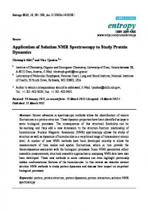
2. Results and Discussion 2.1. Powder X-ray Diffraction Analysis Figure 1 presents the powder X-ray diffraction patterns of pure hydroxyapatite (HA300) and selenium- containing samples (HA300-1.2SeO3 and HA300-1.2SeO4). Each diffractogram matches the International Centre for Diffraction Data (ICDD) standard diffractogram of HA (No. 9.432) well. The results indicate that no other crystalline phase was detected. All the powders exhibit relatively broad reflection lines, especially the HA300-1.2SeO3 sample (see Figure 1). These results indicate that the crystallites are fine and the crystallinity degree is low [21].
Figure 1. PXRD patterns of the analysed samples: HA300, HA300-1.2SeO3 and HA300-1.2SeO4. We have calculated the crystallites’ sizes according to Scherrer’s formula:
D=
0.94 × λ β1/2 × cos θ
(1)
where D is a domain size (crystallite size in nm), λ is the wavelength of the radiation (in nm), β1/2 is the peak full-width at half- maximum (in radians) and θ is the diffraction angle of the corresponding reflex. It was confirmed that all the powders are nanocrystalline with an average crystallite size (in nm) decreasing in the series: HA300 (18) > HA300-1.2SeO4 (15) > HA300-1.2SeO3 (9). In order to estimate the crystallinity degree of the obtained powders the formula below was used [22]: χc = (
K β (002)
)3
(2)
where χc is the crystallinity degree corresponding to the fraction of crystalline phase in powder, K is a constant. (K for a great number of hydroxyapatites is 0.24), β(002) is the peak (002) full-width at half- minimum (in degrees).
Int. J. Mol. Sci. 2015, 16
11455
The calculations have shown that the crystallinity degree dramatically decreases in the series: HA300 (0.7) > HA300-1.2SeO4 (0.5) >> HA300-1.2SeO3 (0.1). Table 1 collects the lattice parameters obtained from Rietveld refinements of the PXRD data. These data show that the incorporation of selenite or selenate ions into hydroxyapatite structure affected the lattice constants; however, SeO32− substitution causes higher increments of both a and c constants. These data are in good agreement with our previous measurements described in [20]. Table 1. Various parameters of the studied samples. The unit cell parameters a and c (in Å), crystallinity index and crystallite size (in nm) were calculated from the PXRD patterns using Rietveld analysis. Chemical composition of the samples was studied using WD-XRF method. Characteristics Cell parameters (Å) Crystallite size (nm) Crystallinity index Se content (wt %) Ca/(P + Se)
HA300 a = 9.4287 c = 6.8772 18 0.7 1.62
HA300-1.2SeO3 a = 9.5084 c = 6.8891 9 0.1 9.64 1.56
HA300-1.2SeO4 a = 9.4333 c = 6.8879 15 0.5 9.55 1.60
The results of WD-XRF elemental analysis are reported in Table 1. The selenium contents are similar to those of the corresponding amount of starting materials. This may be proof of the incorporation of selenite and/or selenate into the crystal lattice. It should be noted that these ions are not eliminated, even through intensive rinsing of the powder with distilled water. 2.2. 31P Solid-State NMR Spectroscopy 31
P solid-state NMR spectra were acquired for all samples using two different techniques: the one-pulse (BD) and 1H→31P cross polarization (CP) techniques. Briefly, the BD signals come from all phosphorus-31 nuclei, while CP lines may be assigned to the 31P nuclei located close to protons (in the case of hydroxyapatite, close to structural hydroxyl groups and to molecules of water in the crystal structure or adsorbed on the crystal surface). All the samples give one signal at about 3 ppm in BD as well as CP experiments; that is a characteristic feature of apatites [23,24] (see Figure 2A,B and Table 2).
Int. J. Mol. Sci. 2015, 16
11456
(A)
(B)
Figure 2. 31P BD (A) and CP (B) MAS NMR spectra of the analysed samples. Table 2. Curve fitting results for the 31P CP NMR spectra (MAS at 7.0 kHz, the CP contact time of 2 ms). The peaks have been assigned according to Pajchel et al. [25]. Peak Characteristics Chemical shift (ppm) FWHM a (Hz) LF b % of total area FWHM ratio: CP/BD Area ratio c: CP/BD a
HA300-1.2SeO3 HA300 Narrow Broad Narrow Broad 3.16 3.44 3.26 3.31 159 897 332 879 0.5 0.0 0.6 0.1 76 24 49 51 Narrow line characteristics 0.95 0.83 0.66 0.40
HA300-1.2SeO4 Narrow Broad 3.17 3.38 247 867 0.6 0.0 66 34 0.92 0.57
Full width in half minimum; b Lorentzian fraction; c Measured at νMAS = 0 kHz.
Both BD and CP signals are significantly broader for the HA300-1.2SeO3 sample. The FWHM parameter (shown in brackets in Hz) measured for BD spectra increased in the following order: HA300 (165) < HA300-1.2SeO4 (267)