ATYPICAL PRESENTATION OF TUBERCULOSIS ... Department of Surgery,
Lokmanya Tilak Municipal General Hospital and Medical College, Sion, Mumbai.
Case Report
ATYPICAL PRESENTATION OF TUBERCULOSIS Dinesh K. Sarda1, Paras Kothari3, Prashani Adivarekar2, Ravikumar1, Raghunath Dipali1 and Bharati Kulkarni4 (Original received on 13.3.2006. Revised version received on 12.4.2006. Accepted on 16.5.2006.) Summary: Incidence of tuberculosis is increasing with emergence of many cases of multi-drug resistance and extrapulmonary manifestation. We report three cases of tuberculosis presenting in very atypical ways.
[Indian J Tuberc 2006; 53-223-226] Key words:
Tuberculosis, Atypical.
CASE HISTORY Case 1 An eight-year old child was brought with complaints of progressive increasing neck pain and restricted neck movements from past fifteen days. There was no history of any constitutional symptoms and exposure to tuberculosis. On examination, all
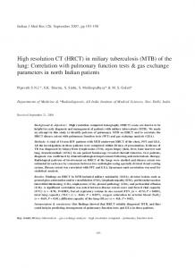
Fig.1: CT scan showing hypoechoic lesion indenting on right pharyngeal wall
neck movements were grossly restricted and were painful. He had mild trismus as well. Intraoral examination revealed bulge on the right lateral pharyngeal wall, just behind the tonsillar fossa. Systemic examination was normal. Investigations revealed lymphocytosis, ESR of 55 mm, positive Mantoux test. X-ray cervical spine and chest were normal. Ultrasound and CT scan demonstrated localized hypoechoic lesion of 32x25x23mm size,
Fig.2: MRI showing tubercular spondolytis affecting craniovertebral junction and C2 vertebra, atlatoaxial dislocation and granulation tissue at C2 level
1. Resident 2. Lecturer 3. Associate Professor 4. Professor and Head Department of Surgery, Lokmanya Tilak Municipal General Hospital and Medical College, Sion, Mumbai. Correspondence: Dr. Dinesh Sarda, Department of Pediatric Surgery, Room No. 440, 4th Floor, College Building, Lokmanya Tilak Municipal General Hospital and Medical College, Sion, Mumbai-400 022 (Maharashtra). E-mail:
[email protected]
Indian Journal of Tuberculosis
224
DINESH K. SARDA ET AL
Fig. 3: X-ray chest showed hyperlucent left lung fields, tracheal shift to right, flattened left hemidiphargm, mediastinal shift to right with pleural hemiation (case 2)
Fig. 4: HRCT showing caseating lymph node compressing on left bronchus with increase in left lung volume
abutting the right lateral pharyngeal wall (Figure1). MRI of cervical spine revealed tubercular spondolytis affecting craniovertebral junction and C2 vertebra, atlantoaxial dislocation and granulation tissue at C2 level. Tip of odontoid process was compressing on cervicomedullary junction (Figure 2). Under anesthesia intraoral aspiration of abscess was done using wide bore needle. 15 ml of thick non-foul smelling yellow coloured fluid was aspirated. Post aspiration ultrasound showed no residual abscess. Ziehl Neilson staining showed presence of tubercle bacilli. In view of absence of neurological symptoms, conservative management was planned for this child. Patient was started on anti-tubercular drugs (Isoniazid, Rifampicin, Ethambutol, Pyrazinamide for 2 months and Isoniazid, Rifampicin for 7 months). Neck was immobilized with external brace and patient was discharged. Patient had improvement in neck pain and movements at six month follow-up.
Case 2
Indian Journal of Tuberculosis
Five-month-old child was admitted with complaints of cough and fever since one month and respiratory distress of two weeks duration. There was no history of foreign body aspiration, exposure to tuberculosis. Patient was tachypneic. Left lung was hyperresonant on percussion and air entry was markedly reduced. There were bilateral ronchi. Hematological investigations were within normal range. Mantoux test was positive. Blood ADA level was 92 IU/ml, IgM TB was positive (1.6 IU/ml). X ray chest showed hyperlucent left lung fields, tracheal shift to right, flattened left hemidiphragm, mediastinal shift to right with pleural herniation (Figure 3). HRCT with mediastinal window revealed caseating lymph node compressing on left bronchus about 6 mm from carina with increase in left lung volume (Figure 4). Bronchoscopy showed slit like opening in left
ATYPICAL PRESENTATION OF TUBERCULOSIS
225
Fig. 5: Repeat X-ray chest at three months follow Fig. 6: Intra-operative photograph showing up showed marked improvement (case 2) abscess on anterior surface of testis with caseous material seen on drainage bronchus only on IPPV. There was no intraluminal obstruction or any foreign body. Patient was started on antitubercular treatment and other supportive treatment. Patient responded well to anti-tubercular treatment. Repeat X-ray chest after three months follow-up showed marked improvement (Figure 5). Case 3 One-year-old male child was brought with complaints of redness of right scrotum and vomiting for past one day. On examination, right hemiscrotum was red and inflammed with loss of cremasteric reflex. Doppler ultrasound test raised suspicion of testicular torsion. On exploration, epidydymis was normal. There was a 5x5 mm abscess on anterior surface of testis. Caseous material was seen on drainage (Figure 6). AFB staining showed tubercle bacilli. Patient was started on anti-tubercular treatment (Isoniazid, Rifampicin, Ethambutol, Pyrazinamide for 2 months and isoniazid, Rifampicin for 7 months). Intravenous pyelography and micturating cystourethrogram done at later date were normal. Repeat ultrasound done after two months was normal. DISCUSSION In spite of the many major advances in understanding the disease, its management, diagnosis
of tuberculosis some times becomes a dilemma. So delay in diagnosis is very common which leads to increase in number of cases of extra-pulmonary tuberculosis. Negative smear for AFB, a lack of granuloma on histopathology, and negative culture do not exclude the diagnosis of tuberculosis. Newer investigations ADA levels, PCR may prove to be useful1. Craniovertebral junction tuberculosis (CVJTB) is rare and occurs in only 0.3 to 1% of patients with tuberculosis spondylitis 2. Dislocation of atlantoaxial dislocation, as in our case, makes it rarer. Management includes occipitocervical fixation, administration of anti-tuberculous medications with or without ventral cervicomedullary decompression3. Mediastinal tubercular lymph nodes may gradually increase in size, caseate and burst inside the endotracheal lumen. Our patient presented with emphysematous changes on one side. Bronchoscopy revealed the presence of extrinsic compression, which was confirmed by HRCT scan. Whenever there is suspicion of foreign body obstruction, other causes such as mediastinal tuberculosis should also be considered4. High index of clinical suspicion, timely judicious use of invasive diagnostic methods and confirmation of the diagnosis, early institution of specific anti-tuberculosis
Indian Journal of Tuberculosis
226
DINESH K. SARDA ET AL
treatment and close clinical monitoring for adverse drug reactions are the key to the successful management of EP atypically presented TB1.
2.
3.
REFERENCES 1.
Golden M.P., Vikram HR Extrapulmonary tuberculosis: an overview. Am Fam Physician. 2005 Nov 1; 72(9): 17611768.
Indian Journal of Tuberculosis
4.
Behari S, Nayak SR, Bhargava V, Banerji D, Chhabra DK, JainVK. Craniocervical tuberculosis: protocol of surgical management. Neurosurgery. 2003 Jan; 52(1): 72-80; discussion 80-1. Sinha S, Singh AK, Gupta V, Singh D, Takayasu M, Yoshida J. Surgical management and outcome of tuberculous atlantoaxial dislocation: a 15-year experience. Neurosurgery. 2003 Feb; 52(2): 331-338; discussion 338-9. Ahmed A, Mirza S, Rothera MP. Mediastinal tuberculosis in a 10-month-old child. J Laryngol Oto .2001 Feb; 115(2): 1613.