Arish A. Qazi*a, Erik B. Damb, Marco Looga,b , Mads Nielsena,b,. Francois Lauzeb, Claus Christiansenc. aImage Group, University of Copenhagen, 2100 ...
A Variational Method for Automatic Localization of the most Pathological ROI in the Knee Cartilage Arish A. Qazi*a, Erik B. Damb, Marco Looga,b , Mads Nielsena,b, Francois Lauzeb, Claus Christiansenc a Image Group, University of Copenhagen, 2100 Copenhagen, Denmark; b Nordic Bioscience Imaging, 2730 Herlev, Denmark; c Center for Clinical and Basic Research, 2750 Ballerup, Denmark. ABSTRACT Osteoarthritis (OA) is a degenerative joint disease characterized by degradation of the articular cartilage, and is a major cause of disability. At present, there is no cure for OA and currently available treatments are directed towards relief of symptoms. Recently it was shown that cartilage homogeneity visualized by MRI and representing the biochemical changes undergoing in the cartilage is a potential marker for early detection of knee OA. In this paper based on homogeneity we present an automatic technique, embedded in a variational framework, for localization of a region of interest in the knee cartilage that best indicates where the pathology of the disease is dominant. The technique is evaluated on 283 knee MR scans. We show that OA affects certain areas of the cartilage more distinctly, and these are more towards the peripheral region of the cartilage. We propose that this region in the cartilage corresponds anatomically to the area covered by the meniscus in healthy subjects. This finding may provide valuable clues in the pathology and the etiology of OA and thereby may improve treatment efficacy. Moreover our method is generic and may be applied to other organs as well. Keywords: image processing, segmentation, level-sets, osteoarthritis, knee cartilage, MRI
1. INTRODUCTION Osteoarthritis (OA) is a degenerative joint disease that is a major cause of disability. Degeneration of the articular cartilage in combination with an altered subchondral compartment are key features of OA [1,2]. At present there is no cure for OA as no drugs have been consistently shown to modify joint structure or even reverse joint pathology in face of the currently available treatments, that are directed towards relief of symptoms [3]. Research is on going to discover disease-modifying anti-osteoarthritis drugs/agents (DMOADs) [4]. However to assess the effectiveness of DMOADs we need to monitor and quantify the structural changes undergoing in the cartilage. The current accepted standard for diagnosing knee OA and monitoring progression is measurement of the joint space width (JSW) (between the femur and tibia on the knee joint) from radiographs [5]. This is an indirect evaluation as the cartilage is not visible in x-rays and is also potentially prone to diagnosing the disease relatively late in its course [6]. Recently, magnetic resonance imaging (MRI) has received much attention in assessment of the articular cartilage. This can be primarily attributed to the fact that MRI is non-invasive, provides excellent soft tissue contrast, high spatial resolution and moreover the articular cartilage can be directly visualized and quantified non-invasively from the MR scans [7,8]. Quantitative measurements from MRI like cartilage volume and thickness measures are now being widely used to monitor progression of OA [9-12]. However, cartilage loss is only secondary to important biochemical changes in the articular cartilage [6,13-15]. Recently, a novel approach for cartilage assessment from MRI was introduced, Cartilage Homogeneity [16], which was based on the distribution of signal intensities inside the cartilage compartment and was affected by the changes in cartilage water content. Homogeneity performed statistically better than other measures specifically volume quantification in detecting early OA and, for separating groups of healthy subjects from those having OA [17]. The technique was preliminarily evaluated on 114 knees. The knee is loaded asymmetrically [18], where the medial compartment of the knee is subject to significantly higher loads relative to the lateral compartment which results in loss of cartilage integrity in the central regions of the medial compartment compared to other regions [18,19]. Thereby, quantification of cartilage thickness is therefore probably most Medical Imaging 2008: Image Processing, edited by Joseph M. Reinhardt, Josien P. W. Pluim, Proc. of SPIE Vol. 6914, 69140T, (2008) 1605-7422/08/$18 · doi: 10.1117/12.769682 Proc. of SPIE Vol. 6914 69140T-1 2008 SPIE Digital Library -- Subscriber Archive Copy
effectively performed in the central weight-bearing regions in the tibio-femoral joint, as they are subjected to most of the load over the gait cycle [20]. However, these studies focus on the cartilage loss, which is secondary to the internal biochemical changes. In this paper we investigate whether internal structure, as measured by cartilage homogeneity, would identify the loadbearing region or alternative regions of interest, as the most dominant, pathological region for discriminating healthy from OA subjects. For this purpose as a first step we present an un-biased statistical framework for localization of a ROI. Next, for the ROI to be simple and anatomically plausible it is then embedded in a variational setting to result in a region that represents the most implicated regions in the cartilage. To our knowledge there has been no study made to date, which involves a fully automatic system for localization of a ROI in the knee cartilage. However variational techniques are quite popular in image segmentation. Pioneering works in this field include [21-23] notably the work by Mumford and Shah. In recent years variational segmentation techniques have often been based on level sets [24] which offer many advantages, among others the implicit representation of regions and their contours. For the same reason the framework presented in this paper will make use of level sets.
2. MATERIALS AND METHODS This section outlines the data acquisition protocol, followed by quantification of cartilage homogeneity and the method for localization of a ROI. 2.1 Imaging and Cartilage Segmentation A total of 283 right and left knees are examined by radiography and MRI. Using radiographs (X-rays) these 283 knees are classified by a radiologist as 0-4 on the Kellgren-Lawrence (KL) [25] index where KL 0 represents healthy and KL 4 severe OA. The number of scans in each category are 140 (KL 0), 87 (KL 1), 31 (KL 2), 24 (KL 3), and 1 (KL4). MRI Image acquisition is done on an Esaote C-Scan low field 0.18T clinical scanner. The imaging sequence consists of 3D, T1 weighted Gradient-Echo acquisition (GRE) (flip angle = 40°, TR = 50 ms, TE = 16 ms). The scans are made through the sagittal plane with the resolution of 0.7mm × 0.7mm in each slice and a slice thickness of 0.8mm. The dimensions of the scans are 256 × 256 pixels with around 110 slices. From the MR scans the medial compartment of tibial cartilage sheets are automatically segmented using voxel classification based on a kNN classifier [9]. Fig. 1 shows a cross section view of a segmented medial tibial cartilage. 2.2 Quantification of Cartilage Homogeneity Homogeneity is quantified by measuring entropy [16]. Entropy is well-known in information theory [26] as a measure of information content (or inversely randomness) present in the data. Entropy is measured from the signal intensity histogram, which represents the distribution of intensities present inside the cartilage compartment. A histogram H can be defined by the following formulation (L is the number of gray levels): H (i ) =ni ; where ni (i = 0,1...L − 1)
(1)
The histogram is normalized by the total number of intensities M such that the histogram represents the probability distribution of the signal intensities – and thereby also becomes invariant to the cartilage volume. Furthermore, the intensity range is divided into B equal sized bins. The bin width is chosen to be 100 using the Freedman-Diaconis rule [27]. Using this normalized binned histogram entropy is calculated as: B −1
E=−
∑ H log( H ) i
i
i =0
Proc. of SPIE Vol. 6914 69140T-2
(2)
Fig. 1. Cross section view of a segmented tibial medial cartilage sheet 2.3 Localizing the ROI Localization of the ROI is a multi-step process where each of the individual steps are outlined below: Partitioning the Knee Cartilages In this step each cartilage sheet is partitioned in squared blocks along the axial plane. A prerequisite to this step is that all cartilages must be aligned. This is achieved by mapping each cartilage on a rectangular grid where the dimensions of the grid are determined by the height and width of the cartilage sheet. This grid ensures that a particular block corresponds to approximately the same anatomical area across all the cartilages. This step can be thought as stacking the cartilage sheets on top of each other vertically and then cutting them from top to bottom. In principle the cartilages could also be aligned using non-rigid registration techniques however due to the small size of the cartilage (few mm thick) and the unknown cartilage loss in diseased subjects this step might not be so trivial, therefore to begin with we settle for a simpler approach. The size of each block is determined by the desired resolution. As an example, Fig. 2(a) shows a partition of 16 blocks (resolution 4 × 4) on a sample medial tibial cartilage sheet. Block-Labeling The block-labeling step searches for the ROI – given by a set of blocks – most indicative of the pathology of the disease. In this paper we propose the “importance” of a set of blocks to be determined by the statistical significance of cartilage homogeneity in separating healthy subjects (KL 0) from the diseased (KL > 0). The phase starts with the entire cartilage sheet included in the ROI, and then shrinks the ROI by excluding blocks such that the significance of homogeneity increases. In principle, all possible block sets could be tested. However, for small block sizes, the number of possible combinations makes exhaustive evaluation infeasible. Therefore to be computationally feasible and to have an un-biased ROI the shrinking is done by randomly picking a block and evaluating whether removing it leads to an increase in the significance of homogeneity. The block is excluded from the ROI whenever the significance increases. The algorithm randomly evaluates the blocks until the number of iterations T has been reached. In this way a block can be evaluated a number of times thus having a more robust and unbiased estimate.
Proc. of SPIE Vol. 6914 69140T-3
Block Significance: Statistical Power Analysis The significance of a block is determined by the statistical power. Statistical power is a measure of the ability (given as a probability) of a statistical test to reject a false null hypothesis H0: that two groups (healthy and diseased subjects in our case) have the same distribution, therefore there is no difference between them [28]. Typically the purpose of power analysis is to estimate the number of individuals N (or the sample size) that will be required to give adequate power level when conducting a clinical trial. This is very crucial as a larger sample size implies more burden both in terms of cost and time. Therefore a feature that requires fewer individuals to indicate a statistical difference between groups such as healthy and diseased would be preferred. Thereby we use the sample size estimate to find the ROI in the cartilage that requires the least number of individuals based on homogeneity. The estimation of sample size depends on the following parameters (assuming that both distributions follow a Gaussian; if not then a non-parametric test can be used): minimum expected difference between the different groups D (also known as the effect size), estimated measurement variability σ 2 , desired statistical power β , significance criterion α , and whether the test is one-or two-tailed. Usually sample size N is computed with a β = 0.8, α = 0.05, and a two-tailed test [28]. The process is illustrated in Figure 2 (a-c) for large blocks. Figure 2 (d-f) illustrates the outcome of the algorithm with decreasing block size. In all the experiments T = 30000, this ensures that each block is inspected a number of times in order to determine whether it should be included. The resolution 80 × 140 is chosen such that the algorithm is executed at sub-voxel level (the mean dimensions of the medial tibial cartilage sheets are 40 × 70 voxels). Regularization for Generalization and Plausibility of the ROI The ROI should bear two characteristics. Firstly, it should generalize to un-seen data i.e. it should not fit to irrelevant details of a particular dataset that are unlikely to appear in other groups. Secondly, it should be regular enough to be anatomically plausible. To evaluate whether the region is generalizable the original set of 283 knees is randomly divided into two subsets, the train set and the test set of 141/142 knees respectively. The algorithm (for a particular block size) is evaluated on the train set and then the resulting ROI is evaluated for a sample size estimate on the test set. This step is repeated a number of times, each time taking a different train-test pair. The variation of sample size on the test set determines how stable or generalizable the solution is. The second characteristic is evaluated more by visual inspection, as it is difficult to evaluate the simplicity and the anatomical plausibility of the region otherwise. In case the ROI does not adhere to the two characteristics, it is then regularized by embedding it in a variational setting. Thereby, the region is enforced to be simpler and more regular which lowers the ability to model minor (likely irrelevant) details and often increases the generalizability. Let I be the set of images, Ω (having same dimensions as V) be the regularized ROI and V be a probability distribution map stating the probability of a block being inside the ROI. The probability of finding Ω given V, I is by P(Ω | V , I ) . Let the relation I → V → Ω hold due to the Markov Assumption which states that values in any state are influenced only by the values in the state that directly precedes it. Thus state V implies that we cannot extract more information from the images I then what is already present in V. We assume that V is a probability map which tells us how probable a certain block is in tracking the pathology of the disease. The ROI is regularized by segmenting V in a variational setting. Segmenting V can be expressed in the framework of Bayesian inference by maximizing the conditional probability
arg max P(Ω | V ) α Ω
P(V | Ω ) P(Ω ) P(V )
(3)
The denominator in (3) does not depend on the estimated quantities and therefore can be neglected in the maximization. Additionally to be computationally more feasible we assume that the probabilities in V are mutually independent (a commonly used assumption). The first term in the numerator can be written
Proc. of SPIE Vol. 6914 69140T-4
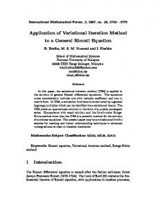
Fig. 2: Illustration of different steps of the algorithm (4 × 4). (a) Cartilage partitioning. (b) After 15 iterations of block labeling. (c) After T iterations. (d-f) Un-regularized ROI localized in a sample cartilage under different resolutions from 283 knees. The areas shaded in light gray belong to the ROI. (d) Resolution 5 × 5. (e) 10 × 20. (f) 80 × 140. P (V | Ω )=
D
∏ P(V
x
| Ωx )
(4)
x =1
where D is the total number of blocks. This conditional probability can be interpreted as the goodness of fit, of Ω to V. Additionally we have if Ω x = 1 ⎧ V (5) P (V x | Ω x ) = ⎨ x ⎩1 − V x if Ω x = 0 where Ω x = 1 is a block belonging to the ROI. Let Ω1 represent these blocks and Ω 2 represent the blocks for which Ω x = 0 . Thus we can write P(V | Ω )=
∏ V ∏1 − V x
x∈Ω1
x
(6)
x∈Ω 2
We assume the second term in the numerator i.e. the prior probability to only depend on the length of the boundary C separating the two regions ( Ω1 , Ω 2 ) P (Ω ) α exp (− υ C )
(7)
Taking the above into account, the a-posteriori segmentation probability of Ω given the vote map V is determined by ⎧⎪ ⎫⎪ −υ C (8) max P (Ω | V )= max ⎨ e Vx 1−Vx ⎬ Ω Ω ,C ⎪⎩ ⎪⎭ x∈Ω1 x∈Ω 2 Equivalently one can minimize the negative logarithm of the above expression which is given by the energy functional
∏ ∏
E (Ω, V )= − log (V ( x ) ) dx −
∫
Ω1
∫ log(1 − V ( x)) dx + υ C
(9)
Ω2
For minimizing this energy a level set function is introduced. Level sets yield a nice representation for functions and their boundaries and can describe topological changes in the segmentation permitting the splitting and merging of the contour [24].
Proc. of SPIE Vol. 6914 69140T-5
900 800
'
Medial Section
. -':'4 t:t.....
13
•1.-•
700 800
Anterior
800 400 400 200 IOU
Fig. 3: (a) Variational regularization: Vote Map (L = 1000). (b) ROI from the Vote Map. (c) Regularized ROI (resolution 80 × 140) in a sample knee. Let boundary C in the functional (9) be represented as the zero level set of a function φ : Ω → R with φ ( x) > 0 for x ∈ Ω1 and φ ( x) < 0 for x ∈ Ω 2 . We also introduce the Heaviside function H (φ ) ⎧1, H (φ ) = ⎨ ⎩0,
φ≥0 φ