A Virtual Reality-Based Exercise Program For Stroke Rehabilitation David Jack 2, 3, Rares Boian 1, Alma Merians 2, 4, Sergei V. Adamovich 2, Marilyn Tremaine 1, Michael Recce 2, 3, Grigore C. Burdea 1 and Howard Poizner 2 * 1
Center for Advanced Information Processing Rutgers University Piscataway, NJ 08854
2
Center for Molecular and Behavioral Neuroscience Rutgers University Newark, NJ 07102
ABSTRACT
A PC based desktop Virtual Reality system was developed for rehabilitating hand function in stroke patients. The system uses two hand input devices, a CyberGlove and a RMII force feedback glove, to allow the user to interact with one of four rehabilitation exercises. Each of which is designed to exercise one specific parameter of hand movement, namely range, speed, fractionation or strength. The therapy program is semi-automated and personalized to each user through the use of performance-based target levels. These are adapted between sessions in order to induce the user to improve. Feedback is provided to each user throughout the exercise sessions. To further motivate the user to continue the exercise program, screen displays are designed as interactive games. The system is described and sample data is presented from preliminary studies performed on control subjects. Keywords
Virtual Reality, Rehabilitation, Stroke, Haptic Glove, CyberGlove, Rutgers Master II. INTRODUCTION
Stroke is the leading cause of adult disability, 65% of the nearly four million people in the United States who have survived a stroke are living with minor to severe impairments (National Stroke Association). Impairments such as muscle weakness, loss of range of motion and impaired force generation create deficits in motor control which affect the stroke survivor’s capacity for independent living and economic self-sufficiency. Many traditional therapeutic interventions have been used in rehabilitation to promote functional recovery with outcome studies yielding inconsistent results [3]. Recent
3
Dept of Comp. and Info. Sci. New Jersey Institute of Technology Newark, NJ 07102
4
University of Medicine and Dentistry of New Jersey Newark, NJ 07103
evidence has demonstrated that intensive massed and repeated practice may be necessary to modify neural organization [7, 13] and effect recovery of functional motor skills [17, 24]. The structure of the current health care system, which provides limited amounts and duration of therapy, challenges us to design innovative rehabilitation programs. These should incorporate intensive and repetitive training, a method which has been shown to be effective in promoting cortical plasticity and behavioral recovery. * Virtual Reality (VR) technology [1] is currently being explored for its potential benefit as a therapeutic intervention for retraining coordinated movement patterns. This technology provides the capability to create an environment in where the intensity of feedback and training can be systematically manipulated and enhanced in order to create the most appropriate, individualized motor learning paradigm. In addition VR-based rehabilitation systems have several other advantages. Similar to computer games VR rehab exercises can be made to be engaging which is important in terms of the patient motivation [14]. VR sensor technology can also be used to fully quantify any progress made by the patient, especially in terms of motor control improvement. Although most neurologic recovery is attained by three months post stroke [8], several studies investigating the outcome of treatment six months after the stroke have shown significant gains in dexterity, strength and function [2, 16, 21]. VR training has the potential to affect patients’ functional outcome by making available new options for additional treatment past the traditional period of inpatient hospitalization and rehabilitation. Stroke Rehabilitation
Several researchers have shown in both animal and human studies that important variables in re-learning motor skills and in changing the underlying neural architecture are the
*
Corresponding author:
[email protected]
quantity, duration and intensity of training sessions. Focal ischemic lesions in monkeys, similar to what occurs in a stroke, usually result in a loss of cortical territory in the motor area adjacent to the infarcted region. However, three-four weeks of intensive, repetitive, hand training prevented this loss and in some instances led to an expansion of this cortical region [13]. A similar phenomenon has been demonstrated in the sensory cortex [7]. Looking at the effects of different intensities of physical therapy treatment, Langhorne [12] reported significant improvement in activities of daily living as a result of higher intensities of treatment. In a further review of the literature, Kwakkel [11] found that in the rehabilitation of patients who had a stroke, there was a small but statistically significant treatment effect related to the intensity of the rehabilitation. When traditional therapy is provided in a hospital or rehabilitation center, the patient is usually seen for half hour sessions, once or twice a day. This is decreased to once or twice a week when seen as an outpatient. The time course from hospital admission to discharge from the rehabilitation center is usually around 42 days [15]. It is evident that in this service delivery model it is difficult to provide the amount or intensity of practice needed to effect neural and functional changes. The proposed benefits of training in the VR environment would be the ability to increase the duration, frequency and intensity of therapy that could be provided to patients by using semiautomated programs. Furthermore, lower cost PC-based VR equipment is now available that will allow the rehab stations to be set up in a patients home or locations other than the rehabilitation center. The internet can be used for data transfer allowing a therapist to remotely monitor the progress of a number of patients simultaneously [14]. Continuous computerized feedback about the patient's performance would permit ongoing modification of the patient’s therapy program. For instance the task difficulty can be set by a patients previous performance or can be altered by a therapists. The rate at which patients can relearn their motor skills, the extent of improvement and the environment in which they are treated, affects the duration, effectiveness and cost of patient care. Therefore, developing new methods to accelerate and improve the level of motor retraining is a very important societal consideration. Virtual Reality and Rehabilitation
The use of a VR technology for the training of motor tasks is in its nascent stage. It is currently being explored in several areas of rehabilitation [19]. Patients with Parkinson’s disease are often aided in overcoming the akinesia that is evident during ambulation by physically placing visual cues in the gait path. The use of VR head mounted displays which permit the continuous presentation of visual cues overlapping the real visual scene during ambulation are being investigated to
determine the effect on these akinetic episodes and the subsequent ability to facilitate a more normal gait pattern [20]. VR training has been used for children with Cerebral Palsy to enhance spatial awareness [4] and to successfully teach these children to operate motorized wheelchairs [6]. The training program used three virtual scenarios that progressed in difficulty level and complexity. The children were motivated to learn and their driving skills subsequently improved. It has been shown in normal subjects, that VR can be a beneficial environment for learning a motor task. Todorov et al. [18] used a VR system for the training of a difficult multi-joint movement. The computer animation of a table tennis task included virtual paddles for the teacher and the subject as well as a virtual ball. Augmented feedback was used to indicate to the trainee the movement variables most relevant for successful performance of the task. The results of this experiment indicated that subjects who received the virtual environment training did better on the task than subjects who received a comparable amount of training in a real environment. However, another experiment comparing VR training and real world training in a pick and place task showed improvement in both groups, but those trained in the real world task did better [9]. Two patients with hemiplegia were trained in a virtual environment on an upper-extremity reaching task that progressed sequentially through six levels of difficulty [5]. Each subject received 16 trials of one to two hours duration. Both patients improved in the task in the virtual environment over the 16 sessions and were able to consistently progress to the fifth and sixth level of difficulty. However, only one of the subjects showed improvement in the clinical motor test and reported that he was able to use his hemiplegic arm in several functional activities that he was not able to perform previously. The second subject showed neither clinical nor functional changes. In addition to sorting out the effects of motor training in a virtual environment, it is important to determine whether the skills gained in that environment transfer to real-world conditions. Wilson et al. [22, 23] studied children with a variety of disabilities and found that internal representations resulting from exploration of simulated space transferred to the real environment. However, although subjects trained on a motor task in a virtual environment demonstrated the ability to improve performance on the task in that environment, the learning did not always transfer to the real-world task [5, 9]. This conflict in findings could be reflective of differences in the learning requirements of perceptual skills and motor skills or it could be reflective of the current paucity of investigations into the use of VR for motor skill training. The experiments on motor task training and transfer of that task to the real world environment indicate that the
effects of training in a virtual environment are not fully understood, nor entirely conclusive. The literature investigating virtual reality as an environment for rehabilitation training does indicate a potential benefit that should be more fully explored in order to ascertain its use as a therapeutic tool. METHODS
The aim of this work is to demonstrate the use of a PCbased VR system that is designed to rehabilitate hand function in stroke patients. The following sections describe the hardware and software that has been developed, and present example data taken from preliminary studies performed with control subjects. The VR System
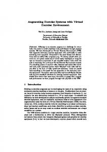
The VR system (Figure 1) uses a PC (Pentium II 400 MHz) with a FireGL 4000 graphics accelerator. Two hand input devices were used, a CyberGlove [10] from Virtual Technologies, Inc. and the Rutgers Master II-ND (RMII) force feedback glove [14]. The CyberGlove is a stretchable data glove with 18 embedded bend sensors that measure the MetaCarpo-Phalangeal (MCP) and Proximal Inter-Phalangeal (PIP) joint angles of the thumb and fingers as well as finger abduction and wrist flexion. The RMII glove is an exoskeleton device that applies force to the user’s fingertips and uses non-contact position sensors to measure the fingertip position in relation to the palm. This data is then used to estimate the MCP, PIP and Distal Inter-Phalageal (DIP) joint angles of three fingers and the thumb. Forces are applied through lightweight pneumatic actuators attached to the tips of the thumb, index, middle and ring fingers. Each piston is capable of delivering 16 Newtons (N) of force although this is currently software-limited to 10 N. Two devices were used as each has advantages for certain types of rehabilitation exercise. The elasticity of the CyberGlove does not restrict the user’s movement but it cannot provide an opposing force in the exercises. Safety concerns affecting the design of the piston displacement in the RMII limits the user’s range of motion. Therefore for exercises that did not require force the CyberGlove was chosen. Both devices are connected to the PC through control interfaces to the standard RS-232 serial port. In the case of the RMII, the glove is attached to the PC through a Haptic Control Interface (HCI) that controls the desired fingertip forces as well as calculating joint angles. The RMII glove also requires an air compressor to provide air to the HCI at 100Psi. High performance drivers have been developed for both devices, allowing the data to be logged to disk at the highest possible rate (100 datasets/sec for both the CyberGlove and the RMII). The VR Exercises
Four VR exercise programs were developed using the commercially available WorldToolkit graphics library (Engineering Animation Inc.). Each program concentrates
Figure 1. The PC based VR rehab system. The user is wearing a CyberGlove that is connected to the interface unit on the right. Also shown to the right of the user is the Haptic Control Interface for the RMII glove.
on one particular parameter of hand movement. The chosen parameters were range, speed, fractionation and strength of movement. Each exercise takes the form of a simple game where the patient performs a number of trials of a particular task. If a certain performance target is reached then the patient achieves the goal which is portrayed through some graphical event. The idea of using target-based trials is that all the exercises are driven by the user’s own performance. The targets for any particular block of trials are set based on the performance in previous sessions. Therefore no matter how limited the user's movement actually is, if their performance falls within their parameter range they will successfully pass the trial. Evaluating the User’s Movement
Implementing target-based exercises requires an initial test to evaluate the patient’s movements. The three parameters of range, speed and fractionation of movement are evaluated for the patient’s right hand using the CyberGlove. First, as the CyberGlove is designed to fit the average hand, large variations in the users hand geometry can cause it to measure erroneous angles. Therefore a calibration step is required. Every joint is placed into two known angles, zero and 60 degrees. From these measurements, two parameters (gain and offset) are obtained that specify the linear relation between the raw sensor output and the corresponding joint angles being measured. To reduce fatigue and tendon strain the fingers are moved together and the thumb is moved alone for all exercises except fractionation. For the range evaluation of the thumb the user is required to move the thumb as far forward and then as far backward as possible. The range of movement is defined as the average angular range of the thumb roll across the palm and the MCP joint. The user then moves all four fingers together as far as possible
(a)
(b)
(c)
(d)
Figure 2. The four VR exercises (a) range of movement, (b) speed of movement, (c) finger fractionation and (d) strength of movement.
forward and then backward. In this case the range of movement for every finger is defined as the average angular range between the MCP and the PIP of that finger. This evaluation exercise is performed ten times in order to get a distribution of joint angle range per finger (mean value and standard deviation). The user’s speed of movement is evaluated by performing the same moves but as fast as possible. Again, ten trials are performed to get the distribution of maximum (forward) angular velocity as well as the minimum (backward) angular velocity. Finger fractionation or independence of finger movement is quantified by the following measure,
movement in this glove is slightly limited so another set of range evaluations are performed which give the user’s mean range whilst wearing the RMII. The user’s finger strength is established by doing a binary search of force levels and comparing the range of movement at each level with the mean obtained from the previous range test. If the range is at least 80% of that previously measured, the test is passed, and the force is increased to the next binary level. If the test is failed, then the force is decreased to the next binary level and so on. Test forces are applied until the maximal force level attainable by the user is found for both the thumb and the combined fingers.
where Favg is the current average joint range of the finger
Once complete, the initial evaluation quantifies the user’s angular range, speed, fractionation and force levels for the thumb and fingers. The total evaluation for a normal subject takes under 15 minutes to complete.
G avg F r = 100% * 1 − Favg
being moved and Gavg is the current average joint range of the other three fingers combined. Moving one finger alone will result in a measure of 100% which decays to zero as more fingers are coupled in the movement. The user is prompted to move only one finger while trying to keep the others still. This is repeated five times for every finger. The last test uses the RMII glove to evaluate the user’s strength of movement. As mentioned above, the range of
VR Session Exercises
One exercise session consists of four blocks of N trials each. Multiple sessions are run on any given day for a number of consecutive days. An individual block concentrates on exercising one of the aforementioned parameters of range, speed, fractionation or strength of movement. The number of trials per block varies as our pilot studies showed that some exercises fatigue the user
more than others. Similar to the evaluation exercise, the user is required to alternate between moving the thumb alone and then moving all the fingers together for every exercise except fractionation. Currently each trial is started and stopped by the therapist pressing the spacebar, though this will be replaced by user initiated trials. As mentioned above, the patient must attain a certain target level of performance in order to successfully complete every trial. For a particular block of trials the first set of targets are drawn from a normal distribution around the mean and standard deviation given by the initial evaluation test. A normal distribution ensures that the majority of the targets will be within the patient’s performance limits but the patient will find some targets easy or difficult depending on whether they come from the low or high end of the target distribution. Initially, the target means are set one standard deviation above the user’s actual measured performance to obtain a target distribution that overlaps the high end of the user’s performance levels. The first training exercise is range of movement (Figure 2a). In this exercise the user manipulates a “window wiper” to wipe a window clean to reveal an attractive landscape behind the window. The higher the measured angular range of movement of the thumb or fingers, the more the wiper rotates and clears the window. The rotation of the wiper is scaled so that if the user achieves the target range for that particular trial the window is cleared completely. During the trial the user is also shown a complete graphical model of their own hand which is updated in real-time to accurately represent the flexion of the user’s fingers and thumb. After every trial is completed the user is shown a graphical digital “performance meter” that shows the target level and their actual performance (Figure 3). This is used to inform the user exactly how close or how far away they were from the desired performance. After the trial, information can be displayed to the therapist that details what the specific actual movement was. In the case of a failed trial this can be used to inform the user about how their movement may be improved to increase their performance. In the second exercise (Figure 2b), speed of movement, the target the user has to achieve is defined as the maximum forward angular velocity during a grasping movement. On a “go” signal (green light on a traffic signal), the user is required to close either the thumb or all the fingers together as fast as possible to catch a red ball. An opponent hand (on the left in the screen) also closes its thumb or fingers around a red ball with an angular velocity equal to that of the target angular velocity. If the user surpasses this target velocity, then they beat the opponent (yellow) hand and get to keep the red ball. Otherwise they lose and their ball drops while the other red ball remains in the opponent’s hand.
Figure 3. The digital “performance meter” shown to the user after every trial. The target level is shown (white bar) as well as the actual performance of the user (black bars).
The fractionation exercise (Figure 2c) is designed around a piano keyboard. The patient is required to move each of the four fingers, in isolation, as far forward as possible. As the finger is moved, the corresponding key on the piano is depressed and shown in green. Nearing the end of the move, the fractionation measure is calculated on-line, and if it is greater than or equal to the trial target measure, then only the one key remains depressed. Otherwise other keys are depressed in red to show which of the other fingers were coupled during the move. The goal of the user is to move so that only one key is depressed for every trial. The last exercise is designed to improve the user’s strength of movement. This exercise uses the RMII glove to apply forces either to the user’s thumb or three fingers (excluding the fifth digit) simultaneously. The forces applied for each individual trial are again taken from a normal distribution around the force level found in the initial evaluation. To successfully complete a trial the user is required to move at least as far as the movement recorded during the evaluation. The user is presented with a graphical representation of their hand showing four pistons (Figure 2d). As each actuator on the RMII is squeezed, the graphical pistons start to fill in a new color. The piston turns yellow and is completely filled if the patient manages to move the desired range at that particular force level. For a control subject the four blocks in one session takes approximately 15-20 minutes to complete. All the parameters such as range, speed, fractionation and strength are estimated on-line in order to drive the graphics display and provide feedback to the patient. After the trial is completed, the data collected on the user’s movement is low-pass filtered at 8 Hz to reduce sensor noise. The parameters are re-evaluated and stored along with the filtered data to disk.
Session: 15:30:00 - 15:55:30 140
120
Incrementing Performance Targets 100
The VR system has been tested on control subjects in order to evaluate fatigue effects, the graphical interfaces and the algorithms we use for setting the performance targets. Figure 4 shows a typical set of trials gathered from a normal pilot subject during one block of the range exercise. The actual performance mean for this block was 119.88° ± 5.6° which lead to the next session’s target mean being increased by one standard deviation to 125°. Similar plots are obtained for the thumb and every finger for all the exercises. What is of more interest is to see how the user’s performance changes over a number of sessions. This is detailed in Figure 5 where the block targets and actual mean performance of the index finger during the range exercise are shown for four sessions taken over a two day period. The first two columns are the result of the initial evaluation, the target being set from the mean actual performance plut one standard deviation. As the exercises proceed, we can see how the targets are being altered based upon the user’s performance. The block target is increased when the user matches or improves upon the target level or is decreased when the user’s performance is less than desired. The trials for the block shown in Figure 4 correspond to session 1.2 in Figure 5. DISCUSSION OF PILOT STUDIES
The VR interface and exercises evolved through a series of pilot studies first on users with no hand deficits and finally with a user who had suffered a stroke but had nearly normal hand function. The obstacles and problems encountered are discussed in the next few paragraphs. The exercises were initially designed to involve single finger movement, but the number of trials per user had to be reduced significantly to counter fatigue. Moving to
80
60
40
20
Target Actual 0
1
2
3
4
5
6 Trial
7
8
9
10
Figure 4. A complete block of the range exercise for the index finger of a control subject. The targets were drawn from a distribution, mean 119° ± 4.5°. The y axis is labeled in degrees. 140
120
100 Joint Range (deg)
PRELIMINARY RESULTS
Joint Range (deg)
After a session is completed the distribution of the patients actual performance for each of the four blocks is compared to the pre-set target mean and standard deviation. If the mean of the patient’s actual performance for any particular block is greater than the target mean then that target is raised by one standard deviation. Likewise if the user’s performance is below the target mean, that block’s target for the next session is lowered by the same amount. To stop the block targets from varying too little or too much between the sessions, lower and upper bounds are placed upon their increment. These parameters allow the therapist to choose how aggressively each training exercise is to proceed. A high upper bound will mean that the targets for the next session may be considerably higher than the previous. Updating the targets in this manner not only pushes the user to improve their performance, but as the targets change through time they provide valuable information to the therapist as to how the user is coping with the rehabilitation program.
80
60
40
20
0
Target Actual Test
1.1
1.2
2.1
2.2
Session (Day.Session)
Figure 5. The mean actual performance and target levels for the range of movement of a control subject’s index finger. The y axis is labeled in degrees.
four fingers and thumb exercises removed this difficulty. Users also exhibit a wide degree of motion when performing the exercises whereas the parameters taken for measuring user performance are more limited. Thus, the user may have the visual perception of accomplishing the task successfully, but program reports that the user has not achieved the required target level. This is particularly problematic with the thumb motion and algorithm adjustments are continuing to be made to correct this problem. The user’s arm is most comfortable and less likely to experience fatigue in these exercises when resting on an armrest with the palm facing the desk surface. If this orientation is used in the screen graphics, the hand occludes the important portions of the task feedback. If the screen graphics present the hand in another orientation, the user tends to orient their hand to conform to the hand displayed on the screen. The screen graphics on the piano key task were adjusted to position the hand correctly since fractionation is one of the tiring exercises
(Figure 2c). A compromise position of a semi-pronated hand was selected for the window clearing task (Figure 2a). For the speed of movement (Figure 2b) and strength (Figure 2d), the feedback portion of the graphics required us to show the hand facing the user. All of our pilot subjects over-pronated their hands for these tasks.
3.
Duncan, P., Synthesis of Intervention Trials to Improve Motor Recovery Following Stroke. Stroke Rehabilitation, 1997. 3(4): p. 1-20.
Finally, an unplanned difficulty was the wide variation in hand sizes of the individuals in the study. To push against the pistons in the RMII glove, the top of the piston has to be positioned at the first digit of the finger. Although three RMII gloves were available (small, medium and large), they did not accommodate the large variation in hand width and individual fingers lengths. This final problem suggests that gloves will need to be fit individually to recovering patients for best results.
4.
Foreman, N., P. Wilson, and D. Stanton, VR and Spatial Awareness in Disabled Children. Communications of the ACM, 1997. 40(8): p. 76-77.
5.
Holden, M., et al., Virtual Environment Training Improves Motor Performance in Two Patients with Stroke: Case Report. Neurology Report, 1999. 23(2): p. 57-67.
6.
Inma, D., et al., Teaching Orthopedically Impaired Children to Drive Motorized Wheelchairs in Virtual Reality, in Center on Disabilities Virtual Reality Conference. 1994.
7.
Jenkins, W. and M. Merzenich, Reorganization of Neocortical Representations After Brain Injury: A Neurophysiological Model of the Bases of Recovery From Stroke., in Progress in Brain, F. Seil, E. Herbert, and B. Carlson, Editors. 1987, Elsivier.
8.
Jorgensen, H., et al., Outcome and Time Course of Recovery in Stroke, Parts I and II. The Copenhagen Stroke Study. Arch Phys Med Rehab, 1995. 76: p. 399-412.
9.
Kozak, J., et al., Transfer of Training From Virtual Reality. Ergonomics, 1993. 36(7): p. 777784.
10.
Kramer, J., P. Lindener, and W. George, Communication System for Deaf, Deaf-Blind, or Nonvocal Individuals using a Instrumented Glove. 1991: USA.
11.
Kwakkel, K.G., et al., Effects of Intensity of Rehabilitation After Stroke, a Research Synthesis. Stroke, 1997. 28(8): p. 1550-1556.
12.
Langhorne, P., R.C. Wagenaar, and C. Partridge, Physiotherapy After Stroke: More is Better? Physiotherapy Research International, 1996. 1: p. 75-88.
13.
Nudo, R.J., Neural Substrates for the Effects of Rehabilitative Training on Motor Recovery After Ischemic Infarction. Science, 1996. 272: p. 1791-1794.
14.
Popescu, V., et al., A Virtual Reality-Based Telerehabilitation System with Force Feedback. IEEE Transactions on Information Technology in Biomedicine, 2000. 4(1): p. 45-51.
15.
Rijken, P. and J. Dekker, Clinical Experience of Rehabilitation Therapists With Chronic
Hemiplegic Patients. Stroke, 1993. 24: p. 11861191.
CONCLUSION AND FUTURE WORK
VR technology can have a large impact on traditional rehabilitation techniques. We have developed a PC based VR system for rehabilitating hand function in stroke patients to explore the potential benefits of such technology. The system exercises four parameters of hand movement; range, speed, fractionation and strength. We have outlined a novel performance driven exercise program where a patient’s own performance dictates future session targets. Currently the VR rehab system is being evaluated with three stroke patients in an intensive therapy program. Four sessions of the four training exercises detailed here will be run every day, five days a week for two week periods. During ongoing development of the system we are adding more elaborate feedback, using auditory cues and improving the content and graphical nature of the four game-like interfaces presented. In addition the system is being combined with an Oracle database that will allow web access for data retrieval, analysis and parameter setting from a remote site. Hardware issues are also being addressed. A left-handed RMII glove is under development to support patients with left-handed deficits. Also, other haptic devices for applying force feedback to the elbow and shoulder are under consideration. ACKNOWLEDGEMENTS
The research reported here was supported by grants from the New Jersey Commission on Science and Technology (R&D Excellence Grant) and from Rutgers University (SROA Grant). REFERENCES
1.
Burdea, G. and P. Coiffet, Virtual Reality Technology. 1994, New York: Wiley.
2.
Dam, M., et al., The Effects of Long-Term Rehabilitation Therapy on Poststroke
Diseases: A Quantitative Approach. Clinical Rehabilitation, 1998. 12(2): p. 143-150. 16.
Tangeman, P., D. Banaitis, and A. Williams, Rehabilitation of Chronic Stroke Patients: Changes in Functional Performance. Arch Phys Med Rehab, 1990. 71: p. 876-880.
17.
Taub, E., et al., Technique to Improve Chronic Motor Deficit After Stroke. Arch Phys Med Rehab, 1993. 74: p. 347-354.
18.
Todorov, E., H. Shadmehr, and E. Bizzi, Augmented Feedback Presented in a Virtual Environment Accelerates Learning of a Difficult Motor Task. Journal of Motor Behavior, 1997. 29(2): p. 147-158.
19.
Wann, P., et al., Rehabilitative Environments For Attention and Movement Disorders. Communications of the ACM, 1997. 40(8): p. 49-52.
20.
Weghorst, S., Augmented Reality and Parkinson's Disease. Communications of the ACM, 1997. 40(8): p. 47-48.
21.
Werner, R. and S. Kessler, Effectiveness of an Intensive Outpatient Rehabilitation Program for Postacute Stroke Patients. American Journal of Physical Medicine Rehabilitation, 1996. 75: p. 114-120.
22.
Wilson, P., N. Foreman, and D. Stanton, Virtual Reality, Disability and Rehabilitation. Disability and Rehabilitation, 1997. 19(6): p. 213-220.
23.
Wilson, P., N. Foreman, and M. Tlauka, Transfer of Spatial Information From a Virtual to a Real Environment in Physically Disabled Children. Disability and Rehabilitation, 1996. 18(12): p. 633-637.
24.
Wolf, S., et al., Forced use of Hemiplegic Upper Extremities to Reverse the Effect of Learned Non-use Among Chronic Stroke and Head Injured Patients. Experimental Neurology, 1989. 104: p. 125-132.