sequences in protein structure database and generates atomic models for the .... by analyzing residue-by-residue geometr
L. Inbathamizh et al. / Journal of Pharmacy Research 2012,5(7),3892-3896
Available online through http://jprsolutions.info
Research Article ISSN: 0974-6943
Ab initio Structure Prediction of Cellular Prostatic Acid Phosphatase and its Interactions with Anti-Cancer Ligands 1
L. Inbathamizh1 and *Dr.E. Padmini2 Research Scholar,Research and Development Centre,Bharathiar University,Coimbatore-641 046 *2 Associate Professor,Department of Biochemistry,Bharathi Women’s College,Chennai-600 108
Received on:07-04-2012; Revised on: 12-05-2012; Accepted on:16-06-2012 ABSTRACT When no suitable homology modeling or experimental analysis could be found, the challenges in protein structure prediction could be explicitly resolved by ab initio methods. Such a de novo protein modeling was essential to determine and differentiate the three dimensional structure of cellular prostatic acid phosphatase, an important drug target in prostate cancer, from its isoform. Insilico tools were adopted for the study. The obtained native structure was validated and docked with the anticancer phytocomponents of Moringa oleifera. The results indicated the impact of protein conformation on docking, with significant interactions between the target and the ligands. This suggested the potent role of cellular prostatic acid phosphatase and the Moringa oleifera ligands in prostate cancer drug design. Key words: Homology modeling, ab initio method, cellular prostatic acid phosphatase, prostate cancer, Moringa oleifera, drug design. INTRODUCTION Structure prediction of protein plays a vital role in determining its function and biological significance. With the discovery of more and more protein sequences, it is highly difficult to totally depend on experiment analysis to determine the three dimensional (3-D) structures of all the proteins. Thus, there is a growing need of computational techniques to predict protein structures.
that the residues from 380-418 in PAP are highly significant in differentiating its isomeric forms.[13] Homology search modeling methods usually adopted require a template with a known structure that suits the sequence range of the target. But these methods are not satisfactory to predict the complete 3D structure of cPAP, as the template with maximum percentage of homology is always the other isoform which shares its sequence till 386. Thus, an alternate method involving the complete sequence of cPAP is required to Human Prostatic acid phosphatase (PAP) is a major phosphatase in prostate predict its tertiary structure. epithelial cells.[1, 2] Two isoforms of PAP have been purified from prostate tissue. One is the cellular form (cPAP) and the other is the secretory form The critical need for tertiary structures of proteins in drug discovery has (sPAP). The two forms vary in their isoelectric point values, biochemical launched a multitude of computational recipes. Molecular dynamics properties and in the structure of carbohydrate moieties.[3, 4] It is suggested simulations have provided extremely high resolution spatial and temporal that these isoforms are the products of the same gene and arise due to data to improve understanding of the protein folding mechanism.[16] In proteolytic processing of a high molecular weight precursor molecule.[5] 1975, Levitt and Warshel simulated and succeeded the folding of Bovine The existence of true isoenzymes consisting of different polypeptide chains Pancreatic Trypsin Inhibitor (BPTI) using a simple representation of protein may be also the reason for the heterogeneity of PAP. The gene encoding conformation, energy minimization and thermalization.[17] Since then PAP is located at chromosome 3q21 qter.[6] The heterogeneity in the length extensive research has been carried and significant progress has been made of the cDNA is explained by multiple polyadenylation signals (AATAAA) towards physics-based computation of protein structure, from the knowledge following two copies of alu-type repetitive sequences in the 3’ non-coding of the amino acid sequence. This approach, commonly referred to as an ab region.[7,8] This is reflected in the differences in the protein length of sPAP initio method is based on the thermodynamic hypothesis formulated by and cPAP with 386 and 418 amino acid residues respectively. Anfinsen, which states that the native structure of a protein corresponds to the global minimum of its free energy under given conditions.[18] This PAP hydrolyses a large variety of phosphomonoesters, with an optimum method compares fragments of query sequence of unknown structure with pH range of 4-6.[9] In spite of extensive research for several years, the sequences in protein structure database and generates atomic models for the physiological role of the enzyme is not fully understood. PAP has been whole protein with varying strategies for the regions with missing matches. reported to dephosphorylate macromolecules, including polynucleotides Success of pharmaceutical industry relies not only on novel drug targets but and phosphoproteins.[10,11] It is now evident that cPAP functions as a also on small molecule compounds which modulate their activity. Moringa tyrosine phosphatase of oncoproteins associated with prostate oleifera is a medicinal plant of high nutritional value.[19] Niazimicin and 4carcinogenesis[12] and is a valuable drug target applicable to prostate cancer (4'-O-acetyl-a-L-rhamnopyranosyloxy)benzyl isothiocyanate, the therapy. phytocomponents of Moringa oleifera have been reported to possess anticancer activity.[20] Earlier studies have involved these ligands for docking Three-dimensional structure of cPAP (Enzyme Commission number with 1CVI being treated as the standard 3-D structure of PAP as given in EC=3.1.3.2) as the drug target is of scientific interest intrinsically due to its PDB.[21] application in structure based anticancer drug design endeavors. PDB (Protein Data Bank) structure of PAP with the highest resolution as given in uniprot This study is a novel attempt to predict the 3-D structure of cPAP involving database(Accession No: P15309)[13] is 1CVI.[14,15] This structure its complete sequence based on ab initio methods and to interpret its comprises of 342 residues with PAP sequence between 33-374. It is found interaction with Moringa oleifera anti-cancer ligands confirming its potency in structure-based drug design.
*Corresponding author. Dr. E. Padmini, Department of Biochemistry, Bharathi Women’s College, Chennai-600 108, Tamilnadu, India
MATERIALS AND METHODS Retrieval of Target Protein sequence The protein sequence of cPAP from Homo sapiens was obtained from the protein sequence database of Uniprot (Accession No: P15309-2).[13]
Journal of Pharmacy Research Vol.5 Issue 7.July 2012
3892-3896
L. Inbathamizh et al. / Journal of Pharmacy Research 2012,5(7),3892-3896 FASTA format of the cPAP sequence was used for the structure prediction analysis. Ab initio Protein structure Prediction Ab initio methods can be used to predict the protein structure from its sequence information in the absence of suitable structure templates. ‘Bhageerath’ is an energy based computer software suite which applies ab initio methods to narrow down the search space of tertiary structures of small globular proteins.[22,23] It is a product of the Supercomputing Facility for Bioinformatics & Computational Biology, Indian Institute of Technology, Delhi and bears the name ‘Bhageerath’ after the great Indian king who could accomplish the impossible task of getting the Ganges from heaven to earth. The amino acid sequence of cPAP in FASTA format was submitted to the Bhageerath web server with process ID: 17415574 and the results were obtained for the query. Validation & comparison of the protein structure The obtained PDB files of 5 candidate structures of cPAP from ‘Bhageerath’ were uploaded to Procheck of Structural Analysis and Verification Server[24] and the validated structure was compared with 1CVI using Dali Lite program.[25] Ligand preparation and Molecular docking Interactions of the ab initio predicted and validated structure of cellular prostatic acid phosphatase with anti cancer Moringa oleifera phytoconstituents namely niazimicin[26,27] and 4-(4'-O-acetyl-a-Lrhamnopyranosyloxy)benzyl isothiocyanate[28] were studied using the docking tool PatchDock Beta 1.3 Version.[29] The results were compared with that of 2 standard therapeutic agents for prostate cancer: a natural compound, Curcumin[30] and a chemotherapeutic drug, Estramustine.[31] Molecular properties of these ligands had been predicted in previous studies.[21] The structures of the ligands were obtained from literature[32] and pubchem database[33], drawn using Chem Sketch tool[34], saved as mol file and converted to PDB files using Open Babel 2.3.1 chemical toolbox.[35] The PDB files of the receptor protein and the ligands were fed as the inputs to PatchDock web server to obtain the docking results. Visualization of the protein-ligand interactions PyMOL is an open source visualization tool used in Structural Biology and is extensible by Python programming language.[36] The PDB outputs of the validated cPAP model and the docked structures were viewed using PYMOL version 0.99 and the interactions were interpreted.
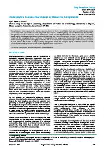
remove steric clashes or overlaps in 3-D space. An atomic level energy optimization was carried out in the fifth module and the structures were scored based on energy in the sixth. Module seven reduced the probable candidates based on the protein regularity index of the (phi)φand (psi) ψdihedral values and module eight further reduced the structures selected to 10 using topological equivalence criterion and accessible surface area. Thus, the software suite combined physics based potentials with biophysical filters to arrive at 5 plausible candidate structures starting from sequence and secondary structure information. The methodology had been validated on 50 small globular proteins consisting of 2–3 helices and strands with known tertiary structures. For each of these proteins, a structure within 3–6Å RMSD (root mean square deviation) of the native had been obtained in the 10 lowest energy structures. Validation & Comparison of the protein structure The stereochemical quality of the 5 candidate protein structures was checked by analyzing residue-by-residue geometry and overall geometry using the tool ‘Procheck’.[24] The protein structures were validated using Ramachandran plots and the plot statistics were obtained for each of these structures as presented in Table1. The fifth structure (pdb5_17415574) with the maximum percentage (86.1%) of residues in the most favoured region was selected as the best model for cPAP and used for further studies. Ramachandran plot of the selected cPAP model and its structure as visualized by PyMOL are shown in Figure 1 and Figure 2 respectively. Comparison of the validated structure to each chain of 1CVI revealed the following results: z-score=1.4, Aligned residues=58, RMSD=2.9, Sequence identity=26% Table 1: Ramachandran plot statistics for the 5 ab initio models of cPAP Details of residues
pdb1
pdb2
pdb3
pdb4
pdb5
Residues in most favoured regions[A,B,L] Residues in additional allowed regions[a,b,l,p] Residues in generously allowed regions[~a,~b,~I,~p] Residues in disallowed regions No. of non-glycine & non-proline residues No.of end residues excluding glycine & proline No.of glycine residues No. of proline residues Total no. of residues % of residues in most favoured regions
314
315
243
217
317
50
49
110
113
36
3
3
10
31
7
2 369
2 369
6 369
8 369
8 368
2
2
2
2
3
21 26 418 85.1
21 26 418 85.4
21 26 418 65.9
21 26 418 58.8
21 26 418 86.1
RESULTS Retrieval of Target Protein sequence The sequence of cellular isoform of PAP varied from the secreted form in the amino acid sequence VLKVIFA (Valine, Leucine, Lysine, Valine, Isoleucine, Phenylalanine, Alanine) instead of GTEDSTD (Glycine, Threonine, Glutamic Acid, Aspartic Acid, Serine, Threonine, Aspartic Acid) from 380-386 and in possessing additional residues (VAFCLISAVLM VLLFIHIRRGLCWQRESYGNI, C-Cysteine, M-Methionine, H-Histidine, R- Arginine, W-Tryptophan, Q-Glutamine, Y-Tyrosine, N-Asparagine ) from 387 to 418.[13] Ab initio Protein structure Prediction The operation mode of ‘Bhageerath’ comprised of eight modules configured to function independently or in a conduit.[22,23] Starting with primary structure(amino acid sequence) and secondary structure information (helix/ sheet/loop) of a protein in the first module, multiple three dimensional atomic level structures were generated sampling the conformational space of the loop dihedrals in the second. In the third module a set of biophysical filters (persistence length, radius of gyration etc.) were applied to screen the trial structures to reduce the sample size. The resultant structures were refined in the fourth module by a Monte Carlo sampling in dihedral space to
Fig.1. Ramachandran plot of pdb5 model of cPAP with details as in Table 1
Journal of Pharmacy Research Vol.5 Issue 7.July 2012
3892-3896
L. Inbathamizh et al. / Journal of Pharmacy Research 2012,5(7),3892-3896
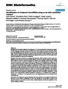
Fig.2. Cartoon representation of pdb5 model of cPAP as displayed by PyMOL
(b)
Protein-Ligand docking and Visualization Docking of the obtained cPAP model with anti cancer Moringa oleifera ligands, niazimicin and 4-(4'-O-acetyl-a-L-rhamnopyranosyloxy)benzyl isothiocyanate and two standard prostate cancer therapeutics, curcumin and estramustine was performed. PatchDock, a molecular docking algorithm based on shape complementarity principles was used.[37] Prediction of active site of the receptor protein was not required and the docking was based on the superimposition of the patched surfaces of the receptor and the ligand. The docking scores were tabulated and evaluated by PyMOL for the polar contacts and the atoms involved in docking, as given in Table 2. The docked conformations of the ligands with the protein as deciphered by PYMOL were obtained as shown in Figure 3. Table 2: cPAP –Ligand docking details as obtained by PatchDock and PyMOL S.No
Ligand
Docking score
cPAP residue
cPAP atom
1
Niazimicin
4846
Glu60 Glu60 Glu324 Glu324
OE1 OE2 OE1 OE1
O O O O
2
Thr291 Lys87 Ser312 Asp290
OG1 NZ N OD1
O O O O
3
2
3
4
4-(4'-O-acetyl-α-Lrhamnopyranosyloxy) benzyl isothiocyanate Curcumin
Estramustine
4346
5256
5240
Ligand No. of Polar atom contacts
(c)
2
1
(d) Fig.3. Docked conformations of cPAP(dark blue) with ligands: (a) Niazimicin(orange) (b) 4-(4'-O-acetyl-a-L-rhamnopyranosyloxy)benzyl isothiocyanate(green) (c) Curcumin(pink) (d) Estramustine(light blue). Dotted lines indicate the polar contacts between the protein and the ligand.
DISCUSSION For years, homology based comparative modeling and fold recognition had been the most successful methods for structure prediction.[38] In the absence of homologous sequences with known structure, determination of secondary (a)
Journal of Pharmacy Research Vol.5 Issue 7.July 2012
3892-3896
L. Inbathamizh et al. / Journal of Pharmacy Research 2012,5(7),3892-3896 structure and local structure motifs was the optional method.[39,40] The prevailing sequence comparison methods could only align a fraction of residues.[41] Better alignment of residues would be essential to improve the accuracy of the structure model. When no suitable structure templates were available, ab initio methods could be used to predict the protein structure from the sequence information only. A protein sequence usually folded to a native conformation at or near the global free energy minimum. Thus, these methods involved development of an efficient energy function and an accurate algorithm for searching the resultant energy landscape.[42] The physical forces acting on the atoms of the protein controlled the folding of the protein sequence. In order to evaluate the accuracy of these methods, Critical Assessment of Structure Prediction (CASP), a biannual, communitywide blind test of prediction methods had been conceived and implemented.[43] Combining Bioinformatics tools and ab initio methodologies, a protocol that was computationally expeditious for tertiary structure prediction of small proteins had been developed through the automated web server, Bhageerath. The physicochemical properties of the constituting amino acids determined the physicochemical properties, molecular interactions and biological activities of the protein. On analyzing the sequences of sPAP and cPAP, a few interesting points were observed. In case of sPAP, out of 7 amino acid residues from 380-386, 6 of them were non-polar while one was polar with a positive side chain(Lysine). But this was opposite in cPAP. One was nonpolar(Glycine) and the other 6 residues were polar with half of them carrying negative side chains(Glutamic acid, Aspartic acid). But the additional 31 residues from 387-418 in cPAP were mostly non-polar(19) and the remaining 12 polar, either neutral or carrying positive(Arginine, Histidine) or negative (Glutamic acid) side chains. The difference in the sequence of the two isoforms of PAP could reflect in their conformations and hence their properties and functions too. Although cPAP decreased during the progression of prostate cancer, its isoform sPAP increased due to the combined effects of enlarged tumor mass, prolonged half-life and loss of membrane polarity.[44,45] A reliable tertiary structure of cPAP was neither available in PDB nor obtained through homology based methods. Thus, ab initio method was applied to achieve the required native conformation of cPAP. This structure varied from 1CVI remarkably. The PDB file of the ab initio structure represented cPAP as a single chained protein with 418 amino acids, starting with Methionine and terminating with Isoleucine. But, PDB file of 1CVI denoted the existence of 4 chains, each with 342 amino acids. The residues from 33(Lysine) to 374(Threonine) alone were considered for conformation. Obviously, these differences were reflected in the RMSD value, number of aligned residues and sequence identity, when these structures were compared. The presence of dimeric subunits and 2 free sulfhydryl groups in the native form of human PAP had already been demonstrated.[46,47] It was also reported that human PAP had a similar 3-D structure to rat PAP.[15] During validation, each conformation was examined for close contacts between atoms. The allowed regions corresponded to conformations where steric clashes were absent. In sterically disallowed conformations, atoms in the polypeptide came closer than the sum of their van der Waals radii.[48] Out of the 5 plausible candidate structures, pdb5_17415574 model possessed the highest percentage in the most favoured region though a few residues were present in the disallowed region. The existence of 8 residues(Leucine31, Threonine145, Tyrosine 214, Arginine236, Alanine311, Leucine342, Glutamine366, Asparagine376) in the disallowed region was mainly due to the steric hindrance between the side chain groups and main chain atoms. The presence of single chain with 3 end residues in the model implied the probable existence of side chains or branching in the structure. This reduced the complexity in conformation and difficulty in docking as with tetrachained 1CVI where chain A and active site 1 were predominantly used for docking analysis.[21] Patch dock calculated the interactions not only at the
active site but also at the other sites on the receptor. Docking was measured as the geometric shape complementarity score which was higher for niazimicin than 4-(4'-O-acetyl-a-L-rhamnopyranosyloxy)benzyl isothiocyanate among the Moringa oleifera ligands and for curcumin than estramustine among the standards. Higher the score, higher was the enhancing effect of the ligand on cPAP. Results indicated that these ligands were amenable for designing drug candidates. Some of the contacts in docking did not meet strict H-bonding criteria. Therefore, the term ‘polar contact’ was used which remained the same for both the Moringa oleifera ligands while it was the highest for curcumin. Upon analyzing the cPAP residues involved in docking, it was observed that all of them were polar with Glumatic acid60 and Glutamic acid324 predominately associated with the binding site for the activating ligands. Histidine12 and Aspartate258, essential for substrate binding were not involved in activator binding.[47] Among the ligands, oxygen atom seemed to be the best interacting atom. Surprisingly, the terminal residues(380-418) demarcating the isoforms of PAP did not seem to be involved in the binding of the ligands studied. Nevertheless, their presence seemed to be significant in characterizing the conformation of cPAP from the other isoform which might affect the docking parameters of these isoforms and hence their applications in structure based drug design. CONCLUSION A predictive model for protein structure could yield invaluable insights into the function of unknown proteins and new drug targets. Ab initio method was used to predict the native conformation of cPAP differentiating it from the other isoform. Docking of the validated structure with the Moringa oleifera anticancer ligands seemed to be effective and accountable for the discovery of novel target and leads in prostate cancer therapy. ACKNOWLEDGEMENT The authors thank Meenakshi College for Women, Chennai and Helixinfosystems, Chennai for their support in carrying out the work. REFERENCES 1. Yam LT, Clinical significance of the human acid phosphatases: a review, American Journal of Medicine, 56, 1974, 604–616. 2. Lin MF, Lee CL, Wojcieszyn JW, Wang MC, Valenzuela LA, Murphy GP, Chu TM, Fundamental biochemical and immunological aspects of prostatic acid phosphatase, Prostate, 1, 1980, 415–425. 3. Vihko P, Human prostatic acid phosphatases: purification of a minor enzyme and comparisons of the enzymes, Investigative Urology, 16, 1979, 349–352. 4. Lad PM, Learn DB, Cooper JF, Reisinger DM, Distribution of prostatic acid phosphatase isoenzymes in normal and cancerous states, Clinica Chimica Acta, 141, 1984, 51–65. 5. Van Etten RL, Waheed A, Biosynthesis of prostatic acid phosphatase in a normal human cell line, Arch Biochem Biophys., 243, 1985, 264-273. 6. Winqvist R, Virkkunen P, Grzeschik KH, Vihko P, Chrosomal localization to 3q21?qter and two TaqI RFLPs of the human prostate-specific acid phosphatase gene (ACCP), Cytogenet Cell Genet., 52, 1989, 68-71. 7. Vihko P, Virkkunen P, Henttu P, Roiko K, Solin T, Huhtala ML, Molecular cloning and sequence analysis of cDNA encoding human prostatic acid phosphatase, FEBS Lett., 236(2), 1988, 275-281. 8. Sharief FS, Li SSL, Structure of human prostatic acid phosphatase gene, Biochem Biophys Res Comm., 184, 1992, 1468-1476. 9. Schmidt G, Nonspecific acid phosphomonoesterases, The Enzymes, 2nd edn., Vol. 5, Academic Press, 1961, pp. 3747. 10. Smith JK, Whitby LG, The heterogeneity of prostatic acid phosphatase, Biochim Biophys Acta, 151, 1968, 607-618. 11. Lin MF, Clinton GM, Human prostatic acid phosphatase has phosphotyrosyl-protein phosphatase activity, Biochem J., 235,
Journal of Pharmacy Research Vol.5 Issue 7.July 2012
3892-3896
L. Inbathamizh et al. / Journal of Pharmacy Research 2012,5(7),3892-3896 12.
13.
14. 15. 16.
17. 18. 19. 20. 21.
22.
23.
24.
25. 26.
27.
28. 29.
1986, 351-357. Suresh Veeramani, Ta-Chun Yuan, Siu-Ju Chen, Fen-Fen Lin, Juliette E Petersen, Syed Shaheduzzaman, Shiv Srivastava, Richard G MacDonald, Ming-Fong Lin, Cellular prostatic acid phosphatase: a protein tyrosine phosphatase involved in androgen-independent proliferation of prostate cancer, Endocrine-Related Cancer, 12 (4), 2005, 805 -822. Apweiler R, Bairoch A, Wu CH, Barker WC, Boeckmann B, Ferro S, Gasteiger E, Huang H, UniProt: The Universal Protein knowledgebase, Nucleic Acids Research, 32, 2004, D115-D119. URL:www.uniprot.org/uniprot/P15309. Berman HM, Westbrook J, Feng Z, Gilliland G, Bhat TN, Weissig H, Shindyalov IN, The Protein Data Bank, Nucl Acids Res., 28, 2000, 235-242. URL: www.rcsb.org/pdb/1CVI Jakob CG, Lewinski K, Kuciel R, Ostrowski W, Lebioda L, Crystal structure of human prostatic acid phosphatase, Prostate, 42(3), 2000, 211-8. Lindorff-Larsen K, Piana S, Palmo K, Maragakis P, Klepeis JL, Dror RO, Shaw DE, Improved side-chain torsion potentials for the Amber ff99SB protein force field, Proteins, 8(8), 2010, 195058. Levitt M, Warshel A, Computer Simulation of Protein Folding, Nature, 253, 1975, 694-698. Anfinsen CB, Principles that govern the folding of protein chains, Science, 181, 1973, 223. Anwar F, Latif S, Ashraf M, Gilani AH, Moringa oleifera: a food plant with multiple medicinal uses, Phytother Res., 21(1), 2007, 17-25. Fuglie LJ, The Miracle Tree: The Multiple Attributes of Moringa, 2001, 172. Inbathamizh L, Padmini E, Insilico studies on the enhancing effect of anti-cancer phytochemicals of Moringa oleifera on cellular prostatic acid phosphatase activity, Drug Invention Today, 3(8), 2011, 186-192. Jayaram B, Bhushan K, Shenoy SR, Narang P, Bose S, Agrawal P, Sahu D, Pandey VS, Bhageerath : An Energy Based Web Enabled Computer Software Suite for Limiting the Search Space of Tertiary Structures of Small Globular Proteins, Nucl Acids Res., 34, 2006, 6195-6204. URL:www.scfbio-iitd.res.in/bhageerath Narang P, Bhushan K, Bose S, Jayaram B, A computational pathway for bracketing native-like structures for small alpha helical globular proteins, Phys. Chem. Chem. Phys., 7, 2005, 2364-2375. Laskowski RA, MacArthur MW, Moss DS, Thornton JM, PROCHECK - a program to check the stereochemical quality of protein structures, J. App. Cryst., 26, 1993, 283-291. URL: nihserver.mbi.ucla.edu/SAVES/ Holm L, Park J, DaliLite workbench for protein structure comparison, Bioinformatics, 16, 2000, 566-567. Guevara AP, Vargas C, Sakurai H, Fujiwara Y, Hashimoto K, Maoka T, Kozuka M, Ito Y, Tokuda H, Nishino H, An antitumor promoter from Moringa oleifera Lam, Mutation Research, 440, 1999,181-188. Murakami A, Kitazono Y, Jiwajinda S, Koshimizu K, Ohigashi H, Niaziminin, a thiocarbamate from the leaves of Moringa oleifera, holds a strict structural requirement for inhibition of tumor-promoter- induced Epstein-Barr virus activation, Planta Medica, 64, 1998, 319-323. Kalkunte S, Swamy N, Dizon DS, Brard L, Benzyl isothiocyanate (BITC) induces apoptosis in ovarian cancer cells in vitro, J Exp Ther Oncol., 5(4), 2006, 287-300. Duhovny D, Nussinov R, Wolfson HJ, Efficient Unbound Docking of Rigid Molecules, Proceedings of the 2'nd Workshop on Algorithms in Bioinformatics(WABI) Rome, Italy, Lecture Notes in Computer Science, 2452, Springer Verlag, 2002, 185-200. URL: bioinfo3d.cs.tau.ac.il/PatchDock/
30. Bharat B Aggarwal, Prostate cancer and curcumin, Cancer Biology & Therapy, 7:9, 2008, 1436-1440. 31. Fizazi K, Le Maitre A, Hudes G, Berry WR, Kelly WK, Eymard JC, Logothetis CJ, Pignon JP, Michiels S, Addition of estramustine to chemotherapy and survival of patients with castrationrefractory prostate cancer: a meta-analysis of individual patient data, Lancet Oncol., 8(11), 2007, 994-1000. 32. Fahey JW, Moringa oleifera: a review of the medical evidence for its nutritional, therapeutic, and prophylactic properties. Part 1, Trees for Life Journal, 1:5, 2005. 33. Bolton E, Wang Y, Thiessen PA, Bryant SH, Pubchem: Integrated Platform of Small Molecules and Biological Activities, Annual Reports in Computational Chemistry, 4(12), 2008, 217-241. URL: pubchem.ncbi.nlm.nih.gov 34. Li Z, Wan H, Shi Y, Ouyang P, Personal Experience with Four Kinds of Chemical Structure Drawing Software: Review on ChemDraw, ChemWindow, ISIS/Draw and ChemSketch, Journal of Chemical Information and Computer Sciences, 44(5), 2004, 1886-1890. URL: www.acdlabs.com 35. O'Boyle NM, Banck M, James CA, Morley C, Vandermeersch T, Hutchison GR, Open Babel: An open chemical toolbox, Journal of Cheminformatics, 3, 2011, 33. URL: http://openbabel.org/ 36. DeLano WL, The PyMOL Molecular Graphics System, DeLano Scientific, San Carlos, CA, USA, 2002. URL: http:// www.pymol.org. 37. Schneidman-Duhovny D, Inbar Y, Nussinov R, Wolfson HJ, PatchDock and SymmDock: servers for rigid and symmetric docking, Nucl. Acids. Res., 33, 2005, W363-367. 38. Moult J, Hubbard T, Fidelis K, Pedersen JT, Critical assessment of methods of protein structure prediction (CASP): round III. Proteins: Struct. Funct. Genet., Suppl. 3, 1999, 2–6. 39. Bystroff C, Baker D, Prediction of local structure in proteins using a library of sequence-structure motifs, J. Mol. Biol., 281, 1998, 565–77. 40. Jones DT, Protein secondary structure prediction based on position-specific scoring matrices, J. Mol. Biol., 292, 1999, 195– 202. 41. Sauder JM, Arthur W, Dunbrack RL, Large-scale comparison of protein sequence alignment algorithms with structure alignments, Proteins: Struct. Func. and Genetics, 40, 2000, 6. 42. Richard Bonneau and David Baker, Ab initio protein structure prediction: Progress and prospects, Annu. Rev. Biophys. Biomol. Struct., 30, 2001, 173–89. 43. Fischer D, Barret C, Bryson K, Elofsson A, Godzik A, CAFASP1: critical assessment of fully automated structure prediction methods, Proteins: Struct. Funct. Genet., Suppl. 3, 1999, 209– 17. 44. Hakalahti L, Vihko P, Henttu P, Autio-Harmainen H, Soini Y, Vihko R, Evaluation of PAP and PSA gene expression in prostatic hyperplasia and prostatic carcinoma using northern-blot analyses, in situ hybridization and immunohistochemical stainings with monoclonal and bispecific antibodies, International Journal of Cancer, 55, 1993, 590–597. 45. Busch C, Hanssen TA, Wagener C, OBrink B, Down-regulation of CEACAM1 in human prostate cancer: correlation with loss of cell polarity, increased proliferation rate, and Gleason grade 3 to 4 transition, Human Pathology, 33, 2002, 290–298. 46. Luchter-Wasyl E, Ostrowski W, Subunit structure of human prostatic acid phosphatase, Biochim Biophys Acta, 365, 1974, 349-359. 47. Ostanin K, Saeed A, Van Etten RL, Heterologous expression of human prostatic acid phosphatase and site-directed mutagenesis of the enzyme active site, J Biol Chem., 269, 1994, 8971-8978. 48. Ramachandran GN, Ramakrishnan C, Sasisekharan V, Stereochemistry of polypeptide chain configurations, Journal of Molecular Biology, 7, 1963, 95–99.
Source of support: Nil, Conflict of interest: None Declared
Journal of Pharmacy Research Vol.5 Issue 7.July 2012
3892-3896