www.nature.com/scientificreports
OPEN
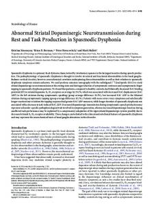
received: 26 November 2015 accepted: 02 March 2016 Published: 18 March 2016
Abnormal proplatelet formation and emperipolesis in cultured human megakaryocytes from gray platelet syndrome patients Christian A. Di Buduo1,2, Maria Adele Alberelli3, Ana C. Glembostky4, Gianmarco Podda5, Paola R. Lev4, Marco Cattaneo5, Raffaele Landolfi3, Paula G. Heller4, Alessandra Balduini1,2,6 & Erica De Candia3 The Gray Platelet Syndrome (GPS) is a rare inherited bleeding disorder characterized by deficiency of platelet α-granules, macrothrombocytopenia and marrow fibrosis. The autosomal recessive form of GPS is linked to loss of function mutations in NBEAL2, which is predicted to regulate granule trafficking in megakaryocytes, the platelet progenitors. We report the first analysis of cultured megakaryocytes from GPS patients with NBEAL2 mutations. Megakaryocytes cultured from peripheral blood or bone marrow hematopoietic progenitor cells from four patients were used to investigate megakaryopoiesis, megakaryocyte morphology and platelet formation. In vitro differentiation of megakaryocytes was normal, whereas we observed deficiency of megakaryocyte α-granule proteins and emperipolesis. Importantly, we first demonstrated that platelet formation by GPS megakaryocytes was severely affected, a defect which might be the major cause of thrombocytopenia in patients. These results demonstrate that cultured megakaryocytes from GPS patients provide a valuable model to understand the pathogenesis of GPS in humans. The gray platelet syndrome (GPS) is a rare inherited platelet disorder characterized by mild to moderate bleeding manifestations, thrombocytopenia, large platelets, increased serum B12 levels, spleen enlargement and progressive myelofibrosis1–3. The distinctive feature of the disease is the deficiency of platelet α -granules responsible for the typical gray appearance of platelets on Wright-stained blood smears1. Platelet α -granules store cargo proteins that mediate platelet adhesion (e.g. von Willebrand Factor), hemostasis (e.g. factor V), inflammation (e.g. IL-1β , IL-8, platelet factor 4) and wound healing and angiogenesis (e.g. VEGF, FGF-2, PDGF)4. Although the GPS displays an autosomal recessive inheritance in most cases3, sporadic families with autosomal dominant inheritance pattern have also been described5,6. Recently, by using a next generation sequencing approach, biallelic mutations in the neurobeachin-like 2 (NBEAL2) gene have been identified in autosomal recessive forms of GPS7–9. NBEAL2 belongs to a family of proteins involved in the membrane dynamics and intracellular vesicle trafficking. One such protein, LYST, is mutated in Chediak-Higashi syndrome, which is characterized by defects in platelet granules and other lysosome-related organelles. The findings indicate that NBEAL2 may be critical for the development of platelet α -granules. However, the mechanisms by which NBEAL2 loss of function contributes to deficiency of platelet α -granules and their cargo proteins and to the macrothrombocytopenic state remain unknown. Three different Nbeal2 deficient (−/−) mouse strains have been generated10–12. All of them recapitulate the typical platelet phenotype observed in GPS patients. Mice have macrothrombocytopenia, deficiency of 1
Department of Molecular Medicine, University of Pavia, Pavia, Italy. 2Biotechnology Research Laboratories, Istituto di Ricovero e Cura a Carattere Scientifico (IRCCS) Policlinico San Matteo Foundation, Pavia, Italy. 3Department of Internal Medicine, Policlinico Agostino Gemelli, Catholic University, Rome, Italy. 4Hematology Research, Instituto de Investigaciones Médicas Alfredo Lanari, University of Buenos Aires, CONICET, Buenos Aires, Argentina. 5Medicina III, Azienda Ospedaliera San Paolo, Dipartimento di Scienze della Salute, Università degli Studi di Milano, Milan, Italy. 6Department of Biomedical Engineering, Tufts University, Medford, MA, USA. Correspondence and requests for materials should be addressed to A.B. (email:
[email protected]) or E.D.C. (email: edecandia@ rm.unicatt.it) Scientific Reports | 6:23213 | DOI: 10.1038/srep23213
1
www.nature.com/scientificreports/ Patients
cDNA (RNA for spicing mutations)
Exon/Intron
Protein
Type of mutation
Transmission
12
p.His418Leufs*54
Frameshift
Maternal
25
p.Arg1195AGln
Missense
Paternal
c.5720+ 1G> A (r.5720_5721ins148)
i35
p.Met1908*
Splice-site Frameshift
Paternal
c.5572C> T
34
p.Arg1858*
Nonsense
Paternal
41
p.Glu2218*
Nonsense
Paternal
45
p.Arg2345Trp
Missense
Maternal
16
p.Tyr729*
Nonsense
Maternal and Paternal
c.1253del Patient 1 Rome (Italy)
Patient 2 Milan (Italy)
c.3584G> A
c.6652G> T
#1
#2
c.7033C> T Patients 3A and 3B Buenos Aires (Argentina)**
c.2187C> A
#3
Table 1. Mutations in NBEAL2 previously identified in three unrelated families and four probands. ** Family from Buenos Aires had two affected brothers with homozygous NBEAL2 mutation. Clinical and laboratory features of the probands have been detailed elsewhere14. platelet α -granules and spleen enlargement. Myelofibrosis was demonstrated in older animals12. Nbeal2−/− mice were used to extensively study the role of platelet α -granules constituents in hemostasis, thrombosis, thrombo-inflammatory disease states, megakaryocyte survival and development, platelet production, tissue reconstitution after injury, development of myelofibrosis and cancer metastasis propagation. However, some differences among the three Nbeal2−/− mouse strains were reported with regard to megakaryocyte development and differentiation, proplatelet formation and α -granules content. Therefore, whether Nbeal2 loss of function in mice affects megakaryopoiesis and/or proplatelet formation, and how it contributes to thrombocytopenia is unclear. To investigate the impact of mutations in NBEAL2 gene on human thrombopoiesis, we enrolled four GPS patients, whose clinical features and NBEAL2 mutations have been previously described13–15. We obtained in vitro differentiated megakaryocytes from peripheral blood or bone marrow hematopoietic progenitor cells of the four GPS patients and five controls and evaluated megakaryocyte maturation and function and proplatelet formation.
Results
NBEAL2 mutations do not affect megakaryocyte differentiation by human hematopoietic progenitors. We differentiated human megakaryocytes in vitro starting from peripheral blood or bone mar-
row hematopoietic progenitor cells of GPS patients with mutated NBEAL2 and healthy controls. Hereinafter, the patient carrying the c.1253del, c.3584G> A and c.5720+ 1G> A (r.5720_5721ins148) mutations will be identified as #1; the patient carrying the c.5572C> T, c.6652G> T and c.7033C> T mutations will be identified as #2; the two patients carrying the c.2187C> A homozygous mutations will be identified as #3 (Table 1). After 14 days of culture, in vitro megakaryocyte differentiation and output of CD61+CD42b+ megakaryocytes were similar to those of healthy control samples (Fig. 1a,b). Further, maturation stages of CD61+ megakaryocytes, classified I to IV according to standard morphological criteria16, were also similar in GPS patients and controls (Fig. 1c). No differences were observed among patients #1, #2 and #3, thus indicating that all the analyzed NBEAL2 mutations did not affect either the differentiation or the maturation of megakaryocytes. The results from both peripheral blood- and bone marrow-derived megakaryocytes were perfectively comparable (not shown).
Reduced α-granule content in human megakaryocytes from GPS patients. Despite normal
differentiation, three of the most abundant proteins normally contained in α -granules, von Willebrand factor, thrombospondin and P-selectin, were markedly reduced in in vitro differentiated human megakaryocytes from both #1 and #2 GPS patients compared to controls (Fig. 2a–c). No differences were observed between patients, thus suggesting that the storage of these proteins was compromised in GPS megakaryocytes regardless of the type of mutation. Consistently, #1 and #2 GPS-derived megakaryocytes stimulated with protease activated receptors (PARs)-activating peptides (APs) exposed less P-selectin compared to control megakaryocytes (Fig. 2d). Interestingly, these results are in agreement with those we have recently observed in platelets from peripheral blood of patients #1 and #215, and demonstrate that defective P-selectin mobilization is a distinctive feature that gray platelets inherit directly from their progenitors megakaryocytes before their release into bloodstream.
Evidence of emperipolesis in human megakaryocytes from GPS patients. Bone marrow mega-
karyocytes from gray platelets syndrome patients display intact cells within their cytoplasm, as a consequence of a rare biological process that is named emperipolesis6,15,17. Emperipolesis, present in less than 2% of megakaryocytes in normal bone marrow biopsies, has been shown in 38% to 65% of megakaryocytes in GPS bone marrow biopsies15. However, whether this abnormality is directly dependent on NBEAL2 mutation rather than a consequence of compromised bone marrow homeostasis, associated with the progressive myelofibrosis, has never been investigated. We found that several megakaryocytes from #1 and #2 GPS patients displayed the presence of intact cells within their cytoplasm, likely as a consequence of emperipolesis, while this abnormality was not observed in any of the mature megakaryocytes from control subjects (Fig. 3). The presence of cells other than megakaryocytes in our in vitro culture conditions is not surprising given the presence of a mixed population of cells in the culture media before megakaryocyte population is enriched through an albumin gradient18–21, which could grow and be engulfed within the megakaryocyte cytoplasm during the course of differentiation. To the best of our knowledge, none of the previously published studies of megakaryocytes from patients with other forms of thrombocytopenia or healthy subjects, grown under similar experimental conditions, revealed the presence of intact cells in cultured megakaryocytes20,22–24. Therefore, our study provides the first evidence that emperipolesis occurs in human GPS Scientific Reports | 6:23213 | DOI: 10.1038/srep23213
2
www.nature.com/scientificreports/
Figure 1. Normal differentiation of human megakaryocytes from patients with GPS. Hematopoietic progenitors from peripheral blood of healthy controls (CTRL) and patients with GPS were differentiated in culture into megakaryocytes in presence of TPO. (a) Representative immunofluorescence staining of plasma membrane CD61 in CTRL- (i) and GPS-derived megakaryocytes (ii–iv) (green = CD61; blue = nuclei; scale bar = 25 μm). (b) Statistical analysis of megakaryocyte (MK) output of GPS patients (#1, #2 and #3) relative to CTRL samples. Data are presented as mean ± SD (p = NS). (c) Statistical analysis of maturation stages in controls (CTRL)- and gray platelet syndrome patients- derived megakaryocytes (MK). Maturation stages were identified according to standard morphological criteria, as specified in the ‘Materials and Methods’ section. Data are presented as means ± SD of CTRL and all GPS patients GPS (p = NS). No differences were observed among patients.
megakaryocytes as a consequence of an intrinsic cellular defect, rather than of abnormalities of the bone marrow microenvironment.
Calcium signaling is maintained in human megakaryocytes with mutated NBEAL2 gene. We have recently demonstrated that constitutively released adenosine diphosphate (ADP) by human mature megakaryocytes promotes the activation of Store-Operated Calcium Entry (SOCE) which in turn is responsible for the regulation of platelet formation and interaction with the components of the extracellular matrix19,25. We explored SOCE functionality in GPS megakaryocytes from #1 and #2 patients by depleting the stores with cyclopiazonic acid (CPA, 10 μM). The treatment evoked a transient rise in intracellular calcium (Ca2+) concentration, because of passive emptying of endoplasmic reticulum, followed by influx of external Ca2+ through activated SOC channels (Fig. 4a). Analysis of the extent of both Ca2+ release and entry demonstrated no significant difference between control- and GPS-derived megakaryocytes (Fig. 4b). Consistent with this data, megakaryocytes from #1 and #2 GPS patients normally responded to ADP stimulation, with no significant differences in the extent of Ca2+ mobilization compared to control megakaryocytes (Fig. 4c,d). All together, these data indicate that Ca2+ homeostasis is normally maintained in GPS megakaryocytes. Human megakaryocytes from GPS patients show dramatically impaired proplatelet formation.
An efficient Ca2+ signaling is fundamental in order to guarantee proper megakaryocyte interaction with extracellular matrix components of the bone marrow environment25, which is in turn relevant for the control of p latelet release26,27. Therefore, based on the demonstration of a normal Ca2+ homeostasis in GPS m egakaryocytes, we expected that they could normally interact with extracellular matrix components. Surprisingly, the analysis of GPS megakaryocyte interaction with type I collagen, an extracellular matrix component that promotes megakaryocyte spreading by supporting cytoskeleton contractility27, revealed abnormal actin stress fibers formation and microtubule assembly and, consequently, defective megakaryocyte spreading (Fig. 5). Similar results were observed in samples from #1 and #2 patients (Fig. 5v–xii). Further, the analysis of proplatelet formation on fibronectin28,29, revealed that megakaryocytes derived from either #1, #2 or #3 GPS patients displayed shorter proplatelet branches compared to control megakaryocytes (Fig. 6a). Similar results were obtained from both peripheral blood- (Fig. 6ai–vi,viii–xiii) and bone marrow-derived megakaryocytes (Fig. 6avii,xiv). The percentage of proplatelet forming megakaryocytes was markedly reduced in diseased megakaryocytes compared to controls, while no significant differences were observed among #1, #2 and #3 samples (Fig. 6b). The tips, terminal ends of proplatelet branches, are the site of the assembly of nascent platelets30. Consequently, the number of platelets that are formed depends on both the percentage of proplatelets and the number of their bifurcations in order to ensure the highest number of tips per single megakaryocyte. Proplatelets from GPS megakaryocytes displayed Scientific Reports | 6:23213 | DOI: 10.1038/srep23213
3
www.nature.com/scientificreports/
Figure 2. Lack of α-granule proteins in GPS cultured megakaryocytes. Fully differentiated megakaryocytes from healthy controls (CTRL) and patients with GPS were harvested and allowed to adhere onto Poly-L-Lysine coated cover-slips by centrifugation, fixed and analyzed. (a) Representative immunofluorescence staining of von Willebrand Factor (vWF) in CTRL- (i–ii) and GPS-derived megakaryocytes (iii–iv) (red = vWF; blue = nuclei; scale bar = 20 μm). Bars report the analysis of fluorescence intensity by Image J software (a.u. = arbitrary units). Data are presented as mean± SD (*p