... Magn Reson Med, in press (2007). [7] D.O. Walsh et al.; Magn Reson Med, V.43, pp.682-690 (2000). Figure 1. Proc. Intl. Soc. Mag. Reson. Med. 16 (2008).
Accelerated Dynamic Imaging by Reconstructing Sparse Differences using Compressed Sensing A. Fischer1,2, F. Breuer2, M. Blaimer2, N. Seiberlich1, and P. M. Jakob1,2 Experimental Physics 5, University of Wuerzburg, Wuerzburg, Bavaria, Germany, 2Research Center for Magnetic Resonance Bavaria e.V., Wuerzburg, Bavaria, Germany
1
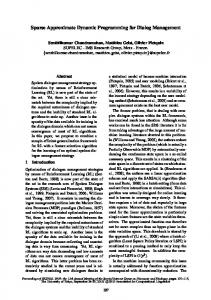
Introduction Dynamic imaging with high spatial and temporal resolution is a demanding task in clinical MR tomography. In case of undersampling in dynamic imaging, radial trajectories are advantageous due to their incoherent artifact behavior. Compressed Sensing (CS) [1,2] is a new technique for reconstructing accelerated datasets without utilizing parallel imaging methods. First applications of CS in the field of MR have been demonstrated [3,4]. CS reconstructs missing data points by optimizing a mathematical functional and depends on a sparse representation (in any basis) of the desired signal. Materials and methods The proposed method aims to recover dynamic information by reconstructing the differences between a composite image of the dynamic dataset and the desired timeframe as suggested in previous work [5]. These differences are sparse in the image domain; therefore, a reconstruction using CS can be utilized. A radial cine dataset of a beating human heart with high spatial and temporal resolution was acquired with a 32channel coil-array (Rapid Biomedical, Rimpar, Germany). Our experiment was performed on a 1.5 T Avanto (Siemens Medical Solutions, Erlangen, Germany) clinical scanner. The dataset contained 21 timeframes, 192 readout points and 224 projections per timeframe, imaging parameters: 2D radial Turbo-FLASH, α = 78°, TR = 47.36 ms, TE = 1.48 ms, FOV 300x300 mm². The data was processed in MATLAB (The MathWorks, Natick, USA) as shown in Fig. 1. Our implementation of CS is based on that from Lustig [3]. Figure 1 Results The results can be seen in Fig. 2. Reconstructions utilizing only 32 projections (15 % of k-space after gridding) show good agreement with the full image (224 projections). Even with only 14 projections (6.7 % of k-space after gridding), the result with CS exhibits only a moderate decline in image quality compared to 32 projections. Discussion The proposed technique achieves high acceleration factors due to the sparse distribution of signal intensities in the difference images and the incoherent artifact nature in undersampled radial images. The CS algorithm employed in this work takes advantage of image denoising techniques. As long as the signal intensities in the difference images are above the noise level, the suggested method is expected to deliver good reconstructions. If the intensities are close to the noise level, signal intensities and noise variances will be equally smoothed. Nonetheless, low differences mean that the desired timeframe is well represented by the composite image; thus, reconstruction errors will not lead to significant artefacts in the resulting timeframe. Although the method suffers from long parameter determination times, it highlights a promising perspective for future real-time dynamic applications. Acknowledgements The authors would like to thank Herbert Köstler and Marcel Gutberlet (Institut für Röntgendiagnostik, University of Würzburg). Figure 2: Reconstruction results. Note the good agreement with the original image and the distinct difference to the composite image References [1] E. J. Candès et al.; IEEE Trans. Inform. Theory, V.52, pp.489-509(2004) [2] D. Donoho; IEEE Trans. Inform. Theory, V.52, pp.1289-1306(2004) [3] M. Lustig et al.: Sparse MRI; Magn Reson Med, in press (2007) [4] K.T. Block et al.; Magn Reson Med, V.57, pp.1086-1098 (2007)
Proc. Intl. Soc. Mag. Reson. Med. 16 (2008)
[5] M. Blaimer et al.; Abstract 749, Proc ISMRM 2007 [6] N. Seiberlich et al.: Non-Cartesian data reconstruction using GRAPPA operator gridding (GROG); Magn Reson Med, in press (2007) [7] D.O. Walsh et al.; Magn Reson Med, V.43, pp.682-690 (2000)
341